



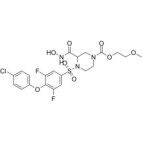
XL-784 free base
Based on 2 publication(s) in Google Scholar
XL-784 free base is a selective matrix metalloproteinases (MMP) inhibitor, with IC50s of ~1900, 0.81, 120, 10.8, 18, 0.56 nM for MMP-1, MMP-2, MMP-3, MMP-8, MMP-9 and MMP-13, respectively.
Nos produits utilisent uniquement pour la recherche. Nous ne vendons pas aux patients.
- CAS No.: 1356992-21-6
- Formule: C21H22ClF2N3O8S
- Masse moléculaire:549.93
-
Stockage:
Please store the product under the recommended conditions in the Certificate of Analysis.

To place orders, for customer services and technical support, please contact: MedChemExpress USA
Tel: 609-228-6898 E-mail: [email protected] [email protected]


-
 Activité biologique
-
Chemical Information
-
Solvant et solubilité
- Protocole
- Pureté et documentation
- Références
-
 Help & FAQs


Publications Citing Use of MedChemExpress (MCE) XL-784 free base
More

Activité biologique
Description
IC50 & Target
|
MMP-2 0.81 nM (IC50) |
MMP-13 0.56 nM (IC50) |
MMP-8 10.8 nM (IC50) |
MMP-9 18 nM (IC50) |
MMP-3 120 nM (IC50) |
MMP-1 1900 nM (IC50) |
In Vitro
XL-784 is a highly potent, low-molecular-weight (1,122 g/mol) inhibitor of MMPs that has very limited aqueous solubility (20 μg/mL). XL-784 potently inhibits MMP-2, MMP-13, and ADAM10 [TNF-α-converting enzyme (TACE)] activity in vitro, with IC50 values in the range of 1-2 nM. XL-784 also inhibits MMP-9 (IC50 ~20 nM) activity and ADAM17 (IC50 ~70 nM) also known as TACE. However, it exhibits low potency for inhibition of MMP-1 (IC50 ~2,000 nM)[1].
MedChemExpress (MCE) has not independently confirmed the accuracy of these methods. They are for reference only.
In Vivo
MedChemExpress (MCE) has not independently confirmed the accuracy of these methods. They are for reference only.
Essai clinique
| NCT Number | Sponsor | Condition | Start Date |
Phase

|
|---|---|---|---|---|
| NCT01329991 | 2011-05 | PHASE1 |


Chemical Information
-
CAS No. 1356992-21-6
-
Masse moléculaire 549.93
-
Formule C21H22ClF2N3O8S
-
SMILES
O=C(N1CC(C(NO)=O)N(S(=O)(C2=CC(F)=C(OC3=CC=C(Cl)C=C3)C(F)=C2)=O)CC1)OCCOC
-
Livraison
Room temperature in continental US; may vary elsewhere.
-
Stockage
Please store the product under the recommended conditions in the Certificate of Analysis.


Publications (2)
-
Journal Impact Factor
-
Most Recent


Solvant et solubilité
In Vitro:
DMSO : 250 mg/mL (454.60 mM; Need ultrasonic; Hygroscopic DMSO has a significant impact on the solubility of product, please use newly opened DMSO)
Concentration (start) × Volume (start) = Concentration (final) × Volume (final)
In Vivo:
Select the appropriate dissolution method based on your experimental animal and administration route.
- For the following dissolution methods, please ensure to first prepare a clear stock solution using an In Vitro approach and then sequentially add co-solvents:
- To ensure reliable experimental results, the clarified stock solution can be appropriately stored based on storage conditions. As for the working solution for In Vivo experiments, it is recommended to prepare freshly and use it on the same day.
- The percentages shown for the solvents indicate their volumetric ratio in the final prepared solution. If precipitation or phase separation occurs during preparation, heat and/or sonication can be used to aid dissolution.
Add each solvent one by one: 10% DMSO 40% PEG300 5% Tween-80 45% Saline
Solubility: ≥ 2.08 mg/mL (3.78 mM); Clear solution
This protocol yields a clear solution of ≥ 2.08 mg/mL (saturation unknown).
Taking 1 mL working solution as an example, add 100 μL DMSO stock solution (20.8 mg/mL) to 400 μL PEG300, and mix evenly; then add 50 μL Tween-80 and mix evenly; then add 450 μL Saline to adjust the volume to 1 mL.
Preparation of Saline: Dissolve 0.9 g sodium chloride in ddH₂O and dilute to 100 mL to obtain a clear Saline solution.
Add each solvent one by one: 10% DMSO 90% (20% SBE-β-CD in Saline)
Solubility: ≥ 2.08 mg/mL (3.78 mM); Clear solution
This protocol yields a clear solution of ≥ 2.08 mg/mL (saturation unknown).
Taking 1 mL working solution as an example, add 100 μL DMSO stock solution (20.8 mg/mL) to 900 μL 20% SBE-β-CD in Saline, and mix evenly.
Preparation of 20% SBE-β-CD in Saline (4°C, storage for one week): 2 g SBE-β-CD powder is dissolved in 10 mL Saline, completely dissolve until clear.
In Vivo Dissolution Calculator
Please enter the basic information of animal experiments:
-
-
-
-
Recommended: Prepare an additional quantity of animals to account for potential losses during experiments.
Please enter your animal formula composition:
-
%DMSO +
Recommended: Keep the proportion of DMSO in working solution below 2% if your animal is weak.
-
%+
-
+%Tween-80 + +
-
%Saline +
The co-solvents required include: DMSO, . All of co-solvents are available by MedChemExpress (MCE). , Tween 80. All of co-solvents are available by MedChemExpress (MCE).
Working solution concentration: 0.22 mg/mL
Method for preparing stock solution: mg drug dissolved in μL DMSO. Stock solution concentration: mg/mL.
1. Take μL DMSO stock solution;
2. Add μL .
μL , mix evenly;
3. Then add μL Tween 80, mix evenly;
4. Then add μL
Please ensure that the stock solution in the first step is dissolved to a clear state, and add co-solvents in sequence. You can use ultrasonic heating (ultrasonic cleaner, recommended frequency 20-40 kHz), vortexing, etc. to assist dissolution.


Pureté et documentation
Références
[1]. Williams JM, et al. Evaluation of metalloprotease inhibitors on hypertension and diabetic nephropathy. Am J Physiol Renal Physiol. 2011 Apr;300(4):F983-98. [Content Brief]
[2]. Ennis T, et al. Effect of novel limited-spectrum MMP inhibitor XL784 in abdominal aortic aneurysms. J Cardiovasc Pharmacol Ther. 2012 Dec;17(4):417-26. [Content Brief]


Complete Stock Solution Preparation Table
| Optional Solvent | Concentration Solvent Mass | 1 mg | 5 mg | 10 mg | 25 mg |
|---|---|---|---|---|---|
| DMSO | 1 mM | 1.8184 mL | 9.0921 mL | 18.1841 mL | 45.4603 mL |
| 5 mM | 0.3637 mL | 1.8184 mL | 3.6368 mL | 9.0921 mL | |
| 10 mM | 0.1818 mL | 0.9092 mL | 1.8184 mL | 4.5460 mL | |
| 15 mM | 0.1212 mL | 0.6061 mL | 1.2123 mL | 3.0307 mL | |
| 20 mM | 0.0909 mL | 0.4546 mL | 0.9092 mL | 2.2730 mL | |
| 25 mM | 0.0727 mL | 0.3637 mL | 0.7274 mL | 1.8184 mL | |
| 30 mM | 0.0606 mL | 0.3031 mL | 0.6061 mL | 1.5153 mL | |
| 40 mM | 0.0455 mL | 0.2273 mL | 0.4546 mL | 1.1365 mL | |
| 50 mM | 0.0364 mL | 0.1818 mL | 0.3637 mL | 0.9092 mL | |
| 60 mM | 0.0303 mL | 0.1515 mL | 0.3031 mL | 0.7577 mL | |
| 80 mM | 0.0227 mL | 0.1137 mL | 0.2273 mL | 0.5683 mL | |
| 100 mM | 0.0182 mL | 0.0909 mL | 0.1818 mL | 0.4546 mL |


XL-784 free base Related Classifications
HY-112160 Related Classifications
 Powered by Bioz