



Marimastat
Based on 22 publication(s) in Google Scholar
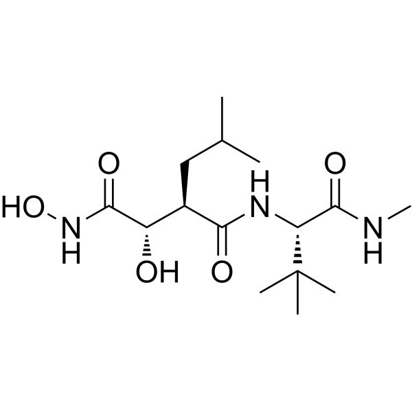
Marimastat (BB2516) is a broad spectrum and orally bioavailable inhibitor of MMPs, with potent activity against MMP-9 (IC50=3 nM), MMP-1 (IC50=5 nM), MMP-2 (IC50=6 nM), MMP-14 (IC50=9 nM) and MMP-7 (IC50=13 nM), used in the treatment of cancer. Marimastat (BB2516) is an angiogenesis and metastasis inhibitor, which limits the growth and production of blood vessels. As an antimetatstatic agent it prevents malignant cells from breaching the basement membranes.
商品は「研究用試薬」です。人や動物の医療用・臨床診断用・食品用の製品ではありません。
研究用途以外に使用した場合、当社は一切の責任を負いかねます。
- 純度 : 99.90%
- CAS 番号: 154039-60-8
- 分子式: C15H29N3O5
- 分子量:331.41
-
保管条件:Powder -20°C, 3 years , 4°C, 2 years ; In solvent -80°C, 6 months , -20°C, 1 month

To place orders, for customer services and technical support, please contact: MedChemExpress USA
Tel: 609-228-6898 E-mail: [email protected] [email protected]


-
 生物活性
生物活性
-
化学情報
-
溶剤 & 溶解度
- プロトコル
- 純度とドキュメンテーション
- 参考文献
-
 Help & FAQs


-
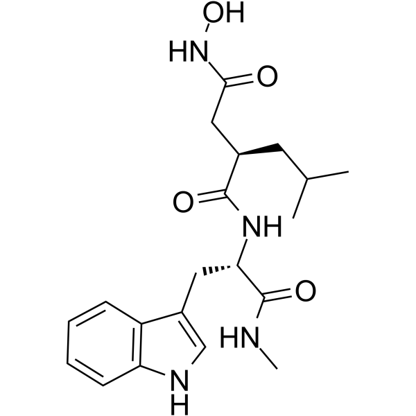
HY-15768
IlomastatMMP Inhibitor -
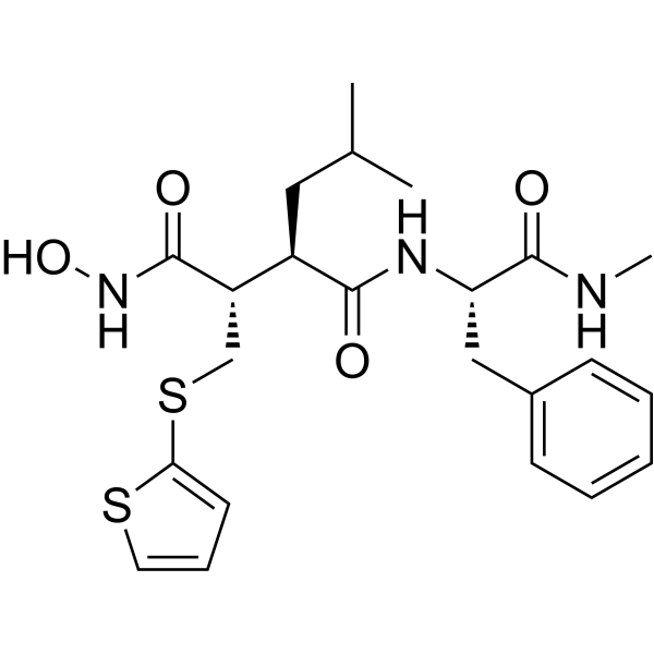
HY-13564
BatimastatMMP Inhibitor -
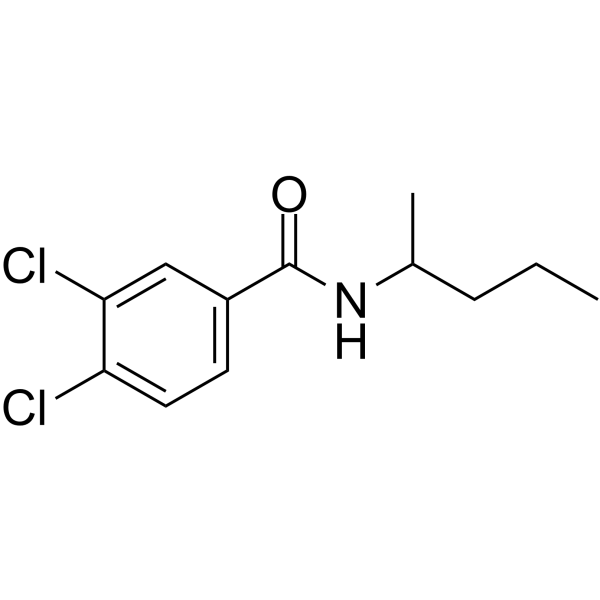
HY-15827
NSC 405020MMP14 Inhibitor -
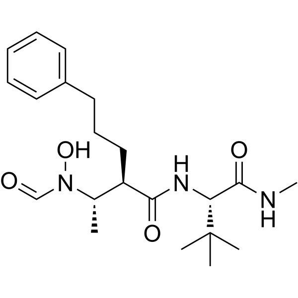
HY-19956
GI254023XADAM10/MMP9 Inhibitor -
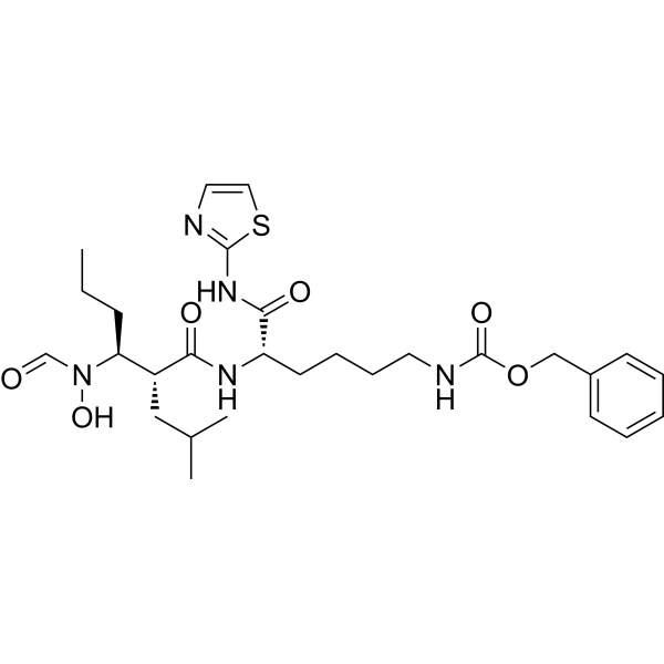
HY-115670
GW280264XADAM10/17 Inhibitor -
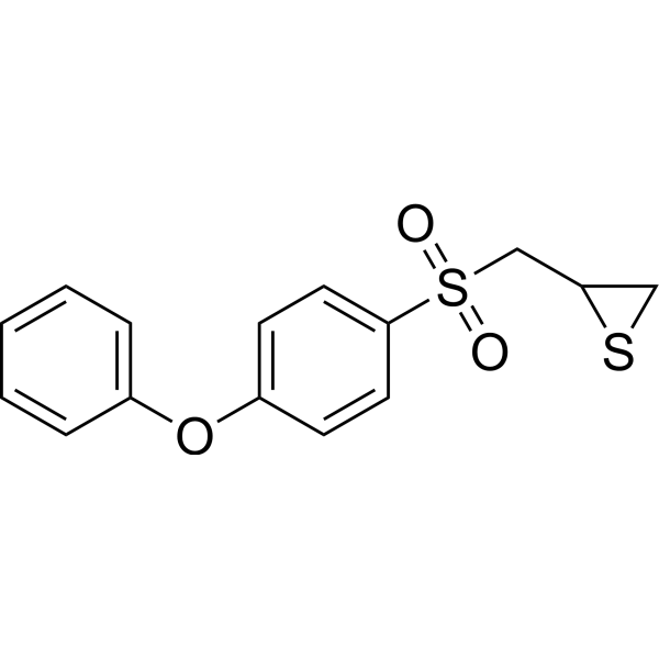
HY-12354
SB-3CTMMP2/9 Inhibitor -
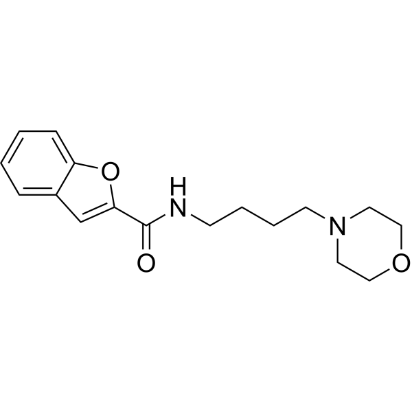
HY-100359
CL-82198MMP-13 Inhibitor -
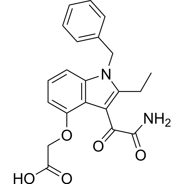
HY-13402
VarespladibsPLA2 Inhibitor -
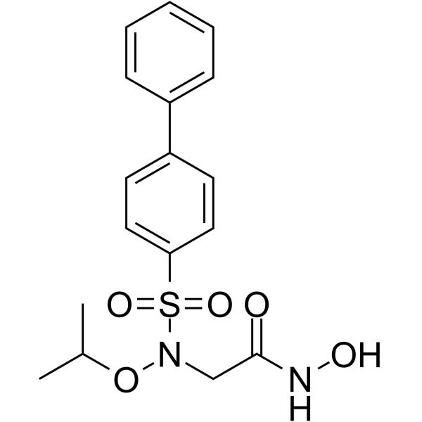
HY-103444
ARP-100MMP-2 Inhibitor -
HY-12170
PrinomastatMMP Inhibitor -
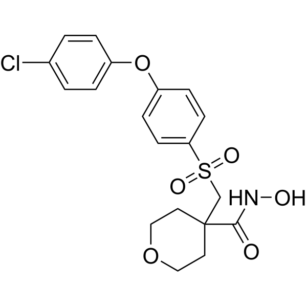
HY-10398
CTS-1027MMP Inhibitor -

HY-10219
RapamycinmTOR Inhibitor
MedChemExpress(MCE)の使用を引用している文献 Marimastat
More- Cell Res. 2020 Sep;30(9):779-793. [Abstract]
- Adv Mater. 2025 Sep 22:e12554. [Abstract]
- Sci Adv. 2025 Mar 14;11(11):eadu7614. [Abstract]
- Mol Ther. 2016 Dec;24(12):2090-2099.
- Cell Prolif. 2025 Dec;58(12):e70060. [Abstract]
- EMBO Rep. 2021 Jul 5;22(7):e51678. [Abstract]
- Int J Mol Sci. 2026 Jun 4;27(11):5104. [Abstract]
- J Cell Mol Med. 2026 Apr;30(7):e71101. [Abstract]
- Rheumatology (Oxford). 2025 Aug 13:keaf437. [Abstract]
- Bioconjug Chem. 2016 Dec 21;27(12):2943-2953. [Abstract]
- Biomed Mater. 2026 Jun 26. [Abstract]
- Biochim Biophys Acta Mol Cell Biol Lipids. 2025 Jun;1870(5):159618. [Abstract]
- Viruses. 2022 Sep 21;14(10):2094. [Abstract]
- Endocrine. 2021 May;72(2):529-538. [Abstract]
- Biol Reprod. 2026 Jul 9:ioag141.
- PLoS One. 2024 Dec 27;19(12):e0315245. [Abstract]
- Eur Biophys J. 2021 May;50(3-4):345-352. [Abstract]
- bioRxiv. 2025 Jul 2:2025.07.01.662195. [Abstract]
- KTH Royal Institute of Technology. 2025.
- SSRN. 2020 Oct 5.
- ACS Comb Sci. 2019 Dec 9;21(12):805-816. [Abstract]
- Chinese Journal of Biochemical and Pharmaceutics. 2017,37(12):1-04.
 Customer Validation & Images

-
Bio/Physico-chemical Assay
-
Bio/Physico-chemical Assay
-
Histological Imaging/Staining
-
WB
-
Bio/Physico-chemical Assay


生物活性
製品説明
IC50 & Target
[1]|
MMP-3 3 nM (IC50) |
MMP-1 5 nM (IC50) |
MMP-2 6 nM (IC50) |
MMP-14 9 nM (IC50) |
MMP-7 13 nM (IC50) |
Cellular Effect
|
Cell Line

|
Type | Value | Description | References |
|---|---|---|---|---|
| THP-1 | IC50 |
2100 nM
Compound: 1Marimastat
|
Selective inhibition of the cellular TNF alpha release from LPS-stimulated THP-1 cells
Selective inhibition of the cellular TNF alpha release from LPS-stimulated THP-1 cells
|
[PMID: 11754593] |
体外実験
Marimastat (BB2516) (1 μM) shows inhibition of vascular outgrowth, and selectively affects angiogenesis[3].
MedChemExpress (MCE) has not independently confirmed the accuracy of these methods. They are for reference only.
体内実験
MedChemExpress (MCE) has not independently confirmed the accuracy of these methods. They are for reference only.
- Mitochondrial membrane-potential fluorescent assay
- Human pluripotent stem cell endothelial-cell differentiation
- Vascular/Branching Fractal Analysis
- Mitochondrial membrane-potential and mitochondrial mass staining
- Endothelial Tube Formation Assay
- Endothelial Cell Migration/Angiogenic Sprouting Assay
- Orthotopic Cell-Line Xenograft
臨床実験
| NCT Number | Sponsor | Condition | Start Date |
Phase
|
|---|---|---|---|---|
| NCT01329991 | 2011-05 | PHASE1 |


化学情報
-
CAS 番号 154039-60-8
-
性状 Solid
-
分子量 331.41
-
分子式 C15H29N3O5
-
Color White to gray
-
SMILES
O=C(NO)[C@@H](O)[C@@H](CC(C)C)C(N[C@H](C(NC)=O)C(C)(C)C)=O
-
別名
BB2516; マリマスタット; TA2516
-
輸送条件
Room temperature in continental US; may vary elsewhere.
-
保管条件
Powder -20°C 3 years 4°C 2 years In solvent -80°C 6 months -20°C 1 month


Publications (22)
-
Journal Impact Factor
-
Most Recent
-
Cell Res
s-HBEGF/SIRT1 circuit-dictated crosstalk between vascular endothelial cells and keratinocytes mediates sorafenib-induced hand-foot skin reaction that can be reversed by nicotinamide. [Abstract]2020 Sep;30(9):779-793. PMID: 32296111
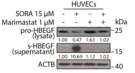
Marimastat purchased from MedChemExpress. Usage Cited in: Cell Res. 2020 Sep;30(9):779-793. [Abstract]
HUVECs were treated with or without sorafenib or marimastat (1 μM) for 24 h. The level of s-HBEGF in the supernatant or pro-HBEGF in total cell lysates was detected by western blot.
-
Adv Mater
Spatiotemporally Engineered Protein Delivery via Integrated Covalent Coupling and Pathological Triggers for Precision Microenvironment Reprogramming in Tissue Injury Repair. [Abstract]2025 Sep 22:e12554. PMID: 40981608 -
Sci Adv
An enzymatic cleavage-triggered minimally invasive nanosensor for urine-based detection of early atherosclerosis. [Abstract]2025 Mar 14;11(11):eadu7614. PMID: 40085714
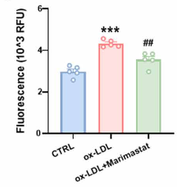
Marimastat purchased from MedChemExpress. Usage Cited in: Sci Adv. 2025 Mar 14;11(11):eadu7614. [Abstract]
Fluorescence analysis of supernatant from foam cells treated with lipopolysaccharide and oxidized low-density lipoprotein, with or without Marimastat (10 μM), followed by incubation with the substrate peptide.
-
-
Cell Prolif
LL-37 Inhibits TMPRSS2-Mediated S2' Site Cleavage and SARS-CoV-2 Infection but Not Omicron Variants. [Abstract]2025 Dec;58(12):e70060. PMID: 40375579
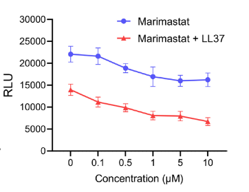
Marimastat purchased from MedChemExpress. Usage Cited in: Cell Prolif. 2025 Dec;58(12):e70060. [Abstract]
293T‐A2 cells were pretreated for 2 h with indicated concentrations of marimastat (0-10 μM), and then SARS‐CoV‐2 spike pseudoparticles pretreated for 2 h with 10 μg/mL of LL‐37 were added to cells. Infection rates were assessed at 2 days post infection by measuring the RLU of luciferase activity.
-
EMBO Rep
Sensing of mycobacterial arabinogalactan by galectin-9 exacerbates mycobacterial infection. [Abstract]2021 Jul 5;22(7):e51678. PMID: 33987949
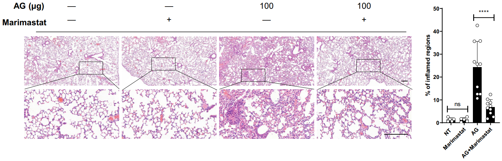
Marimastat purchased from MedChemExpress. Usage Cited in: EMBO Rep. 2021 Jul 5;22(7):e51678. [Abstract]
C57BL/6 mice were left untreated (NT) or were intraperitoneally treated with indicated amounts of AG for 3 days in the absence or presence of the MMP inhibitor marimastat (10 mg/kg) given intraperitoneally prior to AG stimulation. Lung sections stained with H&E and quantification of lung lesion burden from H&E‐stained sections .
-
Int J Mol Sci
Epithelial PCSK6 Promotes Proliferation and Decreases Collagen Deposition by Fibroblasts Potentially via MMP Activation. [Abstract]2026 Jun 4;27(11):5104. PMID: 42278627 -
J Cell Mol Med
2026 Apr;30(7):e71101. PMID: 41896195 -
Rheumatology (Oxford)
Autophagy inhibitors block pathogenic NET release in immune-mediated inflammatory disease without impairing host defence. [Abstract]2025 Aug 13:keaf437. PMID: 40802538 -
Bioconjug Chem
Matrix Metalloproteinase Responsive Nanoparticles for Synergistic Treatment of Colorectal Cancer via Simultaneous Anti-Angiogenesis and Chemotherapy. [Abstract]2016 Dec 21;27(12):2943-2953. PMID: 27998073
Marimastat purchased from MedChemExpress. Usage Cited in: Bioconjug Chem. 2016 Dec 21;27(12):2943-2953. [Abstract]
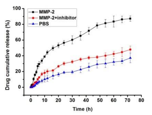
Accumulative release of CPT from nanoparticles in different media: PBS buffer with MMP-2, PBS buffer, PBS buffer with MMP-2 and MMP-2 inhibitor Marimastat (1 mM).
Marimastat purchased from MedChemExpress. Usage Cited in: Bioconjug Chem. 2016 Dec 21;27(12):2943-2953. [Abstract]
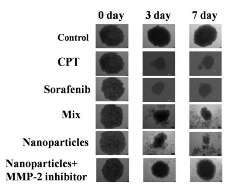
Marimastat (1 μM). Representative photos of tumor spheroids after treatment with various formulations and blank controls, the scale bar is 100 μm.
Marimastat purchased from MedChemExpress. Usage Cited in: Bioconjug Chem. 2016 Dec 21;27(12):2943-2953. [Abstract]
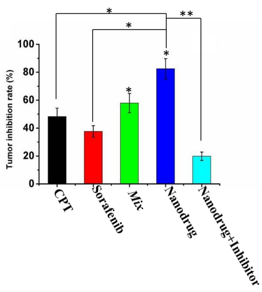
Marimastat (1 μM). The ratio of volume change of HT-29 spheres volume (%) after applying different formulations. Data are represented as average ± standard error (n = 3).
-
Biomed Mater
3-Layer lung cancer invasion model for evaluating MMP-targeted anti-metastatic therapeutics. [Abstract]2026 Jun 26. PMID: 42361847 -
Biochim Biophys Acta Mol Cell Biol Lipids
2025 Jun;1870(5):159618. PMID: 40294697 -
Viruses
Metalloprotease-Dependent S2'-Activation Promotes Cell-Cell Fusion and Syncytiation of SARS-CoV-2. [Abstract]2022 Sep 21;14(10):2094. PMID: 36298651 -
Endocrine
125I-Angiotensin 1-7 binds to a different site than angiotensin 1-7 in tissue membrane preparations. [Abstract]2021 May;72(2):529-538. PMID: 33415576 -
-
PLoS One
Discovery of novel TACE inhibitors using graph convolutional network, molecular docking, molecular dynamics simulation, and Biological evaluation. [Abstract]2024 Dec 27;19(12):e0315245. PMID: 39729480 -
Eur Biophys J
2021 May;50(3-4):345-352. PMID: 33864100 -
bioRxiv
Multimodal profiling of proinflammatory protease activity identifies caspase-1 as a target for lung cancer interception. [Abstract]2025 Jul 2:2025.07.01.662195. PMID: 40631193 -
-
-
ACS Comb Sci
Benzimidazolyl-pyrazolo[3,4- b]pyridinones, Selective Inhibitors of MOLT-4 Leukemia Cell Growth and Sea Urchin Embryo Spiculogenesis: Target Quest. [Abstract]2019 Dec 9;21(12):805-816. PMID: 31689077 -


溶剤 & 溶解度
体外:
DMSO : 100 mg/mL (301.74 mM; Need ultrasonic; Hygroscopic DMSO has a significant impact on the solubility of product, please use newly opened DMSO)
Please refer to the solubility information to select the appropriate solvent. Once prepared, please aliquot and store the solution to prevent product inactivation from repeated freeze-thaw cycles.
Storage method and period of stock solution: -80°C, 6 months; -20°C, 1 month. When stored at -80°C, please use it within 6 months. When stored at -20°C, please use it within 1 month.
Please refer to the solubility information to select the appropriate solvent. Once prepared, please aliquot and store the solution to prevent product inactivation from repeated freeze-thaw cycles.
Storage method and period of stock solution: -80°C, 6 months; -20°C, 1 month. When stored at -80°C, please use it within 6 months. When stored at -20°C, please use it within 1 month.
濃度 (開始) × 体積 (開始) = 濃度 (終了) × 体積 (終了)
体内:
Select the appropriate dissolution method based on your experimental animal and administration route.
- For the following dissolution methods, please ensure to first prepare a clear stock solution using an In Vitro approach and then sequentially add co-solvents:
- To ensure reliable experimental results, the clarified stock solution can be appropriately stored based on storage conditions. As for the working solution for In Vivo experiments, it is recommended to prepare freshly and use it on the same day.
- The percentages shown for the solvents indicate their volumetric ratio in the final prepared solution. If precipitation or phase separation occurs during preparation, heat and/or sonication can be used to aid dissolution.
Add each solvent one by one: 10% DMSO 40% PEG300 5% Tween-80 45% Saline
Solubility: ≥ 2.5 mg/mL (7.54 mM); Clear solution
This protocol yields a clear solution of ≥ 2.5 mg/mL (saturation unknown).
Taking 1 mL working solution as an example, add 100 μL DMSO stock solution (25.0 mg/mL) to 400 μL PEG300, and mix evenly; then add 50 μL Tween-80 and mix evenly; then add 450 μL Saline to adjust the volume to 1 mL.
Preparation of Saline: Dissolve 0.9 g sodium chloride in ddH₂O and dilute to 100 mL to obtain a clear Saline solution.
Add each solvent one by one: 10% DMSO 90% (20% SBE-β-CD in Saline)
Solubility: ≥ 2.5 mg/mL (7.54 mM); Clear solution
This protocol yields a clear solution of ≥ 2.5 mg/mL (saturation unknown).
Taking 1 mL working solution as an example, add 100 μL DMSO stock solution (25.0 mg/mL) to 900 μL 20% SBE-β-CD in Saline, and mix evenly.
Preparation of 20% SBE-β-CD in Saline (4°C, storage for one week): 2 g SBE-β-CD powder is dissolved in 10 mL Saline, completely dissolve until clear.
In Vivo Dissolution Calculator
Please enter the basic information of animal experiments:
-
-
-
-
Recommended: Prepare an additional quantity of animals to account for potential losses during experiments.
Please enter your animal formula composition:
-
%DMSO +
Recommended: Keep the proportion of DMSO in working solution below 2% if your animal is weak.
-
%+
-
+%Tween-80 + +
-
%Saline +
The co-solvents required include: DMSO, . All of co-solvents are available by MedChemExpress (MCE). , Tween 80. All of co-solvents are available by MedChemExpress (MCE).
Working solution concentration: 0.22 mg/mL
Method for preparing stock solution: mg drug dissolved in μL DMSO. Stock solution concentration: mg/mL.
1. Take μL DMSO stock solution;
2. Add μL .
μL , mix evenly;
3. Then add μL Tween 80, mix evenly;
4. Then add μL
Please ensure that the stock solution in the first step is dissolved to a clear state, and add co-solvents in sequence. You can use ultrasonic heating (ultrasonic cleaner, recommended frequency 20-40 kHz), vortexing, etc. to assist dissolution.


純度とドキュメンテーション
-
データシート (280 KB)
-
SDS (396 KB)
- English - EN (396 KB)
- Français - FR (396 KB)
- Deutsch - DE (396 KB)
- Norwegian - NO (396 KB)
- Español - ES (396 KB)
- Swedish - SV (396 KB)
- Italian - IT (396 KB)
- Korean - KR (396 KB)
- Portuguese - PT (396 KB)
-
取扱説明書 (2659 KB)
参考文献
[1]. Rasmussen HS, et al. Matrix metalloproteinase inhibition as a novel anticancer strategy: a review with special focus on batimastat and marimastat. Pharmacol Ther. 1997;75(1):69-75. [Content Brief]
[2]. Yu M, et al. Incorporation of Bulky and Cationic Cyclam-Triazole Moieties into Marimastat Can Generate Potent MMP Inhibitory Activity without Inducing Cytotoxicity. ChemistryOpen. 2013 Jun;2(3):99-105. [Content Brief]
[3]. van Wijngaarden J, et al. An in vitro model that can distinguish between effects on angiogenesis and on established vasculature: actions of TNP-470, marimastat and the tubulin-binding agent Ang-510. Biochem Biophys Res Commun. 2010 Jan 8;391(2):1161-5. [Content Brief]
[4]. Skipper JB, et al. In vivo efficacy of marimastat and chemoradiation in head and neck cancer xenografts. ORL J Otorhinolaryngol Relat Spec. 2009;71(1):1-5. [Content Brief]


Complete Stock Solution Preparation Table
Please refer to the solubility information to select the appropriate solvent. Once prepared, please aliquot and store the solution to prevent product inactivation from repeated freeze-thaw cycles.
Storage method and period of stock solution: -80°C, 6 months; -20°C, 1 month. When stored at -80°C, please use it within 6 months. When stored at -20°C, please use it within 1 month.
| Optional Solvent | Concentration Solvent Mass | 1 mg | 5 mg | 10 mg | 25 mg |
|---|---|---|---|---|---|
| DMSO | 1 mM | 3.0174 mL | 15.0871 mL | 30.1741 mL | 75.4353 mL |
| 5 mM | 0.6035 mL | 3.0174 mL | 6.0348 mL | 15.0871 mL | |
| 10 mM | 0.3017 mL | 1.5087 mL | 3.0174 mL | 7.5435 mL | |
| 15 mM | 0.2012 mL | 1.0058 mL | 2.0116 mL | 5.0290 mL | |
| 20 mM | 0.1509 mL | 0.7544 mL | 1.5087 mL | 3.7718 mL | |
| 25 mM | 0.1207 mL | 0.6035 mL | 1.2070 mL | 3.0174 mL | |
| 30 mM | 0.1006 mL | 0.5029 mL | 1.0058 mL | 2.5145 mL | |
| 40 mM | 0.0754 mL | 0.3772 mL | 0.7544 mL | 1.8859 mL | |
| 50 mM | 0.0603 mL | 0.3017 mL | 0.6035 mL | 1.5087 mL | |
| 60 mM | 0.0503 mL | 0.2515 mL | 0.5029 mL | 1.2573 mL | |
| 80 mM | 0.0377 mL | 0.1886 mL | 0.3772 mL | 0.9429 mL | |
| 100 mM | 0.0302 mL | 0.1509 mL | 0.3017 mL | 0.7544 mL |


Marimastat 関連分類
HY-12169 関連分類
 Powered by Bioz