



Penfluridol
Based on 11 publication(s) in Google Scholar
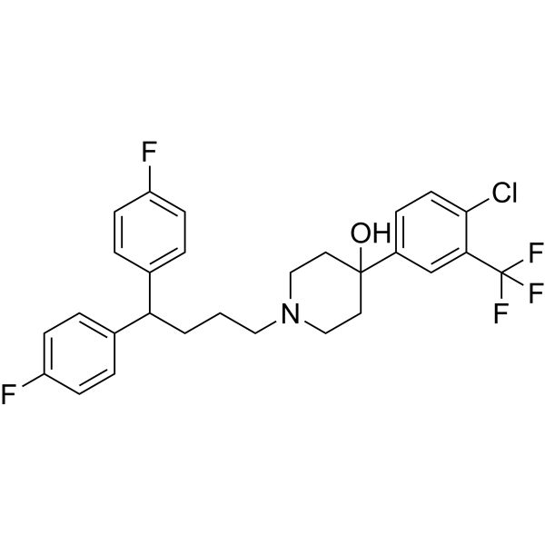
Penfluridol (R-16341) is a potent, long-acting, first-generation, oral diphenylbutylpiperidine antipsychotic agent by targeting D2-like dopamine receptor. Penfluridol effectively inhibits TNFα-induced NF-κB activation and alleviates the severity of arthritis and colitis in vivo. Penfluridol is a Ca2+-calmodulin inhibitor. Penfluridol induces apoptosis and autophagy. Penfluridol is used for chronic schizophrenia, acute psychosis, Tourette syndrome and autoimmune diseases. Penfluridol inhibites the growth of E. faecalis planktonic cells with the MIC of 7.81 μg/ml.
연구목적의 판매만을 진행합니다. 환자를 대상으로 한 판매는 하지 않습니다.
- Purity : 99.84%
- CAS No.: 26864-56-2
- 화학식: C28H27ClF5NO
- 분자량:523.97
-
보관:Powder -20°C, 3 years , 4°C, 2 years ; In solvent -80°C, 1 year , -20°C, 6 months

To place orders, for customer services and technical support, please contact: MedChemExpress USA
Tel: 609-228-6898 E-mail: [email protected] [email protected]


-
 Biological Activity
-
Chemical Information
-
용액&용해도
- Protocol
- 순도&문서
- References
-
 Help & FAQs


-
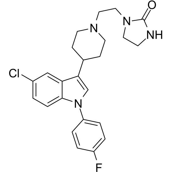
HY-14543
Sertindole5-HT/DA Receptor Antagonist -

HY-B0015
PaclitaxelAntitumor Agent -
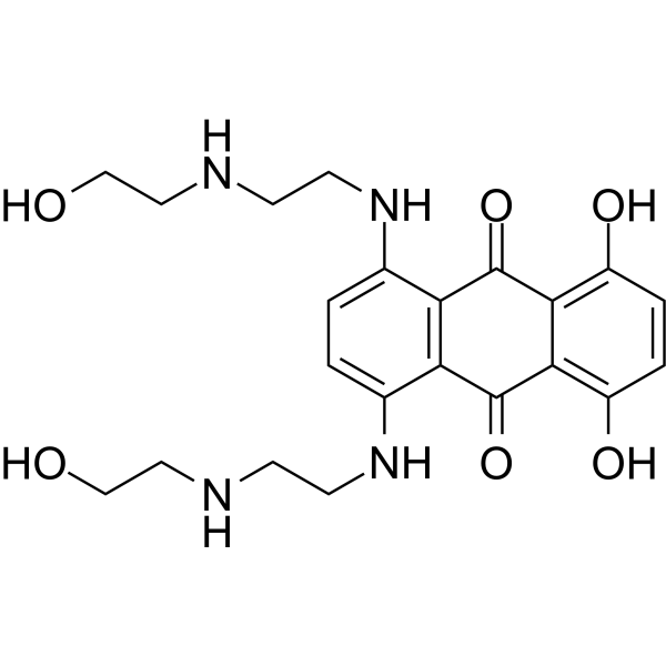
HY-13502
MitoxantroneTop II/PKC Inhibitor -
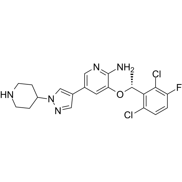
HY-50878
CrizotinibALK/c-Met/ROS1 Inhibitor -
HY-12005
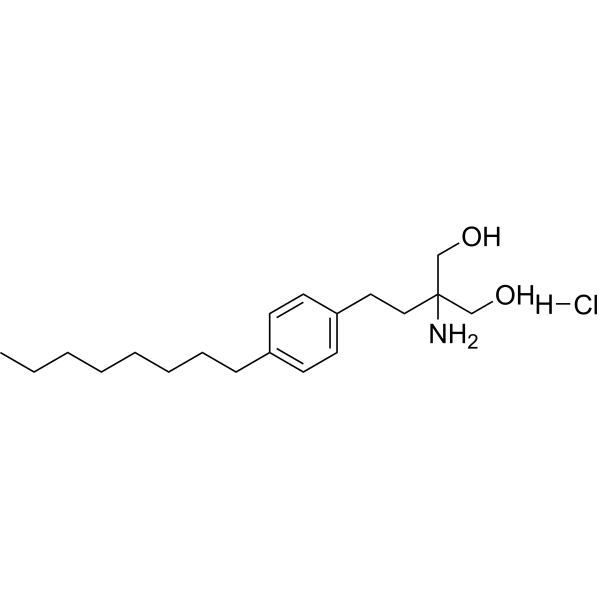
Fingolimod hydrochlorideS1P Receptors Modulator -
HY-12047
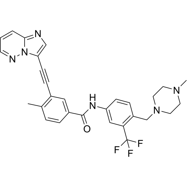
PonatinibMulti-targeted Kinase Inhibitor -
HY-B1123
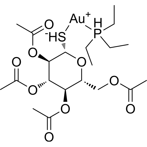
AuranofinBacterial Inhibitor -
HY-90006
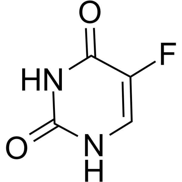
5-FluorouracilThymidylate Synthase Inhibitor -
HY-10211
TanespimycinHSP90 Inhibitor -
HY-16569
ColchicineTubulin Inhibitor -
HY-N0162
LuteolinNrf2 Inhibitor
Publications Citing Use of MedChemExpress (MCE) Penfluridol
More- Adv Sci (Weinh). 2026 Jul 3:e76408. [Abstract]
- Cell Commun Signal. 2022 Jul 16;20(1):105. [Abstract]
- EMBO Mol Med. 2026 Aug;18(8):3200-3225.
- Int Immunopharmacol. 2026 Jan 1;168(Pt 2):115947. [Abstract]
- Arthritis Res Ther. 2022 Jan 19;24(1):27. [Abstract]
- Sci Rep. 2025 May 8;15(1):16114. [Abstract]
- Mediators Inflamm. 2023 Aug 22:2023:9940858. [Abstract]
- Microbiologyopen. 2021 Jan;10(1):e1148. [Abstract]
- J Ocul Pharmacol Ther. 2024 Oct;40(8):536-542. [Abstract]
- Biomed Pharmacother. 2024 Aug:177:117094. [Abstract]
- Research Square Preprint. 2021 Dec.
 Customer Validation & Images

-
Cell Proliferation/Viability Assay
-
Cell Migration/Invasion Assay
-
Flow Cytometry
-
WB
-
Flow Cytometry

All Calcium Channel Isoforms
MoreAll Dopamine Receptor Isoforms
More

Biological Activity
제품 설명
IC50 & Target
[2]|
D2 Receptor |
Cellular Effect
|
Cell Line

|
Type | Value | Description | References |
|---|---|---|---|---|
| H4 | EC50 |
3.2 μM
Compound: 2
|
Induction of autophagy in human H4 cells expressing LC3 using DAPI staining after 4 hrs by fluorescence microscopic analysis
Induction of autophagy in human H4 cells expressing LC3 using DAPI staining after 4 hrs by fluorescence microscopic analysis
|
[PMID: 21126871] |
| Lewis lung carcinoma cell line | IC50 |
4.2 μM
Compound: PFL
|
Cytotoxicity against mouse LLC cells harboring luciferin assessed as reduction in cell viability after 48 hrs by MTT assay
Cytotoxicity against mouse LLC cells harboring luciferin assessed as reduction in cell viability after 48 hrs by MTT assay
|
[PMID: 30389290] |
| Lewis lung carcinoma cell line | IC50 |
4.3 μM
Compound: PFL
|
Cytotoxicity against mouse LLC cells harboring luciferin assessed as reduction in cell viability after 72 hrs by MTT assay
Cytotoxicity against mouse LLC cells harboring luciferin assessed as reduction in cell viability after 72 hrs by MTT assay
|
[PMID: 30389290] |
| Lewis lung carcinoma cell line | IC50 |
6 μM
Compound: PFL
|
Cytotoxicity against mouse LLC cells harboring luciferin assessed as reduction in cell viability after 24 hrs by MTT assay
Cytotoxicity against mouse LLC cells harboring luciferin assessed as reduction in cell viability after 24 hrs by MTT assay
|
[PMID: 30389290] |
| MDA-MB-231 | IC50 |
2.3 μM
Compound: 1
|
Cytotoxicity against human MDA-MB-231 cells assessed as reduction in cell viability incubated for 48 hrs by MTT assay
Cytotoxicity against human MDA-MB-231 cells assessed as reduction in cell viability incubated for 48 hrs by MTT assay
|
[PMID: 33039722] |
| MDA-MB-231 | IC50 |
5 μM
Compound: PFL
|
Cytotoxicity against human MDA-MB-231 cells assessed as reduction in cell viability after 48 hrs by MTT assay
Cytotoxicity against human MDA-MB-231 cells assessed as reduction in cell viability after 48 hrs by MTT assay
|
[PMID: 30389290] |
| MDA-MB-231 | IC50 |
5.1 μM
Compound: PFL
|
Cytotoxicity against human MDA-MB-231 cells assessed as reduction in cell viability after 72 hrs by MTT assay
Cytotoxicity against human MDA-MB-231 cells assessed as reduction in cell viability after 72 hrs by MTT assay
|
[PMID: 30389290] |
| MDA-MB-231 | IC50 |
6.1 μM
Compound: PFL
|
Cytotoxicity against human MDA-MB-231 cells assessed as reduction in cell viability after 24 hrs by MTT assay
Cytotoxicity against human MDA-MB-231 cells assessed as reduction in cell viability after 24 hrs by MTT assay
|
[PMID: 30389290] |
| NIH3T3 | IC50 |
6.01 μM
Compound: PFL
|
Cytotoxicity against mouse NIH/3T3 cells assessed as reduction in cell viability after 72 hrs by MTT assay
Cytotoxicity against mouse NIH/3T3 cells assessed as reduction in cell viability after 72 hrs by MTT assay
|
[PMID: 30389290] |
| NIH3T3 | IC50 |
6.03 μM
Compound: PFL
|
Cytotoxicity against mouse NIH/3T3 cells assessed as reduction in cell viability after 24 hrs by MTT assay
Cytotoxicity against mouse NIH/3T3 cells assessed as reduction in cell viability after 24 hrs by MTT assay
|
[PMID: 30389290] |
| NIH3T3 | IC50 |
6.09 μM
Compound: PFL
|
Cytotoxicity against mouse NIH/3T3 cells assessed as reduction in cell viability after 48 hrs by MTT assay
Cytotoxicity against mouse NIH/3T3 cells assessed as reduction in cell viability after 48 hrs by MTT assay
|
[PMID: 30389290] |
| Vero | CC50 |
8.77 μM
Compound: Penfluridol
|
Cell viability measured by CellTiter-Glo assay in Vero cells at MOI 0.05 after 72hr
Cell viability measured by CellTiter-Glo assay in Vero cells at MOI 0.05 after 72hr
|
10.1101/2020.03.20.999730 |
| Vero | IC50 |
5.01 μM
Compound: Penfluridol
|
Antiviral activity against SARS-CoV-2 (viral titer) measured by plaque assay in Vero cells at MOI 0.0125 after 24 hr
Antiviral activity against SARS-CoV-2 (viral titer) measured by plaque assay in Vero cells at MOI 0.0125 after 24 hr
|
10.1101/2020.03.20.999730 |
In Vitro
Penfluridol (R-16341; 1.25-40 μM; 24, 48 h) reduces cell viability of human AML cells harboring FLT3-WT or the FLT3-ITD mutation[2].
Penfluridol (7.5 μM; 24 h) results in the apoptosis of AML cells harboring FLT3-WT and FLT3-ITD mutation[2].
Penfluridol (1.25-7.5 μM; 24 h) induces ROS-mediated autophagy via triggering LC3 turnover and acidic vesicular organelle (AVO) formation[2].
Penfluridol (1 μM; for 2 h) obviously inhibits TNFα-induced phosphorylation levels of ERK, JNK, and p38[3].
Penfluridol (1 μM; for 2 h) inhibits TNFα-induced mRNA expressions of IL-1β, IL-6, IL-17, and NOS2[3].
Penfluridol suppresses the differentiation of spleen naive CD4+T cells to TH1 and TH17 and inhibits M1 macrophage polarization[3].
MedChemExpress (MCE) has not independently confirmed the accuracy of these methods. They are for reference only.
-
Cell Line:Human AML cell lines, HL-60 (FLT3-WT), U937 (FLT3-WT), and MV4–11 (FLT3-ITD)
-
Concentration:1.25, 2.5, 5, 10, 20, 40 μM
-
Incubation Time:24, 48 h
-
Result:Significantly reduced cell viability in a concentration-dependent manner.
-
Cell Line:HL-60 and U937 cells harboring FLT3-WT
-
Concentration:7.5 μM
-
Incubation Time:24 h
-
Result:Induced concentration-dependent increases in the sub-G1 population.
Triggered caspase-3 activation and corresponding PARP-1 cleavage in concentration- and time-dependent manners.
-
Cell Line:U937 and HL-60 cells
-
Concentration:1.25, 2.5, 5, 7.5 μM
-
Incubation Time:24 h
-
Result:5 μM and 7.5 μM respectively induced dominant LC3B-II formation and caspase-3 activation in U937 and HL-60 cells.
-
Cell Line:BMDMs
-
Concentration:1 μM
-
Incubation Time:Pretream for 2 h
-
Result:Obviously inhibited the increased phosphorylation levels of ERK, JNK, and p38 by TNFα (10 ng/mL; 15, 30, 60 min).
-
Cell Line:BMDMs
-
Concentration:1 μM
-
Incubation Time:Pretream for 2 h
-
Result:Inhibited TNFα-induced mRNA expressions of IL-1β, IL-6, IL-17, and NOS2.
In Vivo
MedChemExpress (MCE) has not independently confirmed the accuracy of these methods. They are for reference only.
-
Animal Model:DBA/1J male mice aged 10–12 weeks with type II chicken collagen-induced arthritis (CIA) model[3]
-
Dosage:10 mg/kg
-
Administration:Daily oral gavage; from the 18th day after the first immunization
-
Result:Inhibited inflammatory cell infiltration, suppressed pannus formation, and protected articular cartilage from damage.
obviously decreased mRNA expressions of CXCL10 and MCP-1 in inflamed joints and statistically reduced production levels of inflammatory cytokines IL-1β and IL-6 in sera.
- Collagen-Induced Arthritis
- Detection of Apoptosis
- TUNEL staining for apoptotic DNA fragmentation
- Annexin V plus membrane-impermeant dye apoptosis staining
- DSS-Induced Colitis
- TNBS-Induced Colitis
- Autophagy
- Apoptosis Solutions
- Research Protocol for Inflammation-related Diseases
- Ca2+ Staining Technique
- Lysosome and acidic-vesicle live-cell staining
- Macroautophagy Solutions
Clinical Trial
| NCT Number | Sponsor | Condition | Start Date |
Phase
|
|---|---|---|---|---|
| NCT01329991 | Plexxikon| | 2011-05 | PHASE1 |


Chemical Information
-
CAS No. 26864-56-2
-
Appearance Solid
-
분자량 523.97
-
화학식 C28H27ClF5NO
-
Color White to off-white
-
SMILES
OC1(C2=CC=C(Cl)C(C(F)(F)F)=C2)CCN(CCCC(C3=CC=C(F)C=C3)C4=CC=C(F)C=C4)CC1
-
Synonyms
R-16341
-
선적
Room temperature in continental US; may vary elsewhere.
-
보관
Powder -20°C 3 years 4°C 2 years In solvent -80°C 1 year -20°C 6 months


Publications (11)
-
Journal Impact Factor
-
Most Recent
-
Adv Sci (Weinh)
Penfluridol Triggers GSDME-Mediated Immunogenic Pyroptosis to Potentiate Antitumor Immunotherapy. [Abstract]2026 Jul 3:e76408. PMID: 42396980 -
Cell Commun Signal
Inhibition of AMPK/PFKFB3 mediated glycolysis synergizes with penfluridol to suppress gallbladder cancer growth. [Abstract]2022 Jul 16;20(1):105. PMID: 35842652
Penfluridol purchased from MedChemExpress. Usage Cited in: Cell Commun Signal. 2022 Jul 16;20(1):105. [Abstract]
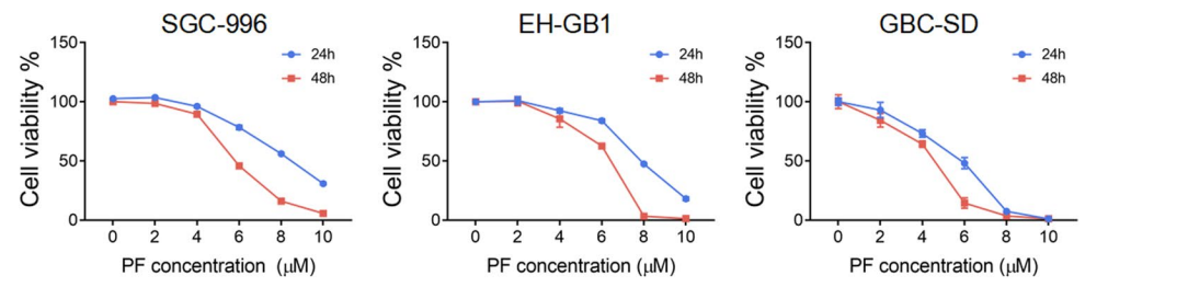
The cell viability measured by the CCK-8 assay of SGC-996, EH-GB1, and GBC-SD cells were treated with Penfluridol (PF, 0-10 μM) as indicated for 24 h/48 h.
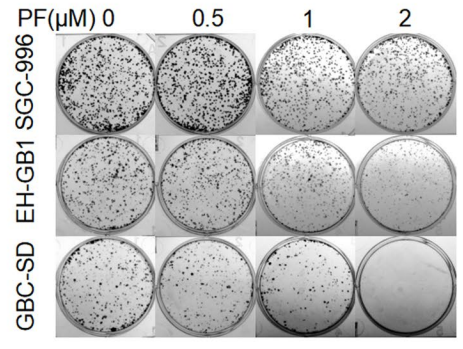
Penfluridol purchased from MedChemExpress. Usage Cited in: Cell Commun Signal. 2022 Jul 16;20(1):105. [Abstract]
The colony formation of SGC-996, EH-GB1, and GBC-SD cells were seeded in 6-well plates and treated with Penfluridol (PF, 0-2 μM) as indicated for 14 days.
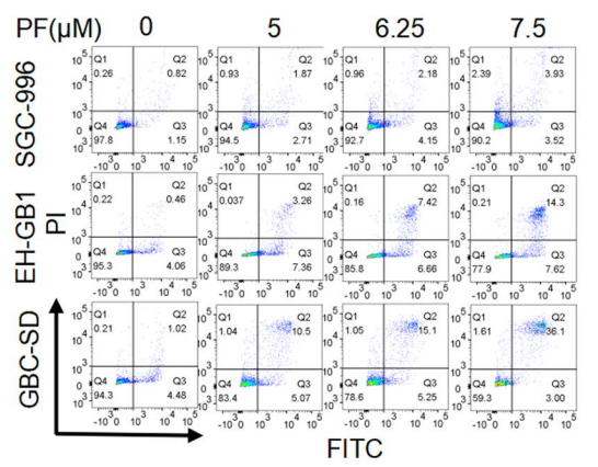
Penfluridol purchased from MedChemExpress. Usage Cited in: Cell Commun Signal. 2022 Jul 16;20(1):105. [Abstract]
Annexin V/PI staining apoptosis assay of SGC-996, EH-GB1, and GBC-SD cells treated with Penfluridol (PF, 0-7.5 μM) as indicated for 24 h.
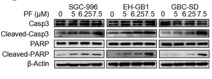
Penfluridol purchased from MedChemExpress. Usage Cited in: Cell Commun Signal. 2022 Jul 16;20(1):105. [Abstract]
The Western blotting of SGC-996, EH-GB1, and GBC-SD were treated with Penfluridol (PF, 0-7.5 μM) as indicated for 24 h.
Penfluridol purchased from MedChemExpress. Usage Cited in: Cell Commun Signal. 2022 Jul 16;20(1):105. [Abstract]
Cell cycle assay of SGC-996, EH-GB1, and GBC-SD cells treated with Penfluridol (PF, 0-7.5 μM) as indicated for 24 h.
-
-
Int Immunopharmacol
Dysregulated neutrophils contribute to bone loss in renal osteodystrophy by enhancing osteoclastogenesis: Insights from integrated bioinformatics and experimental validation. [Abstract]2026 Jan 1;168(Pt 2):115947. PMID: 41314047 -
Arthritis Res Ther
Penfluridol targets acid sphingomyelinase to inhibit TNF signaling and is therapeutic against inflammatory autoimmune diseases. [Abstract]2022 Jan 19;24(1):27. PMID: 35045889 -
Sci Rep
Penfluridol synergizes with colistin to reverse colistin resistance in Gram-negative bacilli. [Abstract]2025 May 8;15(1):16114. PMID: 40341530 -
Mediators Inflamm
Penfluroidol Attenuates the Imbalance of the Inflammatory Response by Repressing the Activation of the NLRP3 Inflammasome and Reduces Oxidative Stress via the Nrf2/HO-1 Signaling Pathway in LPS-Induced Macrophages. [Abstract]2023 Aug 22:2023:9940858. PMID: 37650025 -
Microbiologyopen
Drug repurposing: Antimicrobial and antibiofilm effects of penfluridol against Enterococcus faecalis. [Abstract]2021 Jan;10(1):e1148. PMID: 33345466 -
J Ocul Pharmacol Ther
2024 Oct;40(8):536-542. PMID: 39206555 -
Biomed Pharmacother
Penfluridol regulates p62 / Keap1 / Nrf2 signaling pathway to induce ferroptosis in osteosarcoma cells. [Abstract]2024 Aug:177:117094. PMID: 38996707 -


용액&용해도
In Vitro:
DMSO : ≥ 100 mg/mL (190.85 mM; Hygroscopic DMSO has a significant impact on the solubility of product, please use newly opened DMSO)
* "≥" means soluble, but saturation unknown.
Please refer to the solubility information to select the appropriate solvent. Once prepared, please aliquot and store the solution to prevent product inactivation from repeated freeze-thaw cycles.
Storage method and period of stock solution: -80°C, 1 year; -20°C, 6 months. When stored at -80°C, please use it within 1 year. When stored at -20°C, please use it within 6 months.
Please refer to the solubility information to select the appropriate solvent. Once prepared, please aliquot and store the solution to prevent product inactivation from repeated freeze-thaw cycles.
Storage method and period of stock solution: -80°C, 1 year; -20°C, 6 months. When stored at -80°C, please use it within 1 year. When stored at -20°C, please use it within 6 months.
Concentration (start) × Volume (start) = Concentration (final) × Volume (final)
In Vivo:
Select the appropriate dissolution method based on your experimental animal and administration route.
- For the following dissolution methods, please ensure to first prepare a clear stock solution using an In Vitro approach and then sequentially add co-solvents:
- To ensure reliable experimental results, the clarified stock solution can be appropriately stored based on storage conditions. As for the working solution for In Vivo experiments, it is recommended to prepare freshly and use it on the same day.
- The percentages shown for the solvents indicate their volumetric ratio in the final prepared solution. If precipitation or phase separation occurs during preparation, heat and/or sonication can be used to aid dissolution.
Add each solvent one by one: 10% DMSO 40% PEG300 5% Tween-80 45% Saline
Solubility: ≥ 2.5 mg/mL (4.77 mM); Clear solution
This protocol yields a clear solution of ≥ 2.5 mg/mL (saturation unknown).
Taking 1 mL working solution as an example, add 100 μL DMSO stock solution (25.0 mg/mL) to 400 μL PEG300, and mix evenly; then add 50 μL Tween-80 and mix evenly; then add 450 μL Saline to adjust the volume to 1 mL.
Preparation of Saline: Dissolve 0.9 g sodium chloride in ddH₂O and dilute to 100 mL to obtain a clear Saline solution.
Add each solvent one by one: 10% DMSO 90% (20% SBE-β-CD in Saline)
Solubility: 2.5 mg/mL (4.77 mM); Suspended solution; Need ultrasonic
This protocol yields a suspended solution of 2.5 mg/mL. Suspended solution can be used for oral and intraperitoneal injection.
Taking 1 mL working solution as an example, add 100 μL DMSO stock solution (25.0 mg/mL) to 900 μL 20% SBE-β-CD in Saline, and mix evenly.
Preparation of 20% SBE-β-CD in Saline (4°C, storage for one week): 2 g SBE-β-CD powder is dissolved in 10 mL Saline, completely dissolve until clear.
In Vivo Dissolution Calculator
Please enter the basic information of animal experiments:
-
-
-
-
Recommended: Prepare an additional quantity of animals to account for potential losses during experiments.
Please enter your animal formula composition:
-
%DMSO +
Recommended: Keep the proportion of DMSO in working solution below 2% if your animal is weak.
-
%+
-
+%Tween-80 + +
-
%Saline +
The co-solvents required include: DMSO, . All of co-solvents are available by MedChemExpress (MCE). , Tween 80. All of co-solvents are available by MedChemExpress (MCE).
Working solution concentration: 0.22 mg/mL
Method for preparing stock solution: mg drug dissolved in μL DMSO. Stock solution concentration: mg/mL.
1. Take μL DMSO stock solution;
2. Add μL .
μL , mix evenly;
3. Then add μL Tween 80, mix evenly;
4. Then add μL
Please ensure that the stock solution in the first step is dissolved to a clear state, and add co-solvents in sequence. You can use ultrasonic heating (ultrasonic cleaner, recommended frequency 20-40 kHz), vortexing, etc. to assist dissolution.


순도&문서
-
Data Sheet (282 KB)
-
SDS (479 KB)
- English - EN (479 KB)
- Français - FR (479 KB)
- Deutsch - DE (479 KB)
- Norwegian - NO (479 KB)
- Español - ES (479 KB)
- Swedish - SV (479 KB)
- Italian - IT (479 KB)
- Korean - KR (479 KB)
- Portuguese - PT (479 KB)
-
Handling Instructions (2659 KB)
References
[1]. Eyal Zur, et al. Penfluridol, a Unique Psychiatric Medicine for the Treatment of Chronic Schizophrenia. Int J Pharm Compd. 2019 Mar-Apr;23(2):113-119. [Content Brief]
[2]. Szu-Yuan Wu, et al. Penfluridol triggers cytoprotective autophagy and cellular apoptosis through ROS induction and activation of the PP2A-modulated MAPK pathway in acute myeloid leukemia with different FLT3 statuses. J Biomed Sci. 2019 Aug 31;26(1):63. [Content Brief]
[3]. Yue-Hong Chen, et al. Penfluridol targets acid sphingomyelinase to inhibit TNF signaling and is therapeutic against inflammatory autoimmune diseases. Arthritis Res Ther. 2022 Jan 19;24(1):27. [Content Brief]
[4]. Xianghai Zeng, et al. Drug repurposing: Antimicrobial and antibiofilm effects of penfluridol against Enterococcus faecalis. Microbiologyopen. 2021 Jan;10(1):e1148. [Content Brief]


Complete Stock Solution Preparation Table
Please refer to the solubility information to select the appropriate solvent. Once prepared, please aliquot and store the solution to prevent product inactivation from repeated freeze-thaw cycles.
Storage method and period of stock solution: -80°C, 1 year; -20°C, 6 months. When stored at -80°C, please use it within 1 year. When stored at -20°C, please use it within 6 months.
| Optional Solvent | Concentration Solvent Mass | 1 mg | 5 mg | 10 mg | 25 mg |
|---|---|---|---|---|---|
| DMSO | 1 mM | 1.9085 mL | 9.5425 mL | 19.0851 mL | 47.7127 mL |
| 5 mM | 0.3817 mL | 1.9085 mL | 3.8170 mL | 9.5425 mL | |
| 10 mM | 0.1909 mL | 0.9543 mL | 1.9085 mL | 4.7713 mL | |
| 15 mM | 0.1272 mL | 0.6362 mL | 1.2723 mL | 3.1808 mL | |
| 20 mM | 0.0954 mL | 0.4771 mL | 0.9543 mL | 2.3856 mL | |
| 25 mM | 0.0763 mL | 0.3817 mL | 0.7634 mL | 1.9085 mL | |
| 30 mM | 0.0636 mL | 0.3181 mL | 0.6362 mL | 1.5904 mL | |
| 40 mM | 0.0477 mL | 0.2386 mL | 0.4771 mL | 1.1928 mL | |
| 50 mM | 0.0382 mL | 0.1909 mL | 0.3817 mL | 0.9543 mL | |
| 60 mM | 0.0318 mL | 0.1590 mL | 0.3181 mL | 0.7952 mL | |
| 80 mM | 0.0239 mL | 0.1193 mL | 0.2386 mL | 0.5964 mL | |
| 100 mM | 0.0191 mL | 0.0954 mL | 0.1909 mL | 0.4771 mL |


Penfluridol Related Classifications
HY-B1077 Related Classifications
 Powered by Bioz
Keywords
- Penfluridol
- 26864-56-2
- R-16341
- R16341
- R 16341
- Calcium Channel
- Dopamine Receptor
- Autophagy
- Apoptosis
- Oral
- diphenylbutylpiperidine
- antipsychotic
- D2
- TNFα
- NF-κB
- arthritis
- colitis
- spleen naive CD4+T
- TH1
- TH17
- M1 macrophage
- chronic schizophrenia
- acute psychosis
- Tourette syndrome
- autoimmune
- E. faecalis
- planktonic
- Inhibitor
- inhibitor
- inhibit