



Anti-Mouse CD3ε Antibody (145-2C11)

Based on 1 Customer Validation
Anti-Mouse CD3ε Antibody (145-2C11) is an orally active anti-mouse CD3ε IgG1 monoclonal antibody. Anti-Mouse CD3ε Antibody (145-2C11) can improve the function of regulatory T cells (Tregs). Anti-Mouse CD3ε Antibody (145-2C11) relieves inflammatory response by inhibiting the production of pro-inflammatory cytokines and increasing the secretion of anti-inflammatory factors. Anti-Mouse CD3ε Antibody (145-2C11) can be used for researches on metabolic and immune system conditions such as diabetes and lupus. The recommend isotype control of Anti-Mouse CD3ε Antibody (145-2C11): Armenian hamster IgG, Isotype Control (HY-P990305).
For research use only. We do not sell to patients.
- Purity: 99.30%
- Molecular Weight:150 kDa
-
Storage:
Please store the product under the recommended conditions in the Certificate of Analysis.


To place orders, for customer services and technical support, please contact: MedChemExpress USA
Tel: 609-228-6898 E-mail: [email protected] [email protected]


-
 Biological Activity
-
Chemical Information
- Purity & Documentation
- References
-
 Help & FAQs








Biological Activity
Description
Isotype
Armenian hamster IgG
Recommend Isotype Controls
Species Reactivity
Mouse
IC50 & Target
CD3ε
In Vitro
Anti-Mouse CD3ε Antibody (145-2C11) (0.04-1 μg/mL, 72 h) significantly reduces the proliferation of splenic cells[1].
Anti-Mouse CD3ε Antibody (145-2C11) (1 μg/mL, 48 h) inhibits the production of pro-inflammatory cytokines and increases the production of anti-inflammatory cytokines in splenic cells and pancreatic lymph node cells (pLNs)[1].
Anti-Mouse CD3ε Antibody (145-2C11) (5 μg/mL, 2 h) induces macrophages to engulf T cells[2].
Anti-CD3ε Antibody and Anti-CD28 Antibody Stimulate T Cell Activation[3]:
(1) Anti-CD3ε antibody (Anti-Mouse CD3ε Antibody (145-2C11)) and anti-CD28 antibody (Anti-Mouse CD28 Antibody (PV-1) (HY-P990803)) were pre-coated in 96-well plates at concentrations of 3 µg/mL and 1 µg/mL, respectively.
(2) Sorted T cells were seeded in 96-well plates at a density of 1×10⁵. cells/well, with 200 μL of complete culture medium per well.
(3) Sorted T cells and coated antibodies were co-incubated at 37 ℃ and 5% CO2 for 24 hours.
(4) Collect cells and wash with PBS.
(5) Detect T cell activation by flow cytometry.
MedChemExpress (MCE) has not independently confirmed the accuracy of these methods. They are for reference only.
-
Cell Line:Splenic cells
-
Concentration:0.04, 0.02 and 1 μg/mL
-
Incubation Time:72 h
-
Result:Significantly inhibited the proliferation of splenic cells at 1 μg/mL.
-
Cell Line:Splenic cells and pLNs
-
Concentration:1 μg/mL
-
Incubation Time:48 h
-
Result:Increased the levels of TGF-β and IL-10 and decreased the levels of IFN-γ and IL-17.
In Vivo
Anti-Mouse CD3ε Antibody (145-2C11) (50 μg, p.o., once daily, for 5 days) reduces the pathogenicity of effector T cells and improves T cell function in female NOD-huCD3ε mice[1].
Anti-Mouse CD3ε Antibody (145-2C11) (3 mg/kg, i.p., once weekly, for 20 weeks) prolongs the survival period in female BWF1 mice with a lupus model[2].
MedChemExpress (MCE) has not independently confirmed the accuracy of these methods. They are for reference only.
-
Animal Model:Female NOD mice (6 weeks)[1]
-
Dosage:10 μg
-
Administration:Oral administration (p.o.) or nasal administration, once daily for the first 5 days and then once per week until 20 weeks of age
-
Result:Significantly reduced the incidence rate of diabetes to 50 %.
Reduced pancreatic islet infiltration and inflammation.
-
Animal Model:Female NOD-huCD3ε mice (12 weeks)[1]
-
Dosage:50 μg
-
Administration:Oral administration (p.o.), once daily, for 5 days
-
Result:Reduced the pathogenicity of effector T cells and enhanced the function of regulatory T cells.
Reduced the incidence rate of diabetes.
-
Animal Model:Female BWF1 mice (20 weeks) with a lupus model[2]
-
Dosage:3 mg/kg
-
Administration:Intraperitoneal injection (i.p.), once weekly for 20 weeks
-
Result:Significantly reduced proteinuria in mice.
Extended the survival period of mice.
Gene ID
Accession
Conjugated
Unconjugated
Reconsititution
The product can be reconstituted/diluted with sterile PBS or saline.
Format
-
Product Image
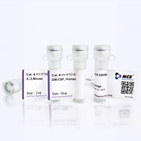
Application
in vivo T cell depletion; in vitro T cell stimulation/activation; Immunofluorescence; Flow cytome


Chemical Information
-
Appearance Liquid
-
Molecular Weight 150 kDa
-
Color Colorless to light yellow
-
SMILES
[Anti-Mouse CD3e Antibody (145-2C11)]
-
Formulation
Please refer to the lot-specific COA for specific buffer information.
-
Storage
Please store the product under the recommended conditions in the Certificate of Analysis.


Purity & Documentation
-
Data Sheet (267 KB)
-
SDS (251 KB)
- English - EN (251 KB)
- Français - FR (251 KB)
- Deutsch - DE (251 KB)
- Norwegian - NO (251 KB)
- Español - ES (251 KB)
- Swedish - SV (251 KB)
- Italian - IT (251 KB)
- Korean - KR (251 KB)
- Portuguese - PT (251 KB)
-
Inhibitory Antibodies User Guide (603 KB)
References
[1]. Kuhn C, et al. Mucosal administration of CD3-specific monoclonal antibody inhibits diabetes in NOD mice and in a preclinical mouse model transgenic for the CD3 epsilon chain. J Autoimmun. 2017 Jan;76:115-122. [Content Brief]
[2]. Morita M, et al. Reduction of Cell Surface T-Cell Receptor by Non-Mitogenic CD3 Antibody to Mitigate Murine Lupus. Front Immunol. 2022 Mar 28;13:855812. [Content Brief]
[3]. Gao R, et al. Protein phosphatase 2A catalytic subunit β suppresses PMA/ionomycin-induced T-cell activation by negatively regulating PI3K/Akt signaling. FEBS J. 2022 Aug;289(15):4518-4535. [Content Brief]

Calculators
Concentration (start) × Volume (start) = Concentration (final) × Volume (final)


Anti-Mouse CD3ε Antibody (145-2C11) Related Classifications
HY-P990802 Related Classifications
 Powered by Bioz