



ACSS2 Antibody (YA645)
(Synonyms: ACAS2, ACSS2, Acetate--CoA ligase, Acetyl-CoA synthetase, Acetyl-CoA synthetase 1, Acyl-CoA synthetase short-chain family member 2, Acyl-activating enzyme, Propionate--CoA ligase, ACS, AceCS, AceCS1)
Based on 1 Customer Validation
ACSS2 Antibody (YA645) is a Rabbit-derived and non-conjugated IgG monoclonal antibody, targeting to ACSS2.
-
Host:
Rabbit
-
Isotype:
IgG
-
Application:
WB, ICC/IF
-
Reactivity :
Human, Mouse, Rat
-
Formulation:
Supplied in 1*TBS (pH7.4), 0.05% BSA and 40% Glycerol. Preservative: 0.05% Sodium Azide.
-
Conjugation:
Non-conjugated


To place orders, for customer services and technical support, please contact: MedChemExpress USA
Tel: 609-228-6898 E-mail: [email protected] [email protected]





Applications
| Application |
WB

WB: Western Blot
|
ICC/IF
ICC/IF: Immunocytochemistry/
Immunofluorescence |
|---|---|---|
| Dilution Ratio | 1:1000 | 1:50-1:200 |


Product Details
ACSS2 Antibody (YA645) is a Rabbit-derived and non-conjugated IgG monoclonal antibody, targeting to ACSS2.
-
Host Rabbit

-
Clonality Recombinant,Monoclonal

-
Species ReactivityHuman, Mouse, Rat
-
Observed Molecular WeightObserved band size: 79 kDa
Note: Due to possible protein modifications or aggregation, the molecular weight should be confirmed by actual measurement, and the predicted value is for reference only.

-
Calculated Molecular Weight Predicted band size: 79 kDa

Entrez Gene: 55902 Human ; 60525 Mouse
SwissProt: Q9NR19 Human ; Q8BK97 Mouse ; Q9QXG4 Mouse ; D3ZFF9 Rat ; D3ZUL6 Rat
OMIM: 605832 Human
Synthetic peptide corresponding to Human Acetyl CoA synthetase.AA range:101-145.
Endogenous
Protein A affinity purified.
Non-conjugated
Unmodified
IgG


Product Properties
-
Appearance
Solution
-
Formulation
Supplied in 1*TBS (pH7.4), 0.05% BSA and 40% Glycerol. Preservative: 0.05% Sodium Azide.
-
Concentration
Batch-dependent, Please check the COA for the concentration of each lot. Check Lot Concentration
-
Storage & Stability
Stored at -20°C for 1 year. Avoid repeated freeze / thaw cycles.
-
Shipping
Shipping with blue ice.


Verification Images
-
 Western blot analysis was performed on protein extracts (25 μg) from A549 (lane 2) and Mouse brain (lane 3) using ACSS2 antibody. Proteins were transferred onto a 0.45 μm PVDF membrane using the Trans-Blot® Turbo™ system for 13 min. The membrane was then blocked with 5% nonfat milk in TBST (HY-K1025) for 1 h at room temperature. The primary antibody (1:1000) and loading control antibody GAPDH Antibody (HRP) (HY-P80954A) (1:5000) were diluted in 5% nonfat milk in TBST and incubated with the membrane overnight at 4°C. After washing, the membrane of primary antibody was incubated with HRP-conjugated goat anti-rabbit/mouse IgG secondary antibody (HY-P8001/HY-P8004) (1:5000) diluted in 5% nonfat milk in TBST for 1 h at room temperature. Protein bands were visualized using an Ultra High Sensitivity ECL detection kit (HY-K1005).
 -
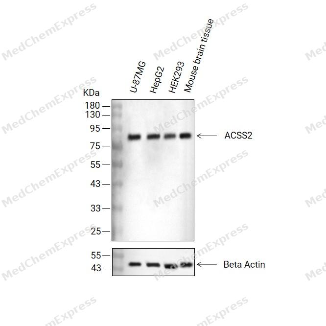 Western blot analysis of extracts from U-87MG(lane 2(20μg) , HepG2(lane 3(20μg) ,HEK293(lane 4(20μg)and Mouse brain tissue( lane 5(20μg) using ACSS2 Antibody (HY-P80483). Proteins were transferred to a PVDF membrane and blocked with 5% non-fat milk in TBST for 2 hour at room temperature. The primary antibody (1/1000) and Loading control antibody (Beta Actin, HY-P80993,1/10000) was used in 5% non-fat milk in TBST at 4°C overnight. Goat Anti-Rabbit IgG-HRP Secondary Antibody (HY-P8001,1/10,000) was used for 1 hour at room temperature.
 -
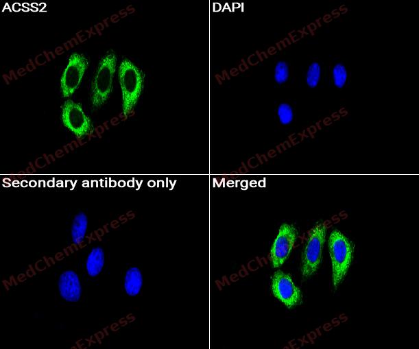 Immunocytochemistry analysis of HepG2 cells labeling ACSS2 with ACSS2 Antibody (HY-P80483) at 1/50 dilution. Cells were fixed in 4% paraformaldehyde for 15 minutes at room temperature, permeabilized with 0.1% Triton X-100 for 10 minutes at room temperature, then blocked with BSA for Immunol Staining for 10 min at room temperature. Cells were then incubated with ACSS2 Antibody (HY-P80483) at 1/50 dilution in BSA for Immunol Staining at 4 ℃ overnight. AF488-conjugated AffiniPure Goat Anti-Rabbit IgG H&L(HY-P8002, Green) was used as the secondary antibody at 1/1,000 dilution. PBS instead of the primary antibody was used as the secondary antibody only control. The Nuclear counterstain was DAPI (Blue).
 -
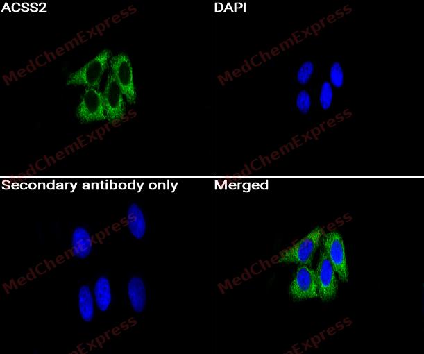 Immunocytochemistry analysis of HepG2 cells labeling ACSS2 with ACSS2 Antibody (HY-P80483) at 1/100 dilution. Cells were fixed in 4% paraformaldehyde for 15 minutes at room temperature, permeabilized with 0.1% Triton X-100 for 10 minutes at room temperature, then blocked with BSA for Immunol Staining for 10 min at room temperature. Cells were then incubated with ACSS2 Antibody (HY-P80483) at 1/100 dilution in BSA for Immunol Staining at 4 ℃ overnight. AF488-conjugated AffiniPure Goat Anti-Rabbit IgG H&L(HY-P8002,Green) was used as the secondary antibody at 1/1,000 dilution. PBS instead of the primary antibody was used as the secondary antibody only control. The Nuclear counterstain was DAPI (Blue).


Background
-
Function
ACSS2 catalyzes the synthesis of acetyl-CoA from short-chain fatty acids. Acetate is the preferred substrate. Can also utilize propionate with a much lower affinity. Nuclear ACSS2 promotes glucose deprivation-induced lysosomal biogenesis and autophagy, tumor cell survival and brain tumorigenesis. Glucose deprivation results in AMPK-mediated phosphorylation of ACSS2 leading to its translocation to the nucleus where it binds to TFEB and locally produces acetyl-CoA for histone acetylation in the promoter regions of TFEB target genes thereby activating their transcription. The regulation of genes associated with autophagy and lysosomal activity through ACSS2 is important for brain tumorigenesis and tumor survival. Acts as a chromatin-bound transcriptional coactivator that up-regulates histone acetylation and expression of neuronal genes. Can be recruited to the loci of memory-related neuronal genes to maintain a local acetyl-CoA pool, providing the substrate for histone acetylation and promoting the expression of specific genes, which is essential for maintaining long-term spatial memory[1][2][3].
-
Subcellular Localization
Cytoplasm, cytosol; Cytoplasm; Nucleus
-
Subunit
Monomer (PubMed:10843999). Interacts with TFEB (PubMed:28552616). AMPK-mediated phosphorylated form at Ser-659 interacts with KPNA1; this interaction results in nuclear translocation of ACSS2 (PubMed:28552616). Interacts with the 'Thr-172' phosphorylated form of PRKAA2 (PubMed:28552616). Interacts with CREBBP (By similarity)
-
SwissProt ID
-
Synonyms
ACAS2, ACSS2, Acetate--CoA ligase, Acetyl-CoA synthetase, Acetyl-CoA synthetase 1, Acyl-CoA synthetase short-chain family member 2, Acyl-activating enzyme, Propionate--CoA ligase, ACS, AceCS, AceCS1
-
Research Field
Signal Transduction


Documentation
-
Data Sheet (261 KB)
-
SDS (251 KB)
- English - EN (251 KB)
- Français - FR (251 KB)
- Deutsch - DE (251 KB)
- Norwegian - NO (251 KB)
- Español - ES (251 KB)
- Swedish - SV (251 KB)
- Italian - IT (251 KB)
- Korean - KR (251 KB)
- Portuguese - PT (251 KB)
-
User Guide for Antibodies (1077 KB)
References
[1]. Luong A, et al. Molecular characterization of human acetyl-CoA synthetase, an enzyme regulated by sterol regulatory element-binding proteins. J Biol Chem. 2000 Aug 25;275(34):26458-66. [Content Brief]
[2]. Yoshimura Y, et al. Molecular cloning of rat acss3 and characterization of mammalian propionyl-CoA synthetase in the liver mitochondrial matrix. J Biochem. 2017 Mar 1;161(3):279-289. [Content Brief]
[3]. Li X, et al. Nucleus-Translocated ACSS2 Promotes Gene Transcription for Lysosomal Biogenesis and Autophagy. Mol Cell. 2017 Jun 1;66(5):684-697.e9. [Content Brief]

 Powered by Bioz
