



Adipose Triglyceride Lipase Antibody (YA637)
(Synonyms: ATGL; Desnutrin; plpl; plpl2; Pnpla2; TTS 2.2; TTS2; TTS2.2; ZETA)Based on 1 publication(s) in Google Scholar
Adipose Triglyceride Lipase Antibody (YA637) is a Rabbit-derived and non-conjugated IgG monoclonal antibody, targeting to Adipose Triglyceride Lipase.
-
Host:
Rabbit
-
Isotype:
IgG
-
Application:
WB
-
Reactivity :
Human, Mouse, Rat
-
Formulation:
Supplied in 50 mM Tris-Glycine (pH 7.4), 0.15 M NaCl, 40% Glycerol and 0.05% BSA. Preservative: 0.01% Sodium azide
-
Conjugation:
Non-conjugated


To place orders, for customer services and technical support, please contact: MedChemExpress USA
Tel: 609-228-6898 E-mail: [email protected] [email protected]



Publications Citing Use of MedChemExpress (MCE) Adipose Triglyceride Lipase Antibody (YA637)
More

Applications
| Application |
WB

WB: Western Blot
|
|---|---|
| Dilution Ratio | 1:500-1:1000 |


Product Details
Adipose Triglyceride Lipase Antibody (YA637) is a Rabbit-derived and non-conjugated IgG monoclonal antibody, targeting to Adipose Triglyceride Lipase.
-
Host Rabbit

-
Clonality Recombinant,Monoclonal

-
Species ReactivityHuman, Mouse, Rat
-
Observed Molecular WeightObserved band size: 55 kDa
Note: Due to possible protein modifications or aggregation, the molecular weight should be confirmed by actual measurement, and the predicted value is for reference only.

-
Calculated Molecular Weight Predicted band size: 55 kDa

Entrez Gene: 57104 Human ; 66853 Mouse ; 361676 Rat
SwissProt: Q96AD5 Human ; Q8BJ56 Mouse ; P0C548 Rat
OMIM: 610717 Human
Synthetic peptide corresponding to Human Adipose Triglyceride Lipase.The exact sequence is proprietary to MCE.
Endogenous
affinity purified
Non-conjugated
Unmodified
IgG


Product Properties
-
Appearance
Solution
-
Formulation
Supplied in 50 mM Tris-Glycine (pH 7.4), 0.15 M NaCl, 40% Glycerol and 0.05% BSA. Preservative: 0.01% Sodium azide
-
Concentration
Batch-dependent, Please check the COA for the concentration of each lot. Check Lot Concentration
-
Storage & Stability
Stored at -20°C for 1 year. Avoid repeated freeze / thaw cycles.
-
Shipping
Shipping with blue ice.


Publications (1)
-
Journal Impact Factor
-
Most Recent
-
Adipocyte
2025 Dec;14(1):2549456. PMID: 41381093


Verification Images
-
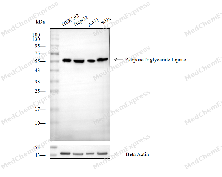 Western blot analysis of extracts from HEK293 (lane 2(20μg), HepG2 (lane 3(20μg), A431 (lane 4(20μg) and SiHa (lane 5(20μg) using Adipose Triglyceride Lipase Antibody (HY-P80533). Proteins were transferred to a PVDF membrane and blocked with 5% non-fat milk in TBST for 2 hour at room temperature. The primary antibody (1/1000) and Loading control antibody (Beta Actin, HY-P80993, 1/10000) was used in 5% non-fat milk in TBST at 4°C overnight. Goat Anti-Mouse IgG-HRP Secondary Antibody (1/10000) was used for 1 hour at room temperature.


Background
-
Function
Adipose Triglyceride Lipase catalyzes the initial step in triglyceride hydrolysis in adipocyte and non-adipocyte lipid droplets. Exhibits a strong preference for the hydrolysis of long-chain fatty acid esters at the sn-2 position of the glycerol backbone and acts coordinately with LIPE/HLS and DGAT2 within the lipolytic cascade. Also possesses acylglycerol transacylase and phospholipase A2 activities. Transfers fatty acid from triglyceride to retinol, hydrolyzes retinylesters, and generates 1,3-diacylglycerol from triglycerides. Regulates adiposome size and may be involved in the degradation of adiposomes. Catalyzes the formation of an ester bond between hydroxy fatty acids and fatty acids derived from triglycerides or diglycerides to generate fatty acid esters of hydroxy fatty acids (FAHFAs) in adipocytes. Acts antagonistically with LDAH in regulation of cellular lipid stores. Inhibits LDAH-stimulated lipid droplet fusion. May play an important role in energy homeostasis. May play a role in the response of the organism to starvation, enhancing hydrolysis of triglycerides and providing free fatty acids to other tissues to be oxidized in situations of energy depletion[1][2][3][4][5][6][7][8][9].
-
Subcellular Localization
Lipid droplet; Cell membrane; Multi-pass membrane protein; Cytoplasm
-
Expression
Tissue_specificity:It is expressed most strongly in adipose tissue. It has also been detected in the heart, skeletal muscle, and parts of the gastrointestinal tract. It can be detected in both normal retina and retinoblastoma cells. It is also detected in the retinal pigment epithelium, with lower expression levels (protein levels) in photoreceptor ganglia and the neuroretinal ganglion cell layer. -
Isoforms & Post-Translational Modification
Q96AD5 has 2 isomers: Q96AD5-1: 55316 Da (predicted); Q96AD5-2: 19875 Da (predicted).
Phosphorylation at Ser-404 by PKA is increased during fasting and moderate intensity exercise, and moderately increases lipolytic activity (By similarity). Phosphorylation at Ser-404 is increased upon beta-adrenergic stimulation (PubMed:22733971);Ubiquitinated by PEX2 in response to reactive oxygen species (ROS), leading to its degradation (PubMed:34903883). Ubiquitination is stimulated by LDAH (PubMed:28578400) -
Subunit
Interacts with ABHD5; this association stimulates PNPLA2 triglyceride hydrolase activity (By similarity). Interacts with SERPINF1; this interaction stimulates the phospholipase A2 activity of PNPLA2 (PubMed:17032652). Despite a colocalization in lipid droplets, it probably does not interact with PLIN (By similarity). Interacts with PLIN5; prevents interaction with ABHD5 (By similarity). Interacts with FAF2 (PubMed:23297223)
-
SwissProt ID
-
Synonyms
ATGL; Desnutrin; plpl; plpl2; Pnpla2; TTS 2.2; TTS2; TTS2.2; ZETA
-
Research Field
Cardiovascular
Documentation
-
Data Sheet (262 KB)
-
SDS (252 KB)
- English - EN (252 KB)
- Français - FR (252 KB)
- Deutsch - DE (252 KB)
- Norwegian - NO (252 KB)
- Español - ES (252 KB)
- Swedish - SV (252 KB)
- Italian - IT (252 KB)
- Korean - KR (252 KB)
- Portuguese - PT (252 KB)
-
User Guide for Antibodies (1077 KB)
References
[1]. Jenkins CM, et al. Identification, cloning, expression, and purification of three novel human calcium-independent phospholipase A2 family members possessing triacylglycerol lipase and acylglycerol transacylase activities. J Biol Chem. 2004 Nov 19;279(47):48968-75. [Content Brief]
[2]. Zimmermann R, et al. Fat mobilization in adipose tissue is promoted by adipose triglyceride lipase. Science. 2004 Nov 19;306(5700):1383-6. [Content Brief]
[3]. Lake AC, et al. Expression, regulation, and triglyceride hydrolase activity of Adiponutrin family members. J Lipid Res. 2005 Nov;46(11):2477-87. [Content Brief]
[4]. Smirnova E, et al. ATGL has a key role in lipid droplet/adiposome degradation in mammalian cells. EMBO Rep. 2006 Jan;7(1):106-13. [Content Brief]
[5]. Gao JG, et al. A comparative study of human GS2, its paralogues, and its rat orthologue. Biochem Biophys Res Commun. 2007 Aug 24;360(2):501-6. [Content Brief]
[6]. Ding L, et al. Peroxisomal β-oxidation acts as a sensor for intracellular fatty acids and regulates lipolysis. Nat Metab. 2021 Dec;3(12):1648-1661. [Content Brief]
[7]. Notari L, et al. Identification of a lipase-linked cell membrane receptor for pigment epithelium-derived factor. J Biol Chem. 2006 Dec 8;281(49):38022-37. [Content Brief]
[8]. Patel R, et al. ATGL is a biosynthetic enzyme for fatty acid esters of hydroxy fatty acids. Nature. 2022 Jun;606(7916):968-975. [Content Brief]
[9]. Goo YH, et al. Lipid Droplet-Associated Hydrolase Promotes Lipid Droplet Fusion and Enhances ATGL Degradation and Triglyceride Accumulation. Sci Rep. 2017 Jun 2;7(1):2743. [Content Brief]

 Powered by Bioz
