



Cannabinoid Receptor I Antibody
(Synonyms: CNR1; CNR; Cannabinoid receptor 1; CB-R; CB1; CANN6)
Based on 1 Customer Validation
Cannabinoid Receptor I Antibody is a Rabbit-derived and non-conjugated IgG polyclonal antibody, targeting to Cannabinoid Receptor I.
-
Host:
Rabbit
-
Isotype:
IgG
-
Application:
WB, IHC-P, IHC-F, ICC/IF, ELISA
-
Reactivity :
Human, Rat
-
Formulation:
Supplied in 0.01M TBS (pH7.4) with 1% BSA, 0.02% Proclin300 and 50% Glycerol.
-
Conjugation:
Non-conjugated


To place orders, for customer services and technical support, please contact: MedChemExpress USA
Tel: 609-228-6898 E-mail: [email protected] [email protected]





Applications
| Application |
WB

WB: Western Blot
|
IHC-P
IHC-P: Immunohistochemistry-Paraffin
|
IHC-F
IHC-F: Immunohistochemistry-Frozen
|
ICC/IF
ICC/IF: Immunocytochemistry/
Immunofluorescence |
ELISA
ELISA: Enzyme Linked Immunosorbent Assay
|
|---|---|---|---|---|---|
| Dilution Ratio | 1:500-2000 | 1:100-500 | 1:100-500 | 1:100-500 | 1:5000-10000 |


Product Details
Cannabinoid Receptor I Antibody is a Rabbit-derived and non-conjugated IgG polyclonal antibody, targeting to Cannabinoid Receptor I.
-
Host Rabbit

-
Clonality Polyclonal

-
Species ReactivityHuman, Rat
-
Observed Molecular WeightObserved band size: 52 kDa
Note: Due to possible protein modifications or aggregation, the molecular weight should be confirmed by actual measurement, and the predicted value is for reference only.

-
Calculated Molecular Weight Predicted band size: 52 kDa

Entrez Gene: 1268 Human ; 12801 Mouse ; 25248 Rat
SwissProt: P21554 Human ; P47746 Mouse ; P20272 Rat
OMIM: 601665 Human
KLH conjugated synthetic peptide derived from human CNR-1: 401-472/472
Endogenous
Affinity Chromatography
Non-conjugated
Unmodified
IgG


Product Properties
-
Appearance
Solution
-
Formulation
Supplied in 0.01M TBS (pH7.4) with 1% BSA, 0.02% Proclin300 and 50% Glycerol.
-
Concentration
Batch-dependent, Please check the COA for the concentration of each lot. Check Lot Concentration
-
Storage & Stability
Stored at -20°C for 1 year. Avoid repeated freeze / thaw cycles.
-
Shipping
Shipping with blue ice.


Verification Images
-
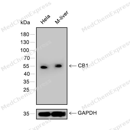 Western blot analysis of extracts from Hela (lane 1) and M-liver (lane 2) using CB1 antibody. Proteins were transferred to a PVDF membrane and blocked with 5% nonfat powdered milk in PBST for 2 hour at room temperature. The primary antibody (1/1000) and loading control antibody (GAPDH, 1/3000) was diluted with 5% nonfat powdered milk in PBST at 4°C overnight. Goat Anti-Rabbit IgG-HRP Secondary Antibody (1/8,000) was incubated for 45min at room temperature.
 -
 Immunocytochemistry analysis of A549 cells labeling Cannabinoid Receptor I with Cannabinoid Receptor I Antibody (HY-P81627) at 1/100 dilution. Cells were fixed in 4% paraformaldehyde for 15 minutes at room temperature, permeabilized with 0.1% Triton X-100 in PBS for 15 minutes at room temperature, then blocked with quick block buffer for 10 minutes at room temperature. Cells were then incubated with Cannabinoid Receptor I Antibody (HY-P81627) at 1/100 dilution in quick block buffer overnight at 4 ℃. AF488-conjugated Goat Anti-Rabbit IgG H&L (HY-P8002, Green) was used as the secondary antibody at 1/1,000 dilution. PBS instead of the primary antibody was used as the secondary antibody only control. The Nuclear counterstain was DAPI (Blue).
 -
 Immunocytochemistry analysis of HepG2 cells labeling Cannabinoid Receptor I with Cannabinoid Receptor I Antibody (HY-P81627) at 1/100 dilution. Cells were fixed in 4% paraformaldehyde for 15 minutes at room temperature, permeabilized with 0.1% Triton X-100 in PBS for 15 minutes at room temperature, then blocked with quick block buffer for 10 minutes at room temperature. Cells were then incubated with Cannabinoid Receptor I Antibody (HY-P81627) at 1/100 dilution in quick block buffer overnight at 4 ℃. AF488-conjugated Goat Anti-Rabbit IgG H&L (HY-P8002, Green) was used as the secondary antibody at 1/1,000 dilution. PBS instead of the primary antibody was used as the secondary antibody only control. The Nuclear counterstain was DAPI (Blue).


Background
-
Function
Cannabinoid Receptor I is a G-protein coupled receptor for endogenous cannabinoids (eCBs), including N-arachidonoylethanolamide (also called anandamide or AEA) and 2-arachidonoylglycerol (2-AG), as well as phytocannabinoids, such as delta(9)-tetrahydrocannabinol (THC). Mediates many cannabinoid-induced effects, acting, among others, on food intake, memory loss, gastrointestinal motility, catalepsy, ambulatory activity, anxiety, cH2O2ic pain. Signaling typically involves reduction in cyclic AMP. In the hypothalamus, may have a dual effect on mitochondrial respiration depending upon the agonist dose and possibly upon the cell type. Increases respiration at low doses, while decreases respiration at high doses. At high doses, CNR1 signal transduction involves G-protein alpha-i protein activation and subsequent inhibition of mitochondrial soluble adenylate cyclase, decrease in cyclic AMP concentration, inhibition of protein kinase A (PKA)-dependent phosphorylation of specific subunits of the mitochondrial electron transport system, including NDUFS2. In the hypothalamus, inhibits leptin-induced reactive oxygen species (ROS) formation and mediates cannabinoid-induced increase in SREBF1 and FASN gene expression. In response to cannabinoids, drives the release of orexigenic beta-endorphin, but not that of melanocyte-stimulating hormone alpha/alpha-MSH, from hypothalamic POMC neurons, hence promoting food intake. In the hippocampus, regulates cellular respiration and energy production in response to cannabinoids. Involved in cannabinoid-dependent depolarization-induced suppression of inhibition (DSI), a process in which depolarization of CA1 postsynaptic pyramidal neurons mobilizes eCBs, which retrogradely activate presynaptic CB1 receptors, transiently decreasing GABAergic inhibitory neurotransmission. Also reduces excitatory synaptic transmission. In superior cervical ganglions and cerebral vascular smooth muscle cells, inhibits voltage-gated Ca(2+) channels in a constitutive, as well as agonist-dependent manner. In cerebral vascular smooth muscle cells, cannabinoid-induced inhibition of voltage-gated Ca(2+) channels leads to vasodilation and decreased vascular tone. Induces leptin production in adipocytes and reduces LRP2-mediated leptin clearance in the kidney, hence participating in hyperleptinemia. In adipose tissue, CNR1 signaling leads to increased expression of SREBF1, ACACA and FASN genes. In the liver, activation by endocannabinoids leads to increased de novo lipogenesis and reduced fatty acid catabolism, associated with increased expression of SREBF1/SREBP-1, GCK, ACACA, ACACB and FASN genes. May also affect de novo cholesterol synthesis and HDL-cholesteryl ether uptake. Peripherally modulates energy metabolism. In high carbohydrate diet-induced obesity, may decrease the expression of mitochondrial dihydrolipoyl dehydrogenase/DLD in striated muscles, as well as that of selected glucose/ pyruvate metabolic enzymes, hence affecting energy expenditure tH2O2gh mitochondrial metabolism. In response to cannabinoid anandamide, elicits a pro-inflammatory response in macrophages, which involves NLRP3 inflammasome activation and IL1B and IL18 secretion. In macrophages infiltrating pancreatic islets, this process may participate in the progression of type-2 diabetes and associated loss of pancreatic beta-cells; Binds both 2-arachidonoylglycerol (2-AG) and anandamide; Only binds 2-arachidonoylglycerol (2-AG) with high affinity. Contrary to its effect on isoform 1, 2-AG behaves as an inverse agonist on isoform 2 in assays measuring GTP binding to membranes; Only binds 2-arachidonoylglycerol (2-AG) with high affinity. Contrary to its effect on isoform 1, 2-AG behaves as an inverse agonist on isoform 3 in assays measuring GTP binding to membranes[1][2][3][4][5][6][7].
-
Subcellular Localization
Cell membrane; Multi-pass membrane protein; Membrane raft; Mitochondrion outer membrane; Cell projection, axon; Presynapse
-
Expression
Tissue_specificity:It is widely expressed, with the highest expression level in the human brain. The expression levels of isoforms 2 and 3 are much lower than those of isoform 1.
Induction:Up-regulated by endocannabinoid anandamide -
Isoforms & Post-Translational Modification
P21554 has 3 isomers: P21554-1: 52858 Da (predicted); P21554-2: 45874 Da (predicted); P21554-3: 49060 Da (predicted).
Palmitoylation at Cys-415 is important for recruitment at plasma membrane and lipid rafts and association with G protein alpha subunits -
Subunit
Interacts (via C-terminus) with CNRIP1; this interaction attenuates constitutive, but not agonist-dependent, inhibition of voltage-gated Ca(2+) channels in neurons (PubMed:17895407). Associates with G protein alpha subunits, including G(i) alpha-1/GNAI1, G(i) alpha-3/GNAI3 and G(o)-alpha/GNAO1; palmitoylation is important for interaction with GNAI3 and GNAO1 (PubMed:12237474)
-
SwissProt ID
-
Synonyms
CNR1; CNR; Cannabinoid receptor 1; CB-R; CB1; CANN6
-
Research Field
Neuroscience


Documentation
-
Data Sheet (236 KB)
-
SDS (251 KB)
- English - EN (251 KB)
- Français - FR (251 KB)
- Deutsch - DE (251 KB)
- Norwegian - NO (251 KB)
- Español - ES (251 KB)
- Swedish - SV (251 KB)
- Italian - IT (251 KB)
- Korean - KR (251 KB)
- Portuguese - PT (251 KB)
-
User Guide for Antibodies (1077 KB)

 Powered by Bioz
