



S1P1/EDG1 Antibody (YA3874)
(Synonyms: S1PR1; EDG1; S1P1; ECGF1; EDG-1; CHEDG1; D1S3362)Based on 1 Customer Validation
S1P1/EDG1 Antibody (YA3874) is a Mouse-derived and non-conjugated IgG2a monoclonal antibody, targeting to S1P1/EDG1.
-
Host:
Mouse
-
Isotype:
IgG2a
-
Application:
WB, IHC-P, FC, ELISA
-
Reactivity :
Human
-
Formulation:
Supplied in PBS with 0.05% sodium azide
-
Conjugation:
Non-conjugated


To place orders, for customer services and technical support, please contact: MedChemExpress USA
Tel: 609-228-6898 E-mail: [email protected] [email protected]





Applications
| Application |
WB

WB: Western Blot
|
IHC-P
IHC-P: Immunohistochemistry-Paraffin
|
FC
FC: Flow Cytometry
|
ELISA
ELISA: Enzyme Linked Immunosorbent Assay
|
|---|---|---|---|---|
| Dilution Ratio | 1:500-1:2000 | 1:200-1:1000 | 1:200-1:400 | 1:10000 |


Product Details
S1P1/EDG1 Antibody (YA3874) is a Mouse-derived and non-conjugated IgG2a monoclonal antibody, targeting to S1P1/EDG1.
-
Host Mouse

-
Clonality Monoclonal

-
Species ReactivityHuman
-
Observed Molecular WeightObserved band size: 43 kDa
Note: Due to possible protein modifications or aggregation, the molecular weight should be confirmed by actual measurement, and the predicted value is for reference only.

-
Calculated Molecular Weight Predicted band size: 43 kDa

Purified recombinant fragment of human CD363 expressed in E. Coli.
affinity purified.
Non-conjugated
Unmodified
IgG2a


Product Properties
-
Appearance
Solution
-
Formulation
Supplied in PBS with 0.05% sodium azide
-
Concentration
Batch-dependent, Please check the COA for the concentration of each lot. Check Lot Concentration
-
Storage & Stability
Stored at -20°C for 1 year. Avoid repeated freeze / thaw cycles.
-
Shipping
Shipping with blue ice.


Verification Images
-
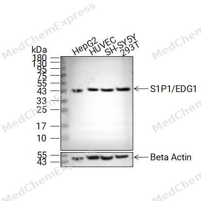 Western blot analysis of extracts from HepG2(lane2(20μg), HUVEC(lane3(20μg), SH-SY5Y(lane4(20μg) and 293T(lane5(20μg) using S1P1/EDG1 Antibody (HY-P84177). Proteins were transferred to a PVDF membrane and blocked with 5% non-fat milk in TBST at 4°C overnight. The primary antibody (1/1000) and Loading control antibody (Beta Actin, HY-P80993, 1/10,000) was used in 5% non-fat milk in TBST for 2 hour at room temperature. Goat Anti-Mouse IgG-HRP Secondary Antibody (HY-P8004, 1/10,000) was used for 1 hour at room temperature.
 -
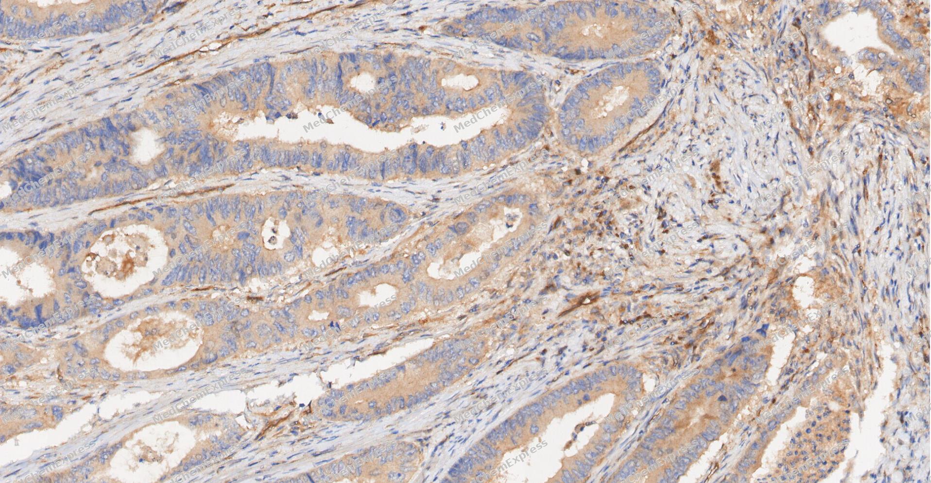 Immunohistochemical analysis of paraffin-embedded human Colon cancer tissue using S1P1/EDG1 antibody was performed. The section was pretreated using high-temperature and high-pressure mediated EDTA antigen retrieval buffer (pH 9.0), for 5 minutes. The tissues were incubated with primary antibody (HY-P84177, 1:50 dilution) at room temperature for 60 minutes. Detection was performed using an HRP conjugated compact polymer system. DAB was used as the chromogen. The tissues were counterstained with hematoxylin and mounted with neutral balsam mounting medium.
 -
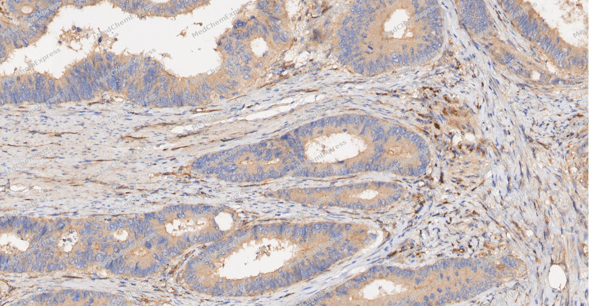 Immunohistochemical analysis of paraffin-embedded human Colon cancer tissue using S1P1/EDG1 antibody was performed. The section was pretreated using high-temperature and high-pressure mediated EDTA antigen retrieval buffer (pH 9.0), for 5 minutes. The tissues were incubated with primary antibody (HY-P84177, 1:50 dilution) at room temperature for 60 minutes. Detection was performed using an HRP conjugated compact polymer system. DAB was used as the chromogen. The tissues were counterstained with hematoxylin and mounted with neutral balsam mounting medium.
 -
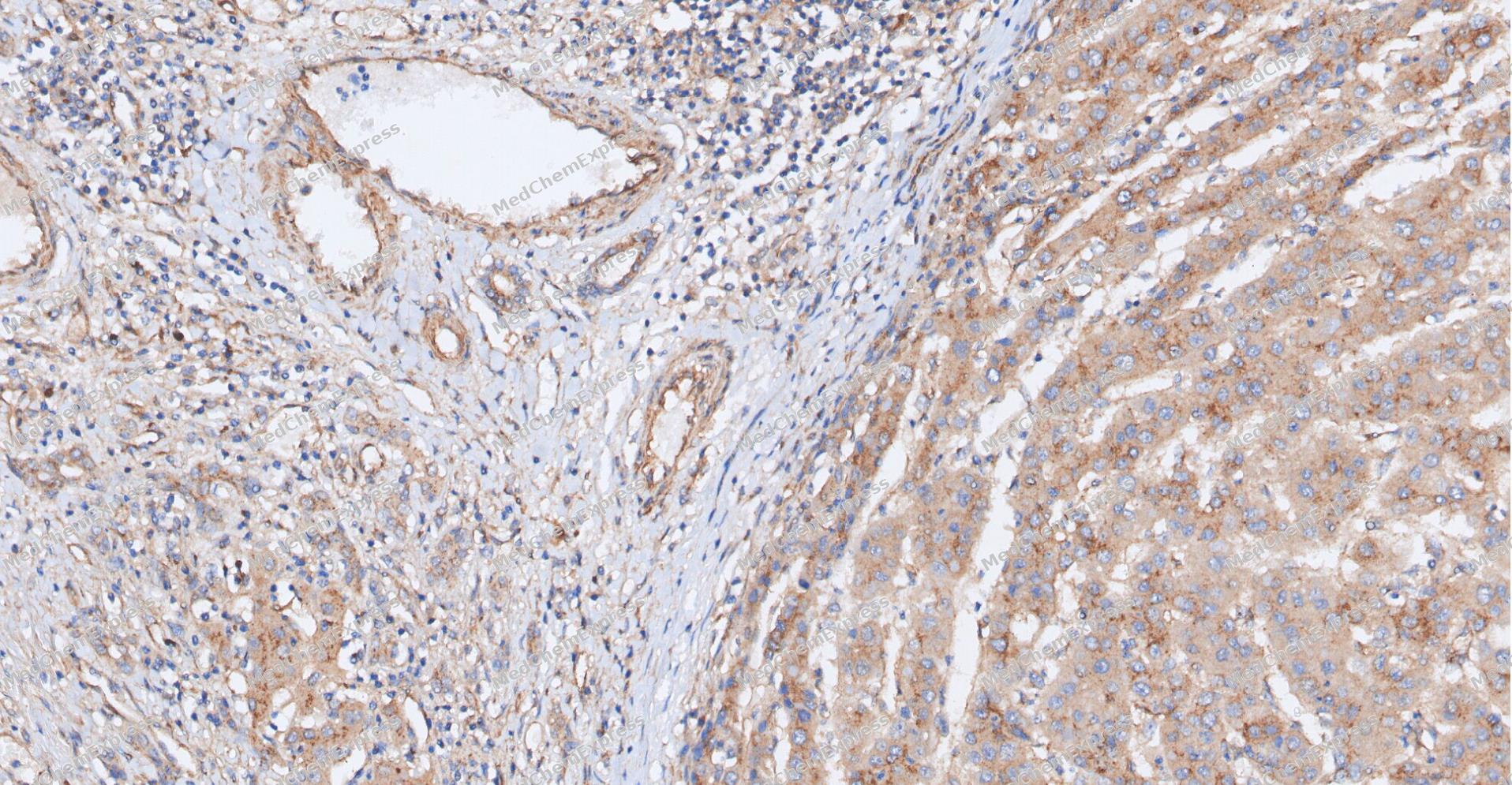 Immunohistochemical analysis of paraffin-embedded human Liver cancer tissue using S1P1/EDG1 antibody was performed. The section was pretreated using high-temperature and high-pressure mediated EDTA antigen retrieval buffer (pH 9.0), for 5 minutes. The tissues were incubated with primary antibody (HY-P84177, 1:50 dilution) at room temperature for 60 minutes. Detection was performed using an HRP conjugated compact polymer system. DAB was used as the chromogen. The tissues were counterstained with hematoxylin and mounted with neutral balsam mounting medium.
 -
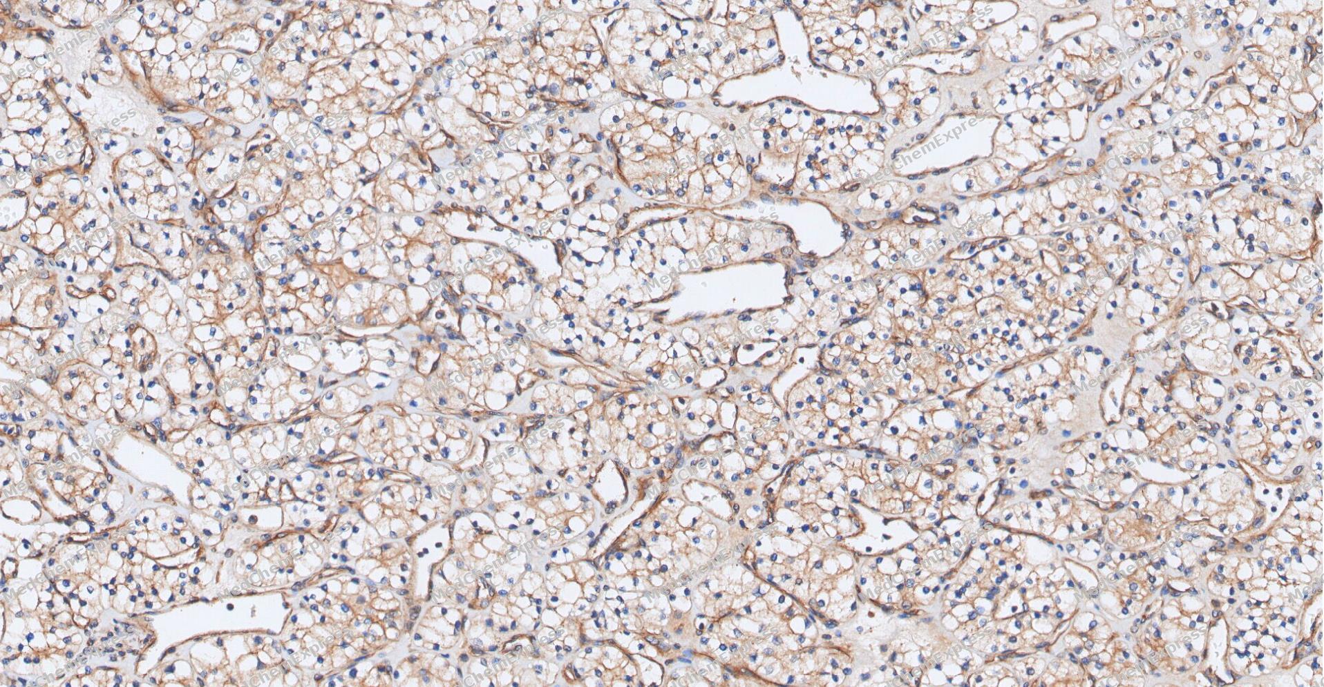 Immunohistochemical analysis of paraffin-embedded human Kidney cancer tissue using S1P1/EDG1 antibody was performed. The section was pretreated using high-temperature and high-pressure mediated EDTA antigen retrieval buffer (pH 9.0), for 5 minutes. The tissues were incubated with primary antibody (HY-P84177, 1:50 dilution) at room temperature for 60 minutes. Detection was performed using an HRP conjugated compact polymer system. DAB was used as the chromogen. The tissues were counterstained with hematoxylin and mounted with neutral balsam mounting medium.
 -
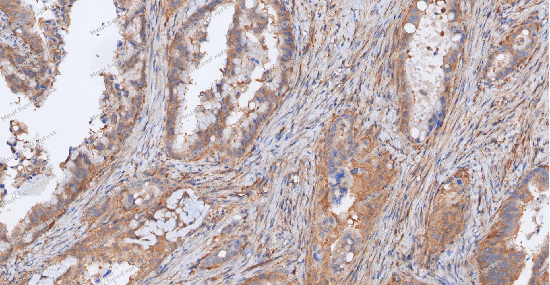 Immunohistochemical analysis of paraffin-embedded human Gastric Cancer tissue using S1P1/EDG1 antibody was performed. The section was pretreated using high-temperature and high-pressure mediated EDTA antigen retrieval buffer (pH 9.0), for 5 minutes. The tissues were incubated with primary antibody (HY-P84177, 1:50 dilution) at room temperature for 60 minutes. Detection was performed using an HRP conjugated compact polymer system. DAB was used as the chromogen. The tissues were counterstained with hematoxylin and mounted with neutral balsam mounting medium.
 -
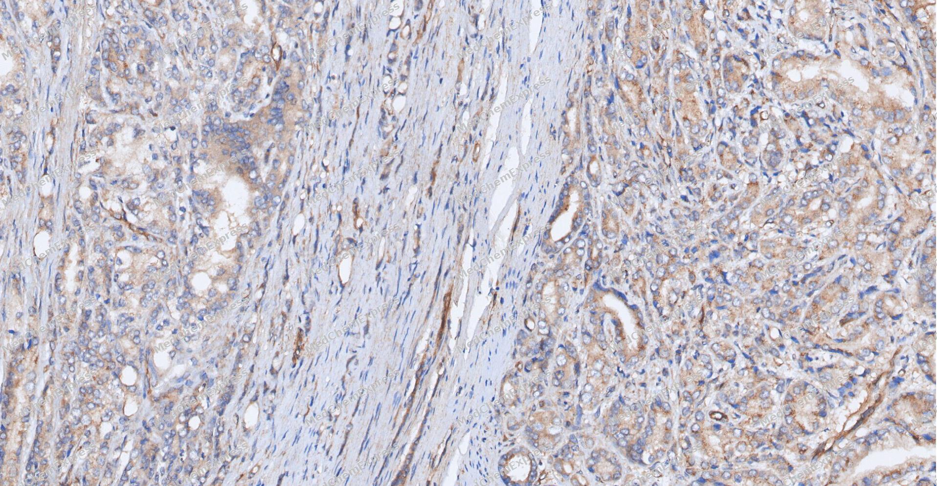 Immunohistochemical analysis of paraffin-embedded human Prostate Cancer tissue using S1P1/EDG1 antibody was performed. The section was pretreated using high-temperature and high-pressure mediated EDTA antigen retrieval buffer (pH 9.0), for 5 minutes. The tissues were incubated with primary antibody (HY-P84177, 1:50 dilution) at room temperature for 60 minutes. Detection was performed using an HRP conjugated compact polymer system. DAB was used as the chromogen. The tissues were counterstained with hematoxylin and mounted with neutral balsam mounting medium.
 -
 Immunohistochemical analysis of paraffin-embedded human glioma tissue using S1P1/EDG1 Antibody (HY-P84177, 1/500). The section was pretreated using heat mediated antigen retrieval with sodium citrate buffer (pH 6.0) for 8 minutes. The tissues were blocked with quick block buffer for 0.5 hours at room temperature, washed with PBS and PBST, and then incubated with the primary antibody overnight at 4℃. The detection was performed using an HRP conjugated compact polymer system. DAB was used as the chromogen. Tissues were counterstained with hematoxylin and mounted with DPX.
 -
 Immunohistochemical analysis of paraffin-embedded human endometrium cancer tissue using S1P1/EDG1 Antibody (HY-P84177, 1/500). The section was pretreated using heat mediated antigen retrieval with sodium citrate buffer (pH 6.0) for 8 minutes. The tissues were blocked with quick block buffer for 0.5 hours at room temperature, washed with PBS and PBST, and then incubated with the primary antibody overnight at 4℃. The detection was performed using an HRP conjugated compact polymer system. DAB was used as the chromogen. Tissues were counterstained with hematoxylin and mounted with DPX.
 -
 Immunohistochemical analysis of paraffin-embedded human stomach tissue using S1P1/EDG1 Antibody (HY-P84177, 1/500). The section was pretreated using heat mediated antigen retrieval with sodium citrate buffer (pH 6.0) for 8 minutes. The tissues were blocked with quick block buffer for 0.5 hours at room temperature, washed with PBS and PBST, and then incubated with the primary antibody overnight at 4℃. The detection was performed using an HRP conjugated compact polymer system. DAB was used as the chromogen. Tissues were counterstained with hematoxylin and mounted with DPX.
 -
 Immunohistochemical analysis of paraffin-embedded human brain tissue using S1P1/EDG1 Antibody (HY-P84177, 1/500). The section was pretreated using heat mediated antigen retrieval with sodium citrate buffer (pH 6.0) for 8 minutes. The tissues were blocked with quick block buffer for 0.5 hours at room temperature, washed with PBS and PBST, and then incubated with the primary antibody overnight at 4℃. The detection was performed using an HRP conjugated compact polymer system. DAB was used as the chromogen. Tissues were counterstained with hematoxylin and mounted with DPX.
 -
 Immunohistochemical analysis of paraffin-embedded human cerebellum tissue using S1P1/EDG1 Antibody (HY-P84177, 1/500). The section was pretreated using heat mediated antigen retrieval with sodium citrate buffer (pH 6.0) for 8 minutes. The tissues were blocked with quick block buffer for 0.5 hours at room temperature, washed with PBS and PBST, and then incubated with the primary antibody overnight at 4℃. The detection was performed using an HRP conjugated compact polymer system. DAB was used as the chromogen. Tissues were counterstained with hematoxylin and mounted with DPX.
 -
 Immunohistochemical analysis of paraffin-embedded human pancreas tissue using S1P1/EDG1 Antibody (HY-P84177, 1/500). The section was pretreated using heat mediated antigen retrieval with sodium citrate buffer (pH 6.0) for 8 minutes. The tissues were blocked with quick block buffer for 0.5 hours at room temperature, washed with PBS and PBST, and then incubated with the primary antibody overnight at 4℃. The detection was performed using an HRP conjugated compact polymer system. DAB was used as the chromogen. Tissues were counterstained with hematoxylin and mounted with DPX.


Background
-
Function
S1P1/EDG1 is a G-protein coupled receptor for the bioactive lysosphingolipid sphingosine 1-phosphate (S1P) that seems to be coupled to the G(i) subclass of heteromeric G proteins. Signaling leads to the activation of RAC1, SRC, PTK2/FAK1 and MAP kinases. Plays an important role in cell migration, probably via its role in the reorganization of the actin cytoskeleton and the formation of lamellipodia in response to stimuli that increase the activity of the sphingosine kinase SPHK1. Required for normal chemotaxis toward sphingosine 1-phosphate. Required for normal embryonic heart development and normal cardiac morphogenesis. Plays an important role in the regulation of sprouting angiogenesis and vascular maturation. Inhibits sprouting angiogenesis to prevent excessive sprouting during blood vessel development. Required for normal egress of mature T-cells from the thymus into the blood stream and into peripheral lymphoid organs. Plays a role in the migration of osteoclast precursor cells, the regulation of bone mineralization and bone homeostasis. Plays a role in responses to oxidized 1-palmitoyl-2-arachidonoyl-sn-glycero-3-phosphocholine by pulmonary endothelial cells and in the protection against ventilator-induced lung injury
-
Subcellular Localization
Cell membrane; Multi-pass membrane protein; Endosome; Membrane raft
-
Expression
Tissue_specificity:Endothelial cells, and to a lesser extent, vascular smooth muscle cells, fibroblasts, melanocytes, and epithelioid cells.
Induction:By the tumor promoter phorbol 12-myristate 13-acetate (PMA) in the presence of cycloheximide -
Subunit
Interacts with GNAI1 and GNAI3. Interacts with CD69; this interaction promotes S1PR1 degradation (PubMed:37039481)
-
SwissProt ID
-
Synonyms
S1PR1; EDG1; S1P1; ECGF1; EDG-1; CHEDG1; D1S3362


Documentation
-
Data Sheet (234 KB)
-
SDS (251 KB)
- English - EN (251 KB)
- Français - FR (251 KB)
- Deutsch - DE (251 KB)
- Norwegian - NO (251 KB)
- Español - ES (251 KB)
- Swedish - SV (251 KB)
- Italian - IT (251 KB)
- Korean - KR (251 KB)
- Portuguese - PT (251 KB)
-
User Guide for Antibodies (1077 KB)

 Powered by Bioz
