



CDK9 Antibody (YA509)
(Synonyms: TAK; C-2k; CTK1; CDC2L4; PITALRE)Based on 1 Customer Validation
CDK9 Antibody (YA509) is a Rabbit-derived and non-conjugated IgG monoclonal antibody, targeting to CDK9.
-
Host:
Rabbit
-
Isotype:
IgG
-
Application:
WB, IHC-F, IHC-P, ICC/IF, IP
-
Reactivity :
Human, Mouse, Rat
-
Formulation:
Supplied in 50 mM Tris-Glycine (pH 7.4), 0.15 M NaCl, 40% Glycerol and 0.05% BSA. Preservative: 0.01% Sodium azide
-
Conjugation:
Non-conjugated


To place orders, for customer services and technical support, please contact: MedChemExpress USA
Tel: 609-228-6898 E-mail: [email protected] [email protected]





Applications
| Application |
WB

WB: Western Blot
|
IHC-P
IHC-P: Immunohistochemistry-Paraffin
|
IHC-F
IHC-F: Immunohistochemistry-Frozen
|
ICC/IF
ICC/IF: Immunocytochemistry/
Immunofluorescence |
IP
IP: Immunoprecipitation
|
|---|---|---|---|---|---|
| Dilution Ratio | 1:500-1:1000 | 1:50-1:100 | 1:50-1:100 | 1:50-1:200 | 1:20 |


Product Details
CDK9 Antibody (YA509) is a Rabbit-derived and non-conjugated IgG monoclonal antibody, targeting to CDK9.
-
Host Rabbit

-
Clonality Recombinant,Monoclonal

-
Species ReactivityHuman, Mouse, Rat
-
Observed Molecular WeightObserved band size: 43 kDa
Note: Due to possible protein modifications or aggregation, the molecular weight should be confirmed by actual measurement, and the predicted value is for reference only.

-
Calculated Molecular Weight Predicted band size: 43 kDa

Entrez Gene: 1025 Human ; 107951 Mouse ; 362110 Rat
SwissProt: P50750 Human ; Q99J95 Mouse ; Q641Z4 Rat
OMIM: 603251 Human
Synthetic peptide corresponding to Human Cdk9.The exact sequence is proprietary to MCE.
Endogenous
affinity purified
Non-conjugated
Unmodified
IgG


Product Properties
-
Appearance
Solution
-
Formulation
Supplied in 50 mM Tris-Glycine (pH 7.4), 0.15 M NaCl, 40% Glycerol and 0.05% BSA. Preservative: 0.01% Sodium azide
-
Concentration
Batch-dependent, Please check the COA for the concentration of each lot. Check Lot Concentration
-
Storage & Stability
Stored at -20°C for 1 year. Avoid repeated freeze / thaw cycles.
-
Shipping
Shipping with blue ice.


Verification Images
-
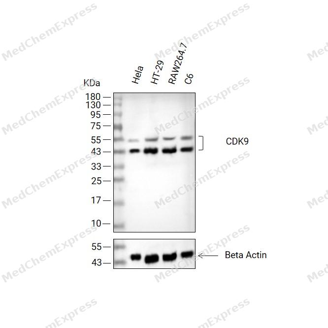 Western blot analysis of extracts from Hela (lane2(20μg), HT-29 (lane3(20μg), RAW264.7 (lane4(20μg) and C6 (lane5(20μg) using CDK9 Antibody (HY-P80614). Proteins were transferred to a PVDF membrane and blocked with 5% non-fat milk in TBST at 4°C overnight. The primary antibody (1/1000) and Loading control antibody (Beta Actin, HY-P80993, 1/10,000) was used in 5% non-fat milk in TBST for 2 hour at room temperature. Goat Anti-Rabbit IgG-HRP Secondary Antibody (HY-P8001, 1/10,000) was used for 1 hour at room temperature.
 -
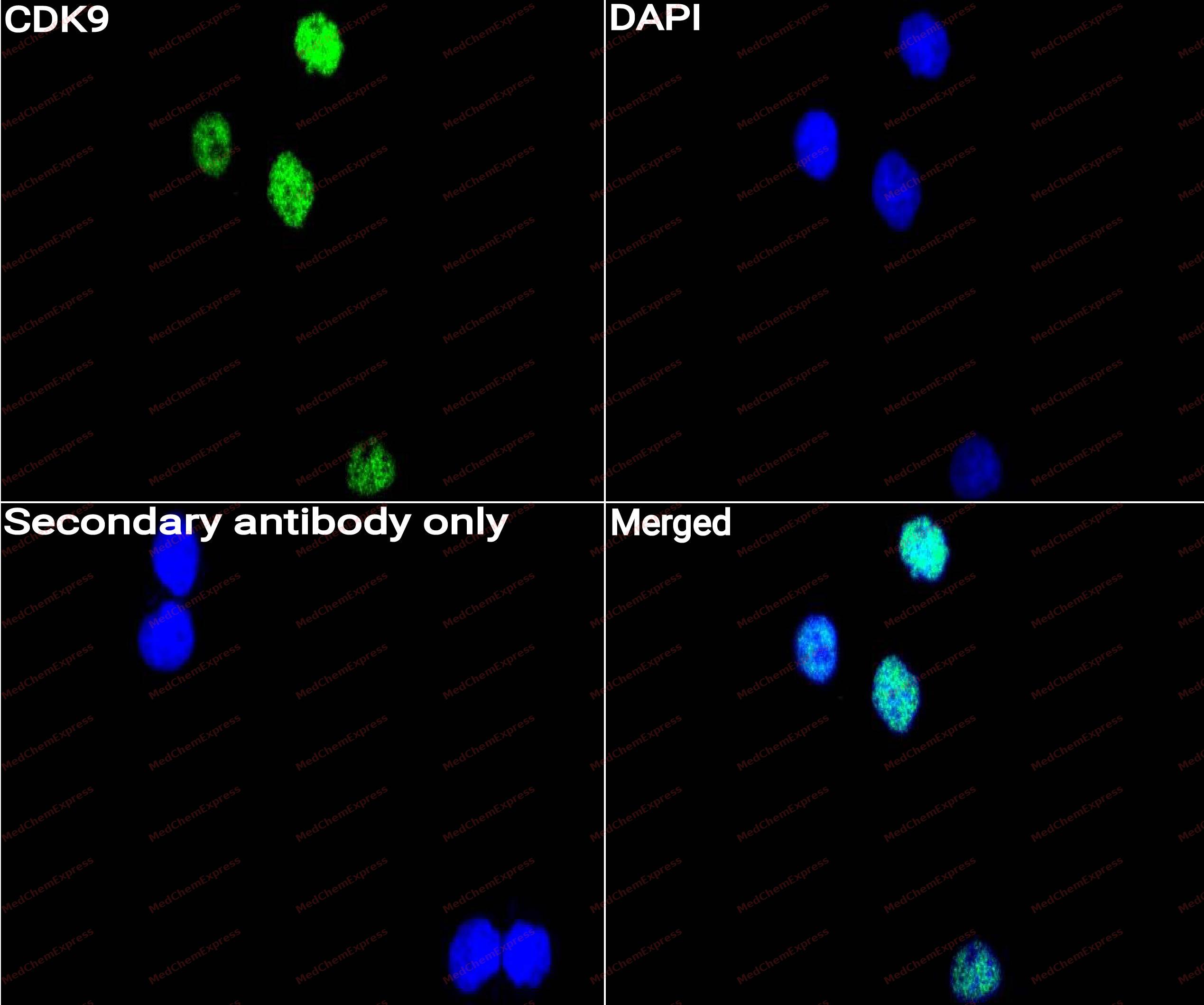 Immunocytochemistry analysis of Hela cells labeling CDK9 with CDK9 Antibody (HY-P80614) at 1/50 dilution. Cells were fixed in 4% paraformaldehyde for 15 minutes at room temperature, permeabilized with 0.1% Triton X-100 for 10 minutes at room temperature, then blocked with QuickBlock™ Blocking Buffer for Immunol Staining for 10 min at room temperature. Cells were then incubated with CDK9 Antibody (HY-P80614) at 1/50 dilution in QuickBlock™ Blocking Buffer for Immunol Staining at 4 ℃. Alexa Fluor® 488-conjugated AffiniPure Goat Anti-Rabbit IgG H&L(HY-P8002, Green) was used as the secondary antibody at 1/1,000 dilution. PBS instead of the primary antibody was used as the secondary antibody only control. The Nuclear counterstain was DAPI (Blue).
 -
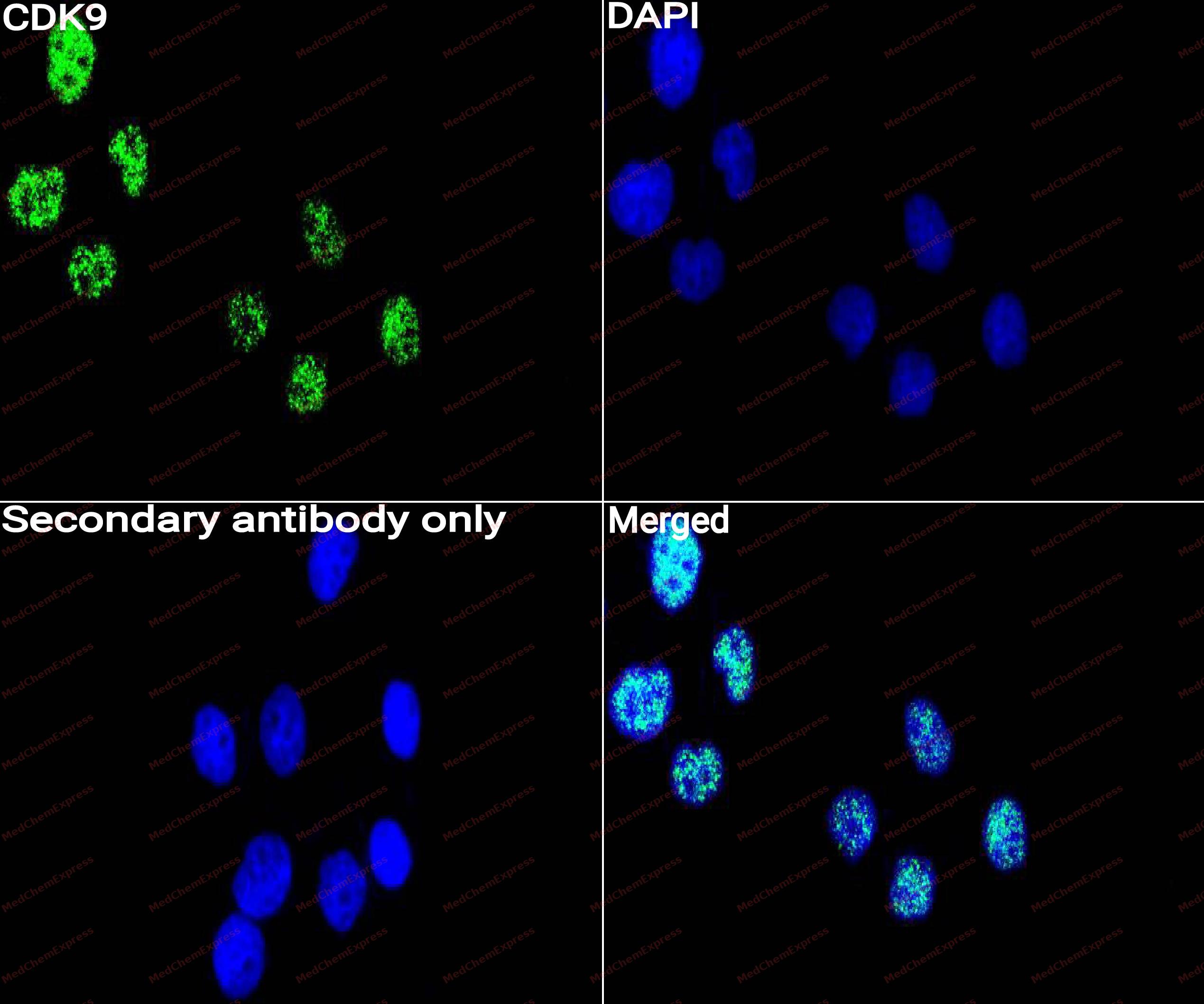 Immunocytochemistry analysis of NIH3T3 cells labeling CDK9 with CDK9 Antibody (HY-P80614) at 1/50 dilution. Cells were fixed in 4% paraformaldehyde for 15 minutes at room temperature, permeabilized with 0.1% Triton X-100 for 10 minutes at room temperature, then blocked with QuickBlock™ Blocking Buffer for Immunol Staining for 10 min at room temperature. Cells were then incubated with CDK9 Antibody (HY-P80614)at 1/50 dilution in QuickBlock™ Blocking Buffer for Immunol Staining at 4 ℃. Alexa Fluor® 488-conjugated AffiniPure Goat Anti-Rabbit IgG H&L(HY-P8002,Green) was used as the secondary antibody at 1/1,000 dilution. PBS instead of the primary antibody was used as the secondary antibody only control. The Nuclear counterstain was DAPI (Blue).


Background
-
Function
CDK9 (cyclin-dependent kinase 9) is a transcription-associated serine/threonine kinase that functions as the catalytic subunit of positive transcription elongation factor b (P-TEFb) and regulates productive RNA polymerase II (RNAPII) elongation through phosphorylation of the RNAPII C-terminal domain and elongation factors.[1][2] Mechanistically, CDK9-cyclin T complexes promote release of promoter-proximally paused RNAPII into gene bodies, thereby controlling transcriptional programs required for cellular responses to developmental and environmental signals.[1][2] Beyond transcriptional elongation, CDK9 contributes to transcription initiation, termination, and maintenance of appropriate transcriptional output, highlighting its central role in gene expression regulation.[1] Dysregulated CDK9 activity has been linked to multiple pathological conditions, particularly cancer, where aberrant transcriptional dependencies create vulnerability to pharmacological CDK9 inhibition.[1][3] In cancer models, activation of P-TEFb-dependent transcription supports oncogenic pathways including MYC, NF-κB, and stress-response signaling, and selective suppression of CDK9 can induce apoptotic programs and impair tumor cell proliferation.[4] Compared with related transcriptional cyclin-dependent kinases such as CDK12 and CDK13, which primarily regulate distinct transcriptional and RNA-processing programs through cyclin K-containing complexes, CDK9 acts as the principal P-TEFb kinase controlling promoter-proximal pause release and rapid transcriptional activation.[1][5] For experimental applications, small-molecule CDK9 inhibitors including flavopiridol have been widely used to suppress RNAPII-dependent transcription, investigate transcriptional addiction, and evaluate therapeutic vulnerabilities associated with transcriptional dysregulation.[1][6]
-
Subcellular Localization
Nucleus; Cytoplasm; Nucleus, PML body
-
Expression
Tissue_specificity:general expression
Induction:By replication stress, in chromatin. Probably degraded by the proteasome upon Thr-186 dephosphorylation -
Isoforms & Post-Translational Modification
P50750 has 2 isomers: P50750-1: 42778 Da (predicted); P50750-2: 53365 Da (predicted).
Autophosphorylation at Thr-186, Ser-347, Thr-350, Ser-353, Thr-354 and Ser-357 triggers kinase activity by promoting cyclin and substrate binding (e.g. HIV TAT) upon conformational changes. Thr-186 phosphorylation requires the calcium Ca(2+) signaling pathway, including CaMK1D and calmodulin. This inhibition is relieved by Thr-29 dephosphorylation. However, phosphorylation at Thr-29 is inhibitory within the HIV transcription initiation complex. Phosphorylation at Ser-175 inhibits kinase activity. Can be phosphorylated on either Thr-362 or Thr-363 but not on both simultaneously (PubMed:18566585);Dephosphorylation of Thr-186 by PPM1A and PPM1B blocks CDK9 activity and may lead to CDK9 proteasomal degradation (PubMed:18483222, PubMed:18829461). However, PPP1CA-mediated Thr-186 dephosphorylation is required to release P-TEFb from its inactive P-TEFb/7SK snRNP complex (PubMed:18483222, PubMed:18829461). Dephosphorylated at Ser-347 by the PNUTS-PP1 complex during RNA polymerase II transcription pause-release (PubMed:39603239). Dephosphorylation of C-terminus Thr and Ser residues by protein phosphatase-1 (PP1) triggers CDK9 activity, contributing to the activation of HIV-1 transcription;N6-acetylation of Lys-44 promotes kinase activity, whereas acetylation of both Lys-44 and Lys-48 mediated by PCAF/KAT2B and GCN5/KAT2A reduces kinase activity (PubMed:17452463, PubMed:18250157). The acetylated form associates with PML bodies in the nuclear matrix and with the transcriptionally silent HIV-1 genome; deacetylated upon transcription stimulation (PubMed:17452463, PubMed:18250157). Deacetylated by SIRT7, promoting the kinase activity and subsequent 'Ser-2' phosphorylation of the C-terminal domain (CTD) of RNA polymerase II (PubMed:28426094);Polyubiquitinated and thus activated by UBR5. This ubiquitination is promoted by TFIIS/TCEA1 and favors 'Ser-2' phosphorylation of RPB1/POLR2A CTD -
Subunit
Component of the super elongation complex (SEC), at least composed of EAF1, EAF2, CDK9, MLLT3/AF9, AFF (AFF1 or AFF4), the P-TEFb complex and ELL (ELL, ELL2 or ELL3). Associates with CCNT1/cyclin-T1, CCNT2/cyclin-T2 (isoform A and isoform B) or CCNK/cyclin-K to form active P-TEFb. P-TEFb forms a complex with AFF4/AF5Q31 and is part of the super elongation complex (SEC). Component of a complex which is composed of at least 5 members: HTATSF1/Tat-SF1, P-TEFb complex, RNA pol II, SUPT5H and NCL/nucleolin. Associates with UBR5 and forms a transcription regulatory complex composed of CDK9, RNAP II, UBR5 and TFIIS/TCEA1 that can stimulate target gene transcription (e.g. gamma fibrinogen/FGG) by recruiting their promoters. Component of the 7SK snRNP inactive complex which is composed of at least 8 members: P-TEFb (composed of CDK9 and CCNT1/cyclin-T1), HEXIM1, HEXIM2, LARP7, BCDIN3, SART3 proteins and 7SK and U6 snRNAs. This inactive 7SK snRNP complex can also interact with NCOR1 and HDAC3, probably to regulate CDK9 acetylation. Release of P-TEFb from P-TEFb/7SK snRNP complex requires both PP2B to transduce calcium Ca(2+) signaling in response to stimuli (e.g. UV or hexamethylene bisacetamide (HMBA)) and PPP1CA to dephosphorylate Thr-186. This released P-TEFb remains inactive in the pre-initiation complex with BRD4 until new Thr-186 phosphorylation occurs after the synthesis of a short RNA (PubMed:10393184, PubMed:10574912, PubMed:11884399, PubMed:12037670, PubMed:12065898, PubMed:12718890, PubMed:15965233, PubMed:16109376, PubMed:17452463, PubMed:17643375, PubMed:18249148, PubMed:18483222, PubMed:18566585, PubMed:20159561, PubMed:20471948, PubMed:21127351, PubMed:21779453, PubMed:22195968, PubMed:30134174, PubMed:9491887). Interacts with BRD4; to target chromatin binding (PubMed:16109376, PubMed:16109377, PubMed:18483222). Interacts with JMJD6 (PubMed:24360279). Interacts with activated nuclear STAT3 and RELA/p65 (PubMed:17956865, PubMed:18362169). Binds to AR and MYOD1 (PubMed:12037670, PubMed:20980437). Forms a complex composed of CDK9, CCNT1/cyclin-T1, EP300 and GATA4 that stimulates hypertrophy in cardiomyocytes (PubMed:20081228). The large PER complex involved in the repression of transcriptional termination is composed of at least PER2, CDK9, DDX5, DHX9, NCBP1 and POLR2A (By similarity). Interacts with HSF1 (PubMed:27189267). Interacts with TBX21 (By similarity). Isoform 3: binds to KU70/XRCC6 (PubMed:20535204). Interacts with WDR43 (By similarity). Interacts with ZMYND8; the association appears to occur between homodimeric ZMYND8 and the activated form of the P-TEFb complex (PubMed:30134174)
-
SwissProt ID
-
Synonyms
TAK; C-2k; CTK1; CDC2L4; PITALRE
-
Research Field
Cell Biology


Documentation
-
Data Sheet (262 KB)
-
SDS (251 KB)
- English - EN (251 KB)
- Français - FR (251 KB)
- Deutsch - DE (251 KB)
- Norwegian - NO (251 KB)
- Español - ES (251 KB)
- Swedish - SV (251 KB)
- Italian - IT (251 KB)
- Korean - KR (251 KB)
- Portuguese - PT (251 KB)
-
User Guide for Antibodies (1077 KB)
References
[1]. Egloff S. CDK9 keeps RNA polymerase II on track. Cell Mol Life Sci. 2021 Jul;78(14):5543-5567. [Content Brief]
[2]. Anshabo AT, et al. CDK9: A Comprehensive Review of Its Biology, and Its Role as a Potential Target for Anti-Cancer Agents. Front Oncol. 2021 May 10;11:678559. [Content Brief]
[3]. Constantin TA, et al. Transcription associated cyclin-dependent kinases as therapeutic targets for prostate cancer. Oncogene. 2022 Jun;41(24):3303-3315. [Content Brief]
[4]. Yuan B, et al. Engineering of cytosine base editors with DNA damage minimization and editing scope diversification. Nucleic Acids Res. 2023 Nov 10;51(20):e105. [Content Brief]
[5]. Fan Z, et al. CDK13 cooperates with CDK12 to control global RNA polymerase II processivity. Sci Adv. 2020 Apr 29;6(18):eaaz5041. [Content Brief]
[6]. Phiel CJ, et al. Histone deacetylase is a direct target of valproic acid, a potent anticonvulsant, mood stabilizer, and teratogen. J Biol Chem. 2001 Sep 28;276(39):36734-41. [Content Brief]

 Powered by Bioz
