



COX IV Antibody (YA5187)
(Synonyms: AL024441; COX 4; COX IV 1; COX IV; COX IV-1; Cox4; COX41_HUMAN; Cox4a; COX4B; COX4I1; COX4I2; COX4L2; COXIV; Cytochrome c oxidase polypeptide IV; Cytochrome c oxidase subunit 4 isoform 1 mitochondrial; Cytochrome c oxidase subunit 4 isoform 1, mitochondrial; Cytochrome C Oxidase subunit IV; Cytochrome c oxidase subunit IV isoform 1; Cytochrome c oxidase subunit IV isoform 2; lung; Cytochrome c oxydase subunit; dJ857M17.2; MGC105470; MGC72016.)
Based on 1 Customer Validation
COX IV Antibody (YA5187) is a Mouse-derived and non-conjugated monoclonal antibody, targeting to COX IV.
-
Host:
Mouse
-
Application:
WB, IHC-P, ICC/IF, IP, FC
-
Reactivity :
Human, Mouse, Rat, Hamster, Goat, Monkey
-
Formulation:
Supplied in PBS containing 50% glycerol, 0.5% BSA and 0.02% sodium azide.
-
Conjugation:
Non-conjugated


To place orders, for customer services and technical support, please contact: MedChemExpress USA
Tel: 609-228-6898 E-mail: [email protected] [email protected]





Applications
| Application |
WB

WB: Western Blot
|
ICC/IF
ICC/IF: Immunocytochemistry/
Immunofluorescence |
FC
FC: Flow Cytometry
|
IHC
|
|---|---|---|---|---|
| Dilution Ratio | 1:1000 | 1:150 | 1:100 | 1:50-200 |


Product Details
COX IV Antibody (YA5187) is a Mouse-derived and non-conjugated monoclonal antibody, targeting to COX IV.
-
Host Mouse

-
Clonality Monoclonal

-
Species ReactivityHuman, Mouse, Rat, Hamster, Goat, Monkey
-
Observed Molecular WeightObserved band size: 17 kDa
Note: Due to possible protein modifications or aggregation, the molecular weight should be confirmed by actual measurement, and the predicted value is for reference only.

A synthetic peptide corresponding to carboxyl terminal residues of human COX IV
affinity chromatography.
Non-conjugated
Unmodified


Product Properties
-
Appearance
Liquid
-
Formulation
Supplied in PBS containing 50% glycerol, 0.5% BSA and 0.02% sodium azide.
-
Concentration
Batch-dependent, Please check the COA for the concentration of each lot. Check Lot Concentration
-
Storage & Stability
Stored at -20°C for 1 year. Avoid repeated freeze / thaw cycles.
-
Shipping
Shipping with blue ice.


Verification Images
-
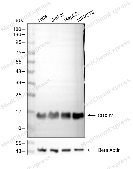 Western blot analysis of extracts from Hela (lane 2(20μg), Jurkat (lane 3(20μg), HepG2 (lane 4(20μg), NIH/3T3 (lane 5(20μg) using COX IV Antibody. Proteins were transferred to a PVDF membrane and blocked with 5% BSA in TBST for 2 hour at room temperature. The primary antibody and Loading control antibody (Beta Actin, HY-P80438, 1/3000) was used in 5% BSA in TBST at 4°C overnight. Goat Anti-Mouse/Rabbit IgG-HRP Secondary Antibody (HY-P8004/HY-P8001, 1/10,000) was used for 1 hour at room temperature.
 -
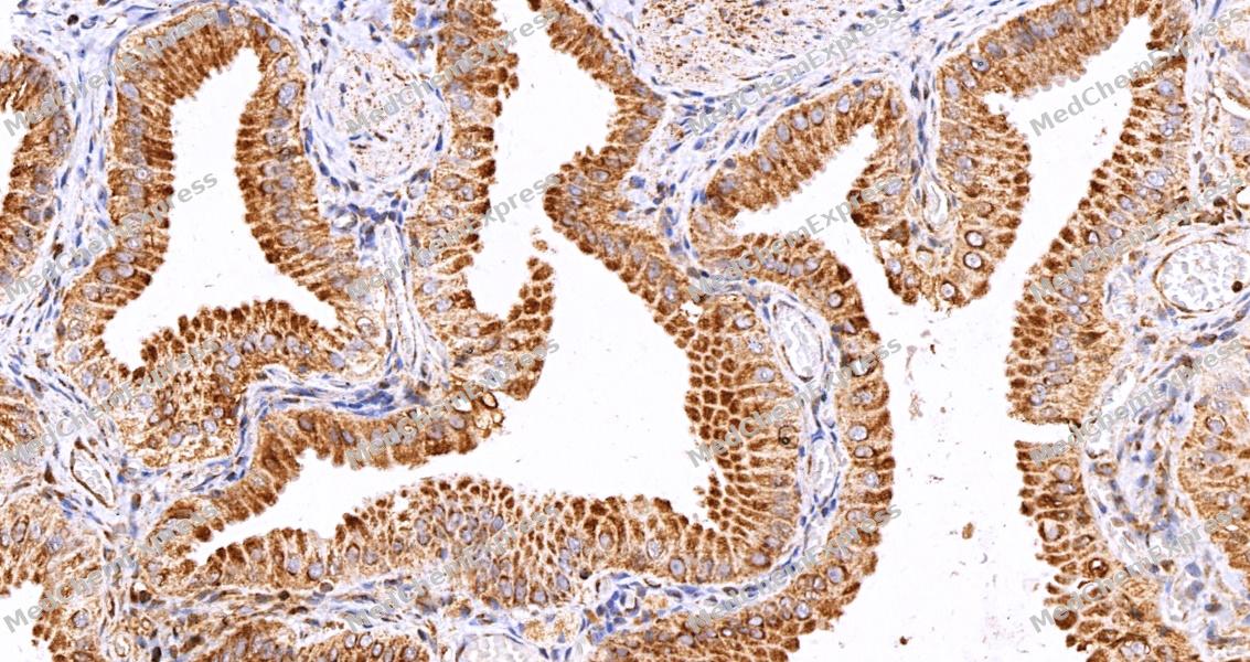 Immunohistochemical analysis of paraffin-embedded human gallbladder tissue using COX IV Antibody (HY-P85495, 1/100). The section was pretreated using heat mediated antigen retrieval with sodium citrate buffer (pH 6.0) for 8 minutes. The tissues were blocked with quick block buffer for 0.5 hours at room temperature, washed with PBS and PBST, and then incubated with the primary antibody overnight at 4℃. The detection was performed using an HRP conjugated compact polymer system. DAB was used as the chromogen. Tissues were counterstained with hematoxylin and mounted with DPX.
 -
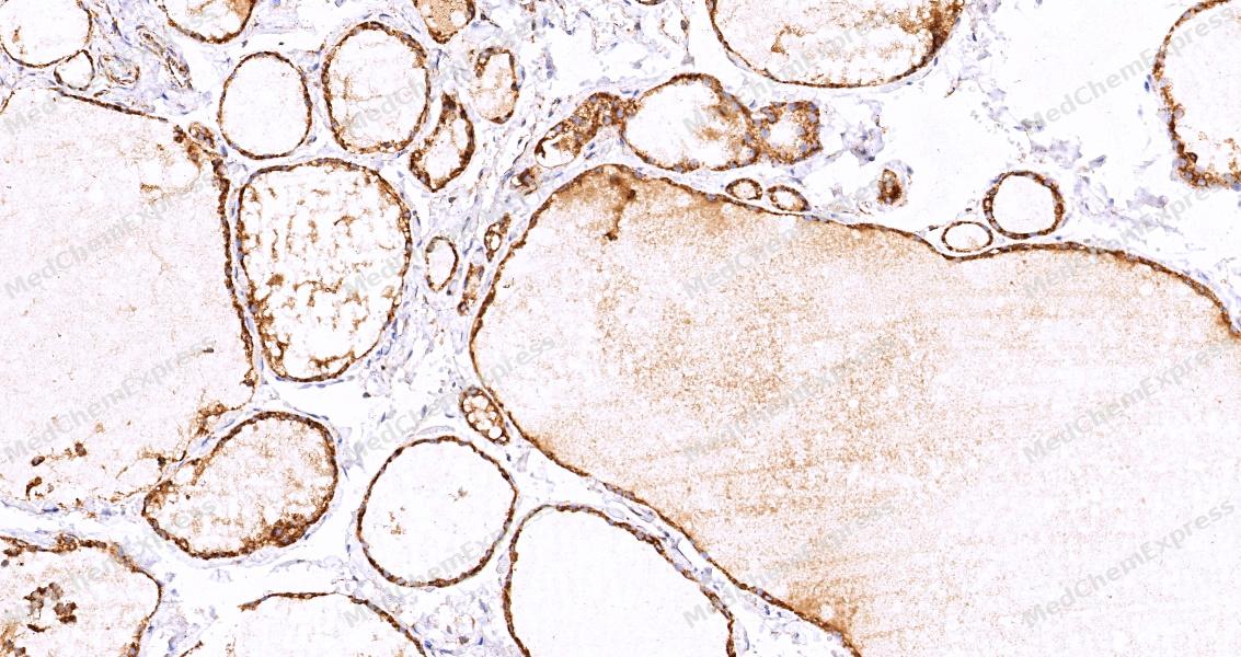 Immunohistochemical analysis of paraffin-embedded human thyroid gland tissue using COX IV Antibody (HY-P85495, 1/100). The section was pretreated using heat mediated antigen retrieval with sodium citrate buffer (pH 6.0) for 8 minutes. The tissues were blocked with quick block buffer for 0.5 hours at room temperature, washed with PBS and PBST, and then incubated with the primary antibody overnight at 4℃. The detection was performed using an HRP conjugated compact polymer system. DAB was used as the chromogen. Tissues were counterstained with hematoxylin and mounted with DPX.
 -
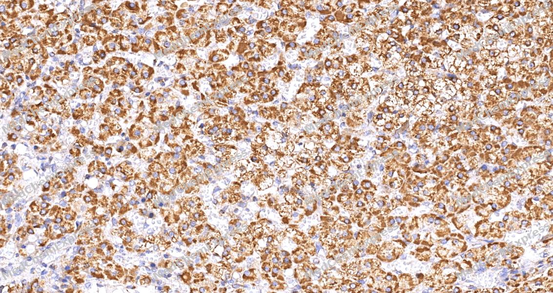 Immunohistochemical analysis of paraffin-embedded human adrenal gland tissue using COX IV Antibody (HY-P85495, 1/100). The section was pretreated using heat mediated antigen retrieval with sodium citrate buffer (pH 6.0) for 8 minutes. The tissues were blocked with quick block buffer for 0.5 hours at room temperature, washed with PBS and PBST, and then incubated with the primary antibody overnight at 4℃. The detection was performed using an HRP conjugated compact polymer system. DAB was used as the chromogen. Tissues were counterstained with hematoxylin and mounted with DPX.
 -
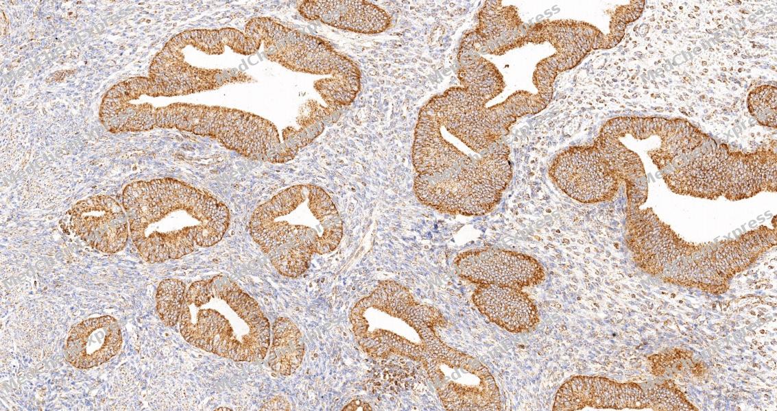 Immunohistochemical analysis of paraffin-embedded human endometrium tissue using COX IV Antibody (HY-P85495, 1/100). The section was pretreated using heat mediated antigen retrieval with sodium citrate buffer (pH 6.0) for 8 minutes. The tissues were blocked with quick block buffer for 0.5 hours at room temperature, washed with PBS and PBST, and then incubated with the primary antibody overnight at 4℃. The detection was performed using an HRP conjugated compact polymer system. DAB was used as the chromogen. Tissues were counterstained with hematoxylin and mounted with DPX.
 -
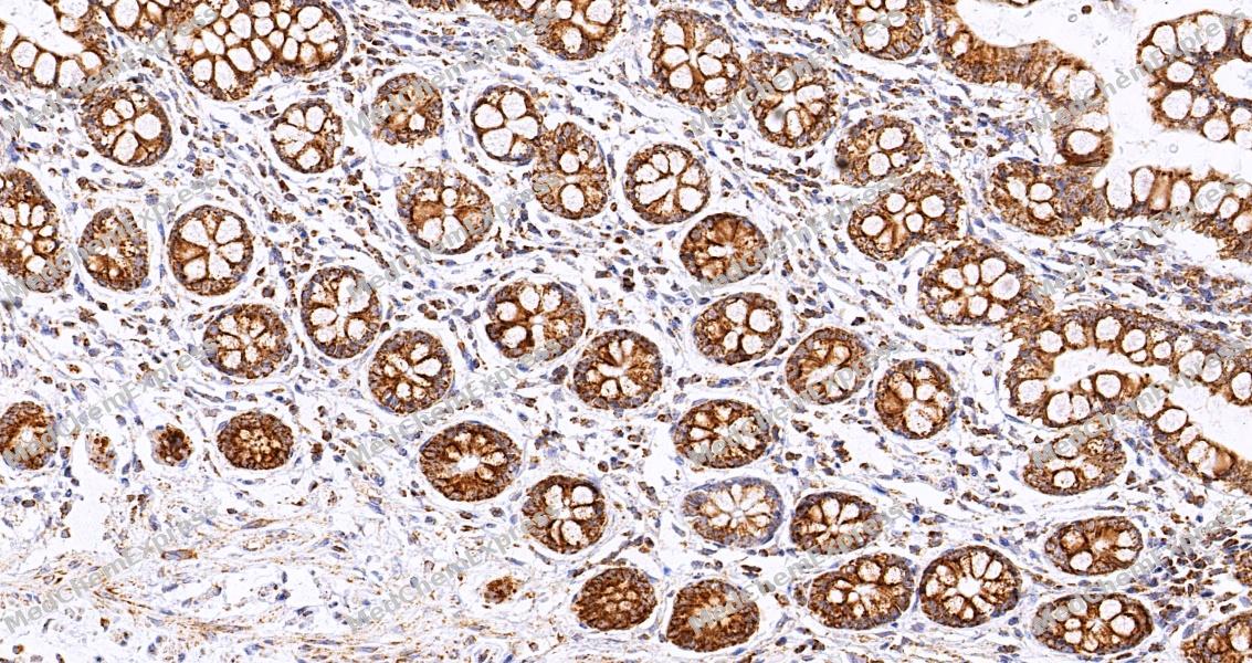 Immunohistochemical analysis of paraffin-embedded human smal lintestine tissue using COX IV Antibody (HY-P85495, 1/100). The section was pretreated using heat mediated antigen retrieval with sodium citrate buffer (pH 6.0) for 8 minutes. The tissues were blocked with quick block buffer for 0.5 hours at room temperature, washed with PBS and PBST, and then incubated with the primary antibody overnight at 4℃. The detection was performed using an HRP conjugated compact polymer system. DAB was used as the chromogen. Tissues were counterstained with hematoxylin and mounted with DPX.
 -
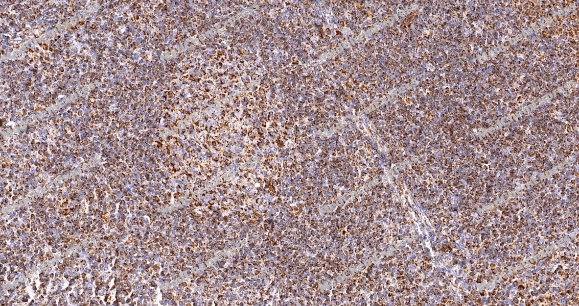 Immunohistochemical analysis of paraffin-embedded human tonsil tissue using COX IV Antibody (HY-P85495, 1/100). The section was pretreated using heat mediated antigen retrieval with sodium citrate buffer (pH 6.0) for 8 minutes. The tissues were blocked with quick block buffer for 0.5 hours at room temperature, washed with PBS and PBST, and then incubated with the primary antibody overnight at 4℃. The detection was performed using an HRP conjugated compact polymer system. DAB was used as the chromogen. Tissues were counterstained with hematoxylin and mounted with DPX.
 -
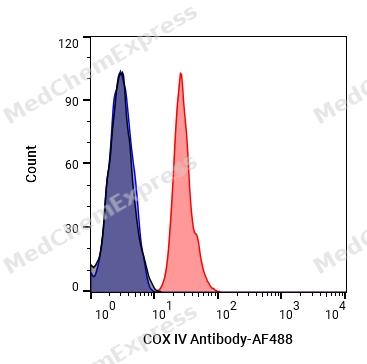 Flow Cytometry analysis of Hela cells labelling COX IV (red) with COX IV Antibody (anti-COX IV) (HY-P85495). Cells were fixed with 4% paraformaldehyde and permeabilised with 90% methanol. Then cells were stained with the primary antibody at 1/200 dilution for an hour at 4℃. Goat Anti-Mouse IgG H&L (AF488) (HY-P8005) was used as the secondary antibody at 1/1,000 dilution for 30 minutes at 4℃. Mouse IgG Isotype Control (HY-P80757, blue) was used as the isotype control, cells without incubation with primary antibody were used as the unlabeled control (black).


Background
-
Function
COX IV component of the cytochrome c oxidase, the last enzyme in the mitochondrial electron transport chain which drives oxidative phosphorylation. The respiratory chain contains 3 multisubunit complexes succinate dehydrogenase (complex II, CII), ubiquinol-cytochrome c oxidoreductase (cytochrome b-c1 complex, complex III, CIII) and cytochrome c oxidase (complex IV, CIV), that cooperate to transfer electrons derived from NADH and succinate to molecular oxygen, creating an electrochemical gradient over the inner membrane that drives transmembrane transport and the ATP synthase. Cytochrome c oxidase is the component of the respiratory chain that catalyzes the reduction of oxygen to water. Electrons originating from reduced cytochrome c in the intermembrane space (IMS) are transferred via the dinuclear copper A center (CU(A)) of subunit 2 and heme A of subunit 1 to the active site in subunit 1, a binuclear center (BNC) formed by heme A3 and copper B (CU(B)). The BNC reduces molecular oxygen to 2 water molecules using 4 electrons from cytochrome c in the IMS and 4 protons from the mitochondrial matrix
-
Subcellular Localization
Mitochondrion inner membrane; Single-pass membrane protein
-
Expression
Tissue_specificity:general expression -
Subunit
Component of the cytochrome c oxidase (complex IV, CIV), a multisubunit enzyme composed of 14 subunits. The complex is composed of a catalytic core of 3 subunits MT-CO1, MT-CO2 and MT-CO3, encoded in the mitochondrial DNA, and 11 supernumerary subunits COX4I1 (or COX4I2), COX5A, COX5B, COX6A1 (or COX6A2), COX6B1 (or COX6B2), COX6C, COX7A2 (or COX7A1), COX7B, COX7C, COX8A and NDUFA4, which are encoded in the nuclear genome (PubMed:30030519). The complex exists as a monomer or a dimer and forms supercomplexes (SCs) in the inner mitochondrial membrane with NADH-ubiquinone oxidoreductase (complex I, CI) and ubiquinol-cytochrome c oxidoreductase (cytochrome b-c1 complex, complex III, CIII), resulting in different assemblies (supercomplex SCI(1)III(2)IV(1) and megacomplex MCI(2)III(2)IV(2)) (PubMed:28844695). Interacts with AFG1L (PubMed:26759378). Interacts with PHB2; the interaction decreases in absence of SPHK2 (By similarity). Interacts with ABCB7; this interaction allows the regulation of cellular iron homeostasis and cellular reactive oxygen species (ROS) levels in cardiomyocytes (By similarity). Interacts with FLVCR2; this interaction occurs in the absence of heme and is disrupted upon heme binding. Interacts with IRGC (By similarity)
-
SwissProt ID
-
Synonyms
AL024441; COX 4; COX IV 1; COX IV; COX IV-1; Cox4; COX41_HUMAN; Cox4a; COX4B; COX4I1; COX4I2; COX4L2; COXIV; Cytochrome c oxidase polypeptide IV; Cytochrome c oxidase subunit 4 isoform 1 mitochondrial; Cytochrome c oxidase subunit 4 isoform 1, mitochondrial; Cytochrome C Oxidase subunit IV; Cytochrome c oxidase subunit IV isoform 1; Cytochrome c oxidase subunit IV isoform 2; lung; Cytochrome c oxydase subunit; dJ857M17.2; MGC105470; MGC72016.


Documentation
-
Data Sheet (235 KB)
-
SDS (254 KB)
- English - EN (254 KB)
- Français - FR (254 KB)
- Deutsch - DE (254 KB)
- Norwegian - NO (254 KB)
- Español - ES (254 KB)
- Swedish - SV (254 KB)
- Italian - IT (254 KB)
- Korean - KR (254 KB)
- Portuguese - PT (254 KB)
-
User Guide for Antibodies (1077 KB)

 Powered by Bioz
