



ERp57 Antibody (YA3300)
(Synonyms: ERp57; ERp60; ERp61; Glucose Regulated Protein 58 Kd; HsT17083; p58; PDIA3; Phospholipase C alpha; PI PLC)
Based on 1 Customer Validation
ERp57 Antibody (YA3300) is a Rabbit-derived and non-conjugated IgG monoclonal antibody, targeting to ERp57.
-
Host:
Rabbit
-
Isotype:
IgG
-
Application:
WB, IHC-P, IP
-
Reactivity :
Human
-
Formulation:
Supplied in 10mM PBS, pH 7.4, 150mM sodium chloride, 0.05% BSA, 0.02% sodium azide and 50% glycerol.
-
Conjugation:
Non-conjugated


To place orders, for customer services and technical support, please contact: MedChemExpress USA
Tel: 609-228-6898 E-mail: [email protected] [email protected]





Applications
| Application |
WB

WB: Western Blot
|
IHC-P
IHC-P: Immunohistochemistry-Paraffin
|
IP
IP: Immunoprecipitation
|
|---|---|---|---|
| Dilution Ratio | 1:500-1:1000 | 1:50-1:100 | 1:50 |


Product Details
ERp57 Antibody (YA3300) is a Rabbit-derived and non-conjugated IgG monoclonal antibody, targeting to ERp57.
-
Host Rabbit

-
Clonality Recombinant,Monoclonal

-
Species ReactivityHuman
-
Observed Molecular WeightObserved band size: 57 kDa
Note: Due to possible protein modifications or aggregation, the molecular weight should be confirmed by actual measurement, and the predicted value is for reference only.

-
Calculated Molecular Weight Predicted band size: 57 kDa

A synthesized peptide derived from human ERp57 aa150-200.
Endogenous
Affinity Chromatography
Non-conjugated
Unmodified
IgG


Product Properties
-
Appearance
Solution
-
Formulation
Supplied in 10mM PBS, pH 7.4, 150mM sodium chloride, 0.05% BSA, 0.02% sodium azide and 50% glycerol.
-
Concentration
Batch-dependent, Please check the COA for the concentration of each lot. Check Lot Concentration
-
Storage & Stability
Stored at -20°C for 1 year. Avoid repeated freeze / thaw cycles.
-
Shipping
Shipping with blue ice.


Verification Images
-
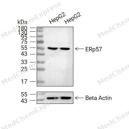 Western blot analysis of extracts from HepG2 (lane 2(20μg) and HepG2 (lane 3(40μg) using ERp57 (HY-P83555) Rabbit mAb. Proteins were transferred to a PVDF membrane and blocked with 5% non-fat milk in TBST for 2 hour at room temperature. The primary antibody (1/1000) and Loading control antibody (Beta Actin, HY-P80438, 1/10000) was used in 5% non-fat milk in TBST at 4°C overnight. Goat Anti-Rabbit IgG-HRP Secondary Antibody (1/10000) was used for 1 hour at room temperature.
 -
 Immunohistochemical analysis of paraffin-embedded human esophagus cancer tissue using ERp57 Antibody (HY-P83555, 1/100). The section was pretreated using heat mediated antigen retrieval with sodium citrate buffer (pH 6.0) for 8 minutes. The tissues were blocked with quick block buffer for 0.5 hours at room temperature, washed with PBS and PBST, and then incubated with the primary antibody overnight at 4℃. The detection was performed using an HRP conjugated compact polymer system. DAB was used as the chromogen. Tissues were counterstained with hematoxylin and mounted with DPX.
 -
 Immunohistochemical analysis of paraffin-embedded human ovarian cancer tissue using ERp57 Antibody (HY-P83555, 1/100). The section was pretreated using heat mediated antigen retrieval with sodium citrate buffer (pH 6.0) for 8 minutes. The tissues were blocked with quick block buffer for 0.5 hours at room temperature, washed with PBS and PBST, and then incubated with the primary antibody overnight at 4℃. The detection was performed using an HRP conjugated compact polymer system. DAB was used as the chromogen. Tissues were counterstained with hematoxylin and mounted with DPX.
 -
 Immunohistochemical analysis of paraffin-embedded human rectum cancer tissue using ERp57 Antibody (HY-P83555, 1/100). The section was pretreated using heat mediated antigen retrieval with sodium citrate buffer (pH 6.0) for 8 minutes. The tissues were blocked with quick block buffer for 0.5 hours at room temperature, washed with PBS and PBST, and then incubated with the primary antibody overnight at 4℃. The detection was performed using an HRP conjugated compact polymer system. DAB was used as the chromogen. Tissues were counterstained with hematoxylin and mounted with DPX.
 -
 Immunohistochemical analysis of paraffin-embedded human prostate cancer tissue using ERp57 Antibody (HY-P83555, 1/100). The section was pretreated using heat mediated antigen retrieval with sodium citrate buffer (pH 6.0) for 8 minutes. The tissues were blocked with quick block buffer for 0.5 hours at room temperature, washed with PBS and PBST, and then incubated with the primary antibody overnight at 4℃. The detection was performed using an HRP conjugated compact polymer system. DAB was used as the chromogen. Tissues were counterstained with hematoxylin and mounted with DPX.
 -
 Immunohistochemical analysis of paraffin-embedded human testis cancer tissue using ERp57 Antibody (HY-P83555, 1/100). The section was pretreated using heat mediated antigen retrieval with sodium citrate buffer (pH 6.0) for 8 minutes. The tissues were blocked with quick block buffer for 0.5 hours at room temperature, washed with PBS and PBST, and then incubated with the primary antibody overnight at 4℃. The detection was performed using an HRP conjugated compact polymer system. DAB was used as the chromogen. Tissues were counterstained with hematoxylin and mounted with DPX.
 -
 Immunohistochemical analysis of paraffin-embedded human liver cancer tissue using ERp57 Antibody (HY-P83555, 1/100). The section was pretreated using heat mediated antigen retrieval with sodium citrate buffer (pH 6.0) for 8 minutes. The tissues were blocked with quick block buffer for 0.5 hours at room temperature, washed with PBS and PBST, and then incubated with the primary antibody overnight at 4℃. The detection was performed using an HRP conjugated compact polymer system. DAB was used as the chromogen. Tissues were counterstained with hematoxylin and mounted with DPX.
 -
 Immunohistochemical analysis of paraffin-embedded human pancreatic cancer tissue using ERp57 Antibody (HY-P83555, 1/100). The section was pretreated using heat mediated antigen retrieval with sodium citrate buffer (pH 6.0) for 8 minutes. The tissues were blocked with quick block buffer for 0.5 hours at room temperature, washed with PBS and PBST, and then incubated with the primary antibody overnight at 4℃. The detection was performed using an HRP conjugated compact polymer system. DAB was used as the chromogen. Tissues were counterstained with hematoxylin and mounted with DPX.
 -
 Immunohistochemical analysis of paraffin-embedded human renal cancer tissue using ERp57 Antibody (HY-P83555, 1/100). The section was pretreated using heat mediated antigen retrieval with sodium citrate buffer (pH 6.0) for 8 minutes. The tissues were blocked with quick block buffer for 0.5 hours at room temperature, washed with PBS and PBST, and then incubated with the primary antibody overnight at 4℃. The detection was performed using an HRP conjugated compact polymer system. DAB was used as the chromogen. Tissues were counterstained with hematoxylin and mounted with DPX.
 -
 Immunohistochemical analysis of paraffin-embedded human skin cancer tissue using ERp57 Antibody (HY-P83555, 1/100). The section was pretreated using heat mediated antigen retrieval with sodium citrate buffer (pH 6.0) for 8 minutes. The tissues were blocked with quick block buffer for 0.5 hours at room temperature, washed with PBS and PBST, and then incubated with the primary antibody overnight at 4℃. The detection was performed using an HRP conjugated compact polymer system. DAB was used as the chromogen. Tissues were counterstained with hematoxylin and mounted with DPX.
 -
 Immunohistochemical analysis of paraffin-embedded human cervical cancer tissue using ERp57 Antibody (HY-P83555, 1/100). The section was pretreated using heat mediated antigen retrieval with sodium citrate buffer (pH 6.0) for 8 minutes. The tissues were blocked with quick block buffer for 0.5 hours at room temperature, washed with PBS and PBST, and then incubated with the primary antibody overnight at 4℃. The detection was performed using an HRP conjugated compact polymer system. DAB was used as the chromogen. Tissues were counterstained with hematoxylin and mounted with DPX.
 -
 Immunohistochemical analysis of paraffin-embedded human thyroid gland tissue using ERp57 Antibody (HY-P83555, 1/100). The section was pretreated using heat mediated antigen retrieval with sodium citrate buffer (pH 6.0) for 8 minutes. The tissues were blocked with quick block buffer for 0.5 hours at room temperature, washed with PBS and PBST, and then incubated with the primary antibody overnight at 4℃. The detection was performed using an HRP conjugated compact polymer system. DAB was used as the chromogen. Tissues were counterstained with hematoxylin and mounted with DPX.
 -
 Immunohistochemical analysis of paraffin-embedded human liver tissue using ERp57 Antibody (HY-P83555, 1/100). The section was pretreated using heat mediated antigen retrieval with sodium citrate buffer (pH 6.0) for 8 minutes. The tissues were blocked with quick block buffer for 0.5 hours at room temperature, washed with PBS and PBST, and then incubated with the primary antibody overnight at 4℃. The detection was performed using an HRP conjugated compact polymer system. DAB was used as the chromogen. Tissues were counterstained with hematoxylin and mounted with DPX.
 -
 Immunohistochemical analysis of paraffin-embedded human spleen tissue using ERp57 Antibody (HY-P83555, 1/100). The section was pretreated using heat mediated antigen retrieval with sodium citrate buffer (pH 6.0) for 8 minutes. The tissues were blocked with quick block buffer for 0.5 hours at room temperature, washed with PBS and PBST, and then incubated with the primary antibody overnight at 4℃. The detection was performed using an HRP conjugated compact polymer system. DAB was used as the chromogen. Tissues were counterstained with hematoxylin and mounted with DPX.


Background
-
Function
ERp57 is a Protein disulfide isomerase that catalyzes the formation, isomerization, and reduction or oxidation of disulfide bonds in client proteins and functions as a protein folding chaperone. Core component of the major histocompatibility complex class I (MHC I) peptide loading complex where it functions as an essential folding chaperone for TAPBP. Through TAPBP, assists the dynamic assembly of the MHC I complex with high affinity antigens in the endoplasmic reticulum. Therefore, plays a crucial role in the presentation of antigens to cytotoxic T cells in adaptive immunity[1][2][3][4][5][6].
-
Subcellular Localization
Endoplasmic reticulum; Endoplasmic reticulum lumen; Melanosome
-
Expression
Tissue_specificity:Detected in the flagellum and head region of spermatozoa (at protein level) (PubMed:20400973) . Expressed in liver, stomach and colon (at protein level) . Expressed in gastric parietal cells and chief cells (at protein level) (PubMed:24188822) -
Subunit
Part of the major histocompatibility complex class I (MHC I) peptide loading complex composed of TAP1, TAP2, B2M, MHC heavy chain, TAPBP, PDIA3, and CALR (PubMed:19119025, PubMed:35948544, PubMed:36104323).
-
SwissProt ID
-
Synonyms
ERp57; ERp60; ERp61; Glucose Regulated Protein 58 Kd; HsT17083; p58; PDIA3; Phospholipase C alpha; PI PLC
-
Research Field
Signal Transduction


Documentation
-
Data Sheet (261 KB)
-
SDS (252 KB)
- English - EN (252 KB)
- Français - FR (252 KB)
- Deutsch - DE (252 KB)
- Norwegian - NO (252 KB)
- Español - ES (252 KB)
- Swedish - SV (252 KB)
- Italian - IT (252 KB)
- Korean - KR (252 KB)
- Portuguese - PT (252 KB)
-
User Guide for Antibodies (1077 KB)
References
[1]. Dick TP, et al. Disulfide bond isomerization and the assembly of MHC class I-peptide complexes. Immunity. 2002 Jan;16(1):87-98. [Content Brief]
[2]. Peaper DR, et al. Tapasin and ERp57 form a stable disulfide-linked dimer within the MHC class I peptide-loading complex. EMBO J. 2005 Oct 19;24(20):3613-23. [Content Brief]
[3]. Gaucci E, et al. Analysis of the interaction of calcitriol with the disulfide isomerase ERp57. Sci Rep. 2016 Nov 29;6:37957. [Content Brief]
[4]. Müller IK, et al. Structure of an MHC I-tapasin-ERp57 editing complex defines chaperone promiscuity. Nat Commun. 2022 Sep 14;13(1):5383. [Content Brief]
[5]. Bourdi M, et al. cDNA cloning and baculovirus expression of the human liver endoplasmic reticulum P58: characterization as a protein disulfide isomerase isoform, but not as a protease or a carnitine acyltransferase. Arch Biochem Biophys. 1995 Nov 10;323(2):397-403. [Content Brief]
[6]. Domnick A, et al. Molecular basis of MHC I quality control in the peptide loading complex. Nat Commun. 2022 Aug 10;13(1):4701. [Content Brief]

 Powered by Bioz
