



FOXO1A Antibody (YA429)
(Synonyms: FKHR, FOXO1A, FOXO1, Forkhead box protein O1, Forkhead box protein O1A, Forkhead in rhabdomyosarcoma)
Based on 1 Customer Validation
FOXO1A Antibody (YA429) is a Rabbit-derived and non-conjugated IgG monoclonal antibody, targeting to FOXO1A.
-
Host:
Rabbit
-
Isotype:
IgG
-
Application:
WB, ICC/IF, IHC-P
-
Reactivity :
Human, Mouse
-
Formulation:
Supplied in 1*TBS (pH7.4), 0.05% BSA and 40% Glycerol. Preservative: 0.05% Sodium Azide.
-
Conjugation:
Non-conjugated


To place orders, for customer services and technical support, please contact: MedChemExpress USA
Tel: 609-228-6898 E-mail: [email protected] [email protected]





Applications
| Application |
WB

WB: Western Blot
|
ICC/IF
ICC/IF: Immunocytochemistry/
Immunofluorescence |
IHC-P
IHC-P: Immunohistochemistry-Paraffin
|
|---|---|---|---|
| Dilution Ratio | 1:1000-1:2000 | 1:50-1:200 | 1:50-1:200 |


Product Details
FOXO1A Antibody (YA429) is a Rabbit-derived and non-conjugated IgG monoclonal antibody, targeting to FOXO1A.
-
Host Rabbit

-
Clonality Recombinant,Monoclonal

-
Species ReactivityHuman, Mouse
-
Observed Molecular WeightObserved band size: 100 kDa
Note: Due to possible protein modifications or aggregation, the molecular weight should be confirmed by actual measurement, and the predicted value is for reference only.

-
Calculated Molecular Weight Predicted band size: 70 kDa

Entrez Gene: 2308 Human ; 56458 Mouse ; 84482 Rat
SwissProt: Q12778 Human ; Q9R1E0 Mouse ; G3V7R4 Rat
OMIM: 268220 Human
Synthetic peptide corresponding to Human FOXO1A.AA range:301-350.
Endogenous
Protein A affinity purified.
Non-conjugated
Unmodified
IgG


Product Properties
-
Appearance
Solution
-
Formulation
Supplied in 1*TBS (pH7.4), 0.05% BSA and 40% Glycerol. Preservative: 0.05% Sodium Azide.
-
Concentration
Batch-dependent, Please check the COA for the concentration of each lot. Check Lot Concentration
-
Storage & Stability
Stored at -20°C for 1 year. Avoid repeated freeze / thaw cycles.
-
Shipping
Shipping with blue ice.


Verification Images
-
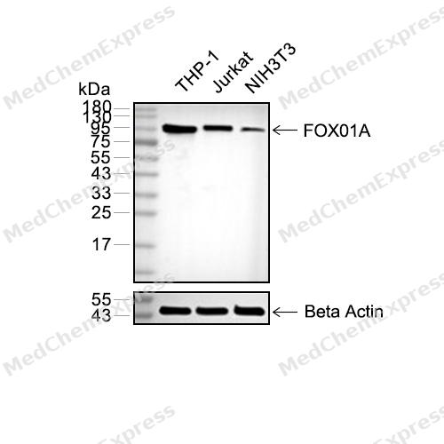 Western blot analysis of extracts from THP-1(lane 2(20μg), Jurkat (lane 3(20μg) and NIH3T3(lane 4(20μg) using FOXO1A (HY-P80132) Rabbit mAb. Proteins were transferred to a PVDF membrane and blocked with 5% non-fat milk in TBST for 2 hour at room temperature. The primary antibody (1/1000) and Loading control antibody (Beta Actin, HY-P80438, 1/10000) was used in 5% non-fat milk in TBST at 4°C overnight. Goat Anti-Mouse/Rabbit IgG-HRP Secondary Antibody (1/10000) was used for 1 hour at room temperature.
 -
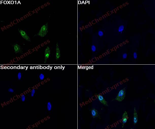 Immunocytochemistry analysis of A549 cells labeling FOXO1A with FOXO1A antibody (HY-P80132) at 1/50 dilution. Cells were fixed in 4% paraformaldehyde for 15 minutes at room temperature, permeabilized with 0.1% Triton X-100 for 10 minutes at room temperature, then blocked with QuickBlock™ Blocking Buffer for Immunol Staining for 10 min at room temperature. Cells were then incubated with FOXO1A antibody (HY-P80132) at 1/50 dilution in QuickBlock™ Blocking Buffer for Immunol Staining at 4 ℃. Alexa Fluor® 488-conjugated AffiniPure Goat Anti-Rabbit IgG H&L(HY-P8002, Green) was used as the secondary antibody at 1/1,000 dilution. PBS instead of the primary antibody was used as the secondary antibody only control. The Nuclear counterstain was DAPI (Blue).
 -
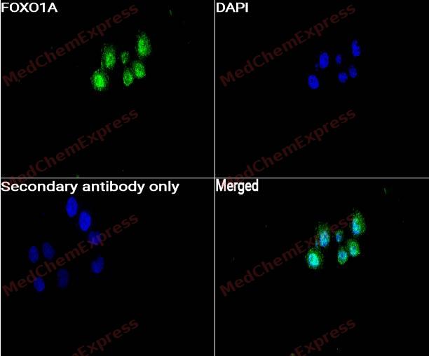 Immunocytochemistry analysis of HeLa cells labeling FOXO1A with FOXO1A antibody (HY-P80132) at 1/50 dilution. Cells were fixed in 4% paraformaldehyde for 15 minutes at room temperature, permeabilized with 0.1% Triton X-100 for 10 minutes at room temperature, then blocked with QuickBlock™ Blocking Buffer for Immunol Staining for 10 min at room temperature. Cells were then incubated with FOXO1A antibody (HY-P80132) at 1/50 dilution in QuickBlock™ Blocking Buffer for Immunol Staining at 4 ℃. Alexa Fluor® 488-conjugated AffiniPure Goat Anti-Rabbit IgG H&L(HY-P8002, Green) was used as the secondary antibody at 1/1,000 dilution. PBS instead of the primary antibody was used as the secondary antibody only control. The Nuclear counterstain was DAPI (Blue).
 -
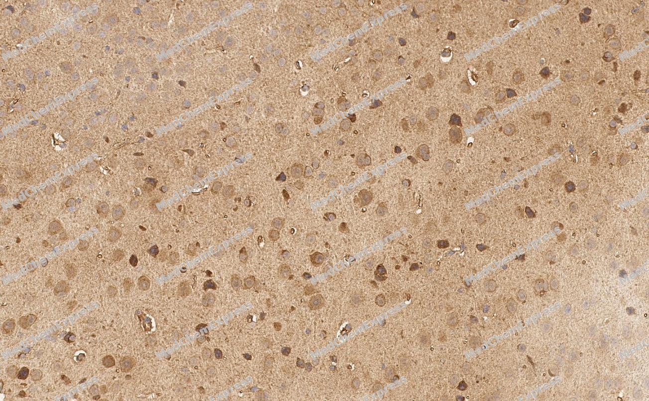 Immunohistochemical analysis of paraffin-embedded Mouse brain tissue using FOXO1A Antibody. The section was pre-treated using heat mediated antigen retrieval with sodium citrate buffer (pH 6.0) for 8 minutes. The tissues were blocked in QuickBlock for 20 minutes at room temperature, washed with ddH2O and PBS, and then probed with the primary antibody (HY-P80132, 1/400) in 4℃ overnight. The detection was performed using an HRP conjugated compact polymer system. DAB was used as the chromogen. Tissues were counterstained with hematoxylin and mounted with DPX.
 -
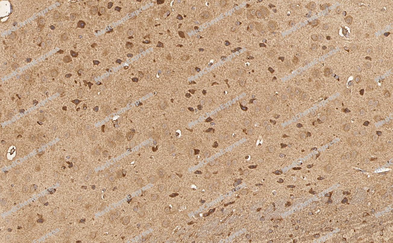 Immunohistochemical analysis of paraffin-embedded Mouse brain tissue using FOXO1A Antibody. The section was pre-treated using heat mediated antigen retrieval with sodium citrate buffer (pH 6.0) for 8 minutes. The tissues were blocked in QuickBlock for 20 minutes at room temperature, washed with ddH2O and PBS, and then probed with the primary antibody (HY-P80132, 1/400) in 4℃ overnight. The detection was performed using an HRP conjugated compact polymer system. DAB was used as the chromogen. Tissues were counterstained with hematoxylin and mounted with DPX.


Background
-
Function
FOXO1A transcription factor that is the main target of insulin signaling and regulates metabolic homeostasis in response to oxidative stress. Binds to the insulin response element (IRE) with consensus sequence 5'-TT[G/A]TTTTG-3' and the related Daf-16 family binding element (DBE) with consensus sequence 5'-TT[G/A]TTTAC-3'. Activity suppressed by insulin. Main regulator of redox balance and osteoblast numbers and controls bone mass. Orchestrates the endocrine function of the skeleton in regulating glucose metabolism. Also acts as a key regulator of chondrogenic commitment of skeletal progenitor cells in response to lipid availability: when lipids levels are low, translocates to the nucleus and promotes expression of SOX9, which induces chondrogenic commitment and suppresses fatty acid oxidation. Acts synergistically with ATF4 to suppress osteocalcin/BGLAP activity, increasing glucose levels and triggering glucose intolerance and insulin insensitivity. Also suppresses the transcriptional activity of RUNX2, an upstream activator of osteocalcin/BGLAP. Acts as an inhibitor of glucose sensing in pancreatic beta cells by acting as a transcription repressor and suppressing expression of PDX1. In hepatocytes, promotes gluconeogenesis by acting together with PPARGC1A and CEBPA to activate the expression of genes such as IGFBP1, G6PC1 and PCK1. Also promotes gluconeogenesis by directly promoting expression of PPARGC1A and G6PC1. Important regulator of cell death acting downstream of CDK1, PKB/AKT1 and STK4/MST1. Promotes neural cell death. Mediates insulin action on adipose tissue. Regulates the expression of adipogenic genes such as PPARG during preadipocyte differentiation and, adipocyte size and adipose tissue-specific gene expression in response to excessive calorie intake. Regulates the transcriptional activity of GADD45A and repair of nitric oxide-damaged DNA in beta-cells. Required for the autophagic cell death induction in response to starvation or oxidative stress in a transcription-independent manner. Mediates the function of MLIP in cardiomyocytes hypertrophy and cardiac remodeling. Positive regulator of apoptosis in cardiac smooth muscle cells as a result of its transcriptional activation of pro-apoptotic genes. Regulates endothelial cell (EC) viability and apoptosis in a PPIA/CYPA-dependent manner via transcription of CCL2 and BCL2L11 which are involved in EC chemotaxis and apoptosis[1][2][3][4][5][6][7][8][9][10][11].
-
Subcellular Localization
Cytoplasm; Nucleus
-
Expression
Tissue_specificity:It is expressed in umbilical cord endothelial cells (protein level) (PubMed: 19483080) . It is highly expressed in skeletal muscle and ovaries, and less so in the heart, placenta, lungs, liver, pancreas, spleen, testes, and small intestine (PubMed: 9479491) . It is weakly expressed in the brain, thymus, prostate, and colonic mucosa (PubMed: 9479491) .
Induction:Expression is regulated by KRIT1. Levels of expression also regulated by FOXC1 which binds to a conserved element in the FOXO1 promoter -
Subunit
Interacts with LRPPRC. Interacts with RUNX2; the interaction inhibits RUNX2 transcriptional activity and mediates the IGF1/insulin-dependent BGLAP expression in osteoblasts Interacts with PPP2R1A; the interaction regulates the dephosphorylation of FOXO1 at Thr-24 and Ser-256 leading to its nuclear import. Interacts (acetylated form) with PPARG. Interacts with XBP1 isoform 2; this interaction is direct and leads to FOXO1 ubiquitination and degradation via the proteasome pathway (By similarity). Interacts with NLK. Interacts with SIRT1; the interaction results in the deacetylation of FOXO1 leading to activation of FOXO1-mediated transcription of genes involved in DNA repair and stress resistance. Binds to CDK1. Interacts with the 14-3-3 proteins, YWHAG and YWHAZ; the interactions require insulin-stimulated phosphorylation on Thr-24, promote nuclear exit and loss of transcriptional activity. Interacts with SKP2; the interaction ubiquitinates FOXO1 leading to its proteasomal degradation. The interaction requires the presence of KRIT1. Interacts (via the C-terminal half) with ATF4 (via its DNA-binding domain); the interaction occurs in osteoblasts, regulates glucose homeostasis via suppression of beta-cell proliferation and subsequent decrease in insulin production. Interacts with PRMT1; the interaction methylates FOXO1, prevents PKB/AKT1 phosphorylation and retains FOXO1 in the nucleus. Interacts with EP300 and CREBBP; the interactions acetylate FOXO1. Interacts with SIRT2; the interaction is disrupted in response to oxidative stress or serum deprivation, leading to increased level of acetylated FOXO1, which promotes stress-induced autophagy by stimulating E1-like activating enzyme ATG7. Interacts (acetylated form) with ATG7; the interaction is increased in response to oxidative stress or serum deprivation and promotes the autophagic process leading to cell death. Interacts (via the Fork-head domain) with CEBPA; the interaction increases when FOXO1 is deacetylated. Interacts with WDFY2. Forms a complex with WDFY2 and AKT1 (By similarity). Interacts with CRY1 (By similarity). Interacts with PPIA/CYPA; the interaction promotes FOXO1 dephosphorylation, nuclear accumulation and transcriptional activity (PubMed:31063815). Interacts with TOX4; FOXO1 is required for full induction of TOX4-dependent activity and the interaction is inhibited by insulin (By similarity). Interacts (when phosphorylated on Ser-256) with STUB1/CHIP (PubMed:19483080)
-
SwissProt ID
-
Synonyms
FKHR, FOXO1A, FOXO1, Forkhead box protein O1, Forkhead box protein O1A, Forkhead in rhabdomyosarcoma
-
Research Field
Epigenetics and Nuclear Signaling


Documentation
-
Data Sheet (264 KB)
-
SDS (251 KB)
- English - EN (251 KB)
- Français - FR (251 KB)
- Deutsch - DE (251 KB)
- Norwegian - NO (251 KB)
- Español - ES (251 KB)
- Swedish - SV (251 KB)
- Italian - IT (251 KB)
- Korean - KR (251 KB)
- Portuguese - PT (251 KB)
-
User Guide for Antibodies (1077 KB)
References
[1]. Guo S, et al. Phosphorylation of serine 256 by protein kinase B disrupts transactivation by FKHR and mediates effects of insulin on insulin-like growth factor-binding protein-1 promoter activity through a conserved insulin response sequence. J Biol Chem. 1999 Jun 11;274(24):17184-92. [Content Brief]
[2]. Zhang X, et al. Phosphorylation of serine 256 suppresses transactivation by FKHR (FOXO1) by multiple mechanisms. Direct and indirect effects on nuclear/cytoplasmic shuttling and DNA binding. J Biol Chem. 2002 Nov 22;277(47):45276-84. [Content Brief]
[3]. Daitoku H, et al. Silent information regulator 2 potentiates Foxo1-mediated transcription through its deacetylase activity. Proc Natl Acad Sci U S A. 2004 Jul 6;101(27):10042-7. [Content Brief]
[4]. Perrot V, et al. The coactivator p300 directly acetylates the forkhead transcription factor Foxo1 and stimulates Foxo1-induced transcription. Mol Endocrinol. 2005 Sep;19(9):2283-98. [Content Brief]
[5]. Yuan Z, et al. Activation of FOXO1 by Cdk1 in cycling cells and postmitotic neurons. Science. 2008 Mar 21;319(5870):1665-8. [Content Brief]
[6]. Yuan Z, et al. Regulation of neuronal cell death by MST1-FOXO1 signaling. J Biol Chem. 2009 Apr 24;284(17):11285-92. [Content Brief]
[7]. Zhao Y, et al. Cytosolic FoxO1 is essential for the induction of autophagy and tumour suppressor activity. Nat Cell Biol. 2010 Jul;12(7):665-75. [Content Brief]
[8]. Valis K, et al. Hippo/Mst1 stimulates transcription of the proapoptotic mediator NOXA in a FoxO1-dependent manner. Cancer Res. 2011 Feb 1;71(3):946-54. [Content Brief]
[9]. Schilling MM, et al. Gluconeogenesis: re-evaluating the FOXO1-PGC-1alpha connection. Nature. 2006 Oct 5;443(7111):E10-1. [Content Brief]
[10]. Li F, et al. C terminus of Hsc70-interacting protein promotes smooth muscle cell proliferation and survival through ubiquitin-mediated degradation of FoxO1. J Biol Chem. 2009 Jul 24;284(30):20090-8. [Content Brief]
[11]. Xie Y, et al. Cyclophilin A-FoxO1 signaling pathway in endothelial cell apoptosis. Cell Signal. 2019 Sep;61:57-65. [Content Brief]

 Powered by Bioz
