



FOXP3 Antibody (YA6250)
(Synonyms: FOXP3; IPEX; JM2; Forkhead box protein P3; Scurfin)
Based on 1 Customer Validation
FOXP3 Antibody (YA6250) is a Rabbit-derived and non-conjugated IgG monoclonal antibody, targeting to FOXP3.
-
Host:
Rabbit
-
Isotype:
IgG
-
Application:
WB, IHC-P, ICC/IF, IP, ELISA
-
Reactivity :
Human, Mouse, Rat
-
Formulation:
Supplied in PBS, 50% glycerol, 0.05% Proclin 300, 0.05%BSA
-
Conjugation:
Non-conjugated


To place orders, for customer services and technical support, please contact: MedChemExpress USA
Tel: 609-228-6898 E-mail: [email protected] [email protected]





Applications
| Application |
IHC-P

IHC-P: Immunohistochemistry-Paraffin
|
WB
WB: Western Blot
|
ICC/IF
ICC/IF: Immunocytochemistry/
Immunofluorescence |
ELISA
ELISA: Enzyme Linked Immunosorbent Assay
|
IP
IP: Immunoprecipitation
|
|---|---|---|---|---|---|
| Dilution Ratio | 1:200-1:1000 | 1:1000-1:5000 | 1:200-1:1000 | 1:5000-1:20000 | 1:50-1:200 |


Product Details
FOXP3 Antibody (YA6250) is a Rabbit-derived and non-conjugated IgG monoclonal antibody, targeting to FOXP3.
-
Host Rabbit

-
Clonality Monoclonal

-
Species ReactivityHuman, Mouse, Rat
-
Observed Molecular WeightObserved band size: 47 kDa
Note: Due to possible protein modifications or aggregation, the molecular weight should be confirmed by actual measurement, and the predicted value is for reference only.

-
Calculated Molecular Weight Predicted band size: 47 kDa

Protein A
Non-conjugated
Unmodified
IgG


Product Properties
-
Appearance
Solution
-
Formulation
Supplied in PBS, 50% glycerol, 0.05% Proclin 300, 0.05%BSA
-
Concentration
Batch-dependent, Please check the COA for the concentration of each lot. Check Lot Concentration
-
Storage & Stability
Stored at -20°C for 1 year. Avoid repeated freeze / thaw cycles.
-
Shipping
Shipping with blue ice.


Verification Images
-
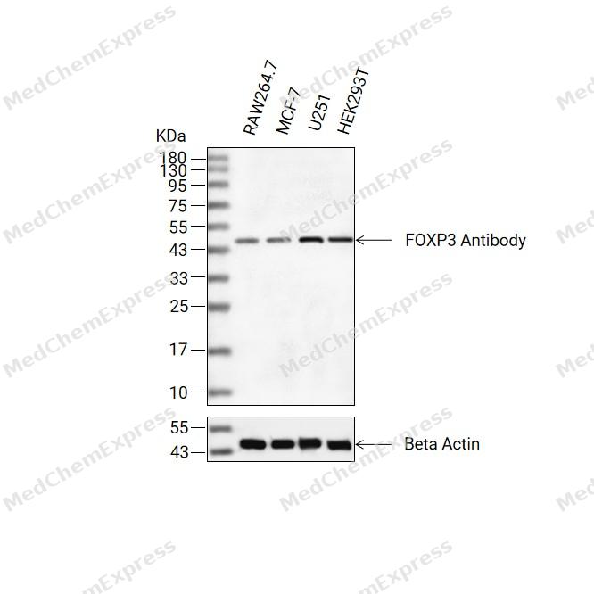 Western blot analysis of extracts from RAW264.7(lane 2(20ug) , MCF-7(lane 3(20ug) and U251(lane 4(20ug) , HEK293T(lane 5(20ug) using FOXP3 Antibody(HY-P86558) Rabbit mAb. Proteins were transferred to a PVDF membrane and blocked with 5% non-fat milk in TBST for 2 hour at room temperature. The primary antibody (1/1000) and Loading control antibody (Beta Actin, HY-P83730, 1/10000) was used in 5% non-fat milk in TBST at 4°C overnight. Goat Anti-Mouse/Rabbit IgG-HRP Secondary Antibody (1/10000) was used for 1 hour at room temperature.
 -
 Immunohistochemical analysis of paraffin-embedded human tonsil tissue using FOXP3 Antibody (HY-P86558, 1/1000). The section was pretreated using heat mediated antigen retrieval with sodium citrate buffer (pH 6.0) for 8 minutes. The tissues were blocked with quick block buffer for 0.5 hours at room temperature, washed with PBS and PBST, and then incubated with the primary antibody overnight at 4℃. The detection was performed using an HRP conjugated compact polymer system. DAB was used as the chromogen. Tissues were counterstained with hematoxylin and mounted with DPX.
 -
 Immunohistochemical analysis of paraffin-embedded human lymph node tissue using FOXP3 Antibody (HY-P86558, 1/1000). The section was pretreated using heat mediated antigen retrieval with sodium citrate buffer (pH 6.0) for 8 minutes. The tissues were blocked with quick block buffer for 0.5 hours at room temperature, washed with PBS and PBST, and then incubated with the primary antibody overnight at 4℃. The detection was performed using an HRP conjugated compact polymer system. DAB was used as the chromogen. Tissues were counterstained with hematoxylin and mounted with DPX.
 -
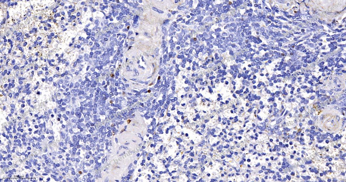 Immunohistochemical analysis of paraffin-embedded human spleen tissue using FOXP3 Antibody (HY-P86558, 1/1000). The section was pretreated using heat mediated antigen retrieval with sodium citrate buffer (pH 6.0) for 8 minutes. The tissues were blocked with quick block buffer for 0.5 hours at room temperature, washed with PBS and PBST, and then incubated with the primary antibody overnight at 4℃. The detection was performed using an HRP conjugated compact polymer system. DAB was used as the chromogen. Tissues were counterstained with hematoxylin and mounted with DPX.
 -
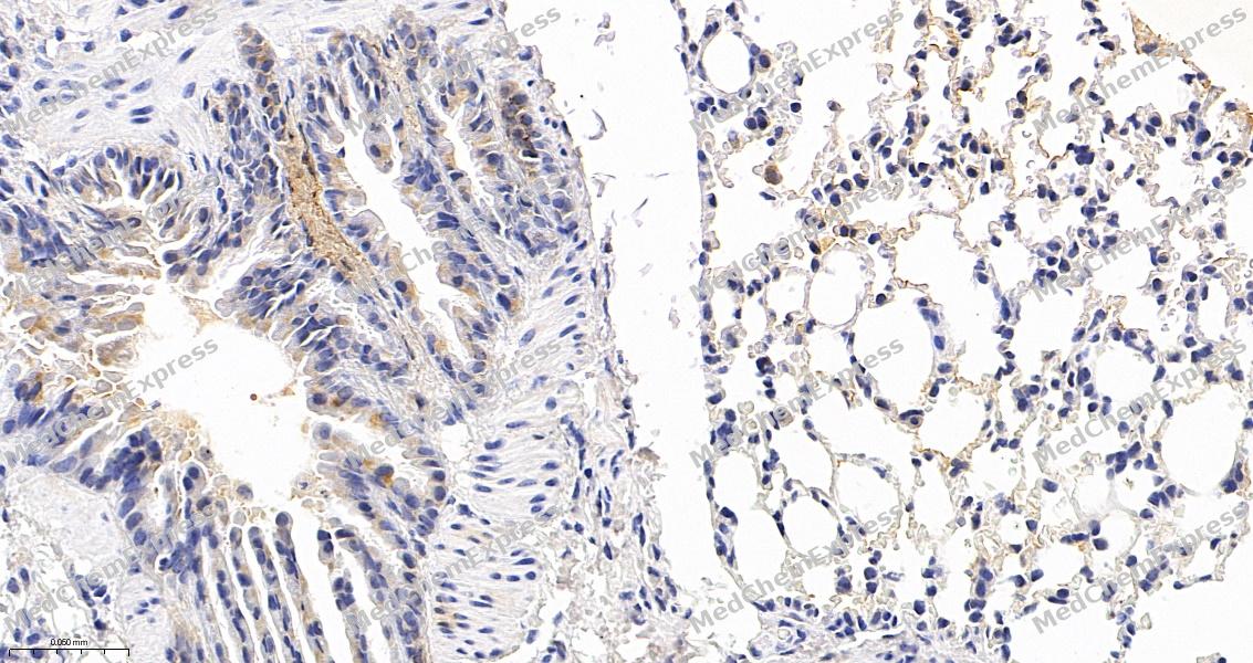 Immunohistochemical analysis of paraffin-embedded mouse lung tissue using FOXP3 Antibody (HY-P86558, 1/1000). The section was pretreated using heat mediated antigen retrieval with sodium citrate buffer (pH 6.0) for 8 minutes. The tissues were blocked with quick block buffer for 0.5 hours at room temperature, washed with PBS and PBST, and then incubated with the primary antibody overnight at 4℃. The detection was performed using an HRP conjugated compact polymer system. DAB was used as the chromogen. Tissues were counterstained with hematoxylin and mounted with DPX.
 -
 Immunohistochemical analysis of paraffin-embedded mouse spleen tissue using FOXP3 Antibody (HY-P86558, 1/1000). The section was pretreated using heat mediated antigen retrieval with sodium citrate buffer (pH 6.0) for 8 minutes. The tissues were blocked with quick block buffer for 0.5 hours at room temperature, washed with PBS and PBST, and then incubated with the primary antibody overnight at 4℃. The detection was performed using an HRP conjugated compact polymer system. DAB was used as the chromogen. Tissues were counterstained with hematoxylin and mounted with DPX.


Background
-
Function
FOXP3, so called scurfin, belongs to the forkhead box protein P (FOXP) family. FOXP3 is critically involved in differentiation and function of regulatory T cells or CD4+/CD25+ regulatory T (Treg) cells for cancer immunotherapy. FOXP3 modulates Treg development and functions by immune evasion of tumor cells through imbalance of immunoediting and immunosurveillance in some cancers. FOXP3 is associated with IL-17, RUNX1, STAT3, NF-κB, FOXO3 and other cofactors such as EOS (Ikzf4), interferon regulatory factor 4 (IRF4), special AT-rich sequence-binding protein-1 (SATB1) and GATA1.
-
Subcellular Localization
Nucleus,Cytoplasm
-
Expression
Induction: Down-regulated in regulatory T-cells (Treg) during inflammation. Up-regulated by FOXO3
-
Isoforms & Post-Translational Modification
FOXP3 has four isomers: Q9BZS1-1: 47244 Da (predicted); Q9BZS1-2: 43410 Da (predicted); Q9BZS1-3: 49843 Da (predicted); Q9BZS1-4: 44407 Da (predicted).
Polyubiquitinated, leading to its proteasomal degradation in regulatory T-cells (Treg) which is mediated by STUB1 in a HSPA1A/B-dependent manner. -
Subunit
Homodimer
-
SwissProt ID
-
Synonyms
FOXP3; IPEX; JM2; Forkhead box protein P3; Scurfin


Documentation
-
Data Sheet (261 KB)
-
SDS (251 KB)
- English - EN (251 KB)
- Français - FR (251 KB)
- Deutsch - DE (251 KB)
- Norwegian - NO (251 KB)
- Español - ES (251 KB)
- Swedish - SV (251 KB)
- Italian - IT (251 KB)
- Korean - KR (251 KB)
- Portuguese - PT (251 KB)
-
User Guide for Antibodies (1077 KB)
References
[1]. Kim JH, et al. Molecular networks of FOXP family: dual biologic functions, interplay with other molecules and clinical implications in cancer progression. Mol Cancer. 2019 Dec 9;18(1):180. [Content Brief]
[2]. Co M, et al. FOXP transcription factors in vertebrate brain development, function, and disorders. Wiley Interdiscip Rev Dev Biol. 2020 Sep;9(5):e375. [Content Brief]

 Powered by Bioz
