



Galectin 1 Antibody (YA422)
(Synonyms: Galectin-1, Gal-1, 14 kDa laminin-binding protein, 14 kDa lectin, Beta-galactoside-binding lectin L-14-I, Galaptin, HBL, HPL, Lactose-binding lectin 1, Lectin galactoside-binding soluble 1, Putative MAPK-activating protein PM12, S-Lac lectin 1, HLBP14, LGALS1)
Based on 1 Customer Validation
Galectin 1 Antibody (YA422) is a Rabbit-derived and non-conjugated IgG monoclonal antibody, targeting to Galectin 1.
-
Host:
Rabbit
-
Isotype:
IgG
-
Application:
WB, ICC/IF, IHC-P, IP, FC
-
Reactivity :
Human, Mouse, Rat
-
Formulation:
Supplied in 1*TBS (pH7.4), 0.05% BSA and 40% Glycerol. Preservative: 0.05% Sodium Azide.
-
Conjugation:
Non-conjugated


To place orders, for customer services and technical support, please contact: MedChemExpress USA
Tel: 609-228-6898 E-mail: [email protected] [email protected]





Applications
| Application |
WB

WB: Western Blot
|
ICC/IF
ICC/IF: Immunocytochemistry/
Immunofluorescence |
IHC-P
IHC-P: Immunohistochemistry-Paraffin
|
FC
FC: Flow Cytometry
|
IP
IP: Immunoprecipitation
|
|---|---|---|---|---|---|
| Dilution Ratio | 1:500-1:1000 | 1:50-1:200 | 1:400-1:800 | 1:50-1:100 | Use at an assay dependent concentration. |


Product Details
Galectin 1 Antibody (YA422) is a Rabbit-derived and non-conjugated IgG monoclonal antibody, targeting to Galectin 1.
-
Host Rabbit

-
Clonality Recombinant,Monoclonal

-
Species ReactivityHuman, Mouse, Rat
-
Observed Molecular WeightObserved band size: 14 kDa
Note: Due to possible protein modifications or aggregation, the molecular weight should be confirmed by actual measurement, and the predicted value is for reference only.

-
Calculated Molecular Weight Predicted band size: 15 kDa

Entrez Gene: 3956 Human ; 16852 Mouse ; 56646 Rat
SwissProt: P09382 Human ; P16045 Mouse ; P11762 Rat
OMIM: 150570 Human
Synthetic peptide corresponding to Human Galectin 1.AA range:1-50.
Endogenous
Protein A affinity purified.
Non-conjugated
Unmodified
IgG


Product Properties
-
Appearance
Solution
-
Formulation
Supplied in 1*TBS (pH7.4), 0.05% BSA and 40% Glycerol. Preservative: 0.05% Sodium Azide.
-
Concentration
Batch-dependent, Please check the COA for the concentration of each lot. Check Lot Concentration
-
Storage & Stability
Stored at -20°C for 1 year. Avoid repeated freeze / thaw cycles.
-
Shipping
Shipping with blue ice.


Verification Images
-
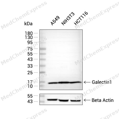 Western blot analysis of extracts from A549 (lane 2(20μg), NIH3T3 (lane 3(20μg) and HCT116 (lane 4(20μg) using Galectin 1(HY-P80403) Rabbit mAb. Proteins were transferred to a PVDF membrane and blocked with 5% non-fat milk in TBST for 2 hour at room temperature. The primary antibody (1/1000) and Loading control antibody (Beta Actin, HY-P80438, 1/10000) was used in 5% non-fat milk in TBST at 4°C overnight. Goat Anti-Mouse IgG-HRP Secondary Antibody (1/10000) was used for 1 hour at room temperature.
 -
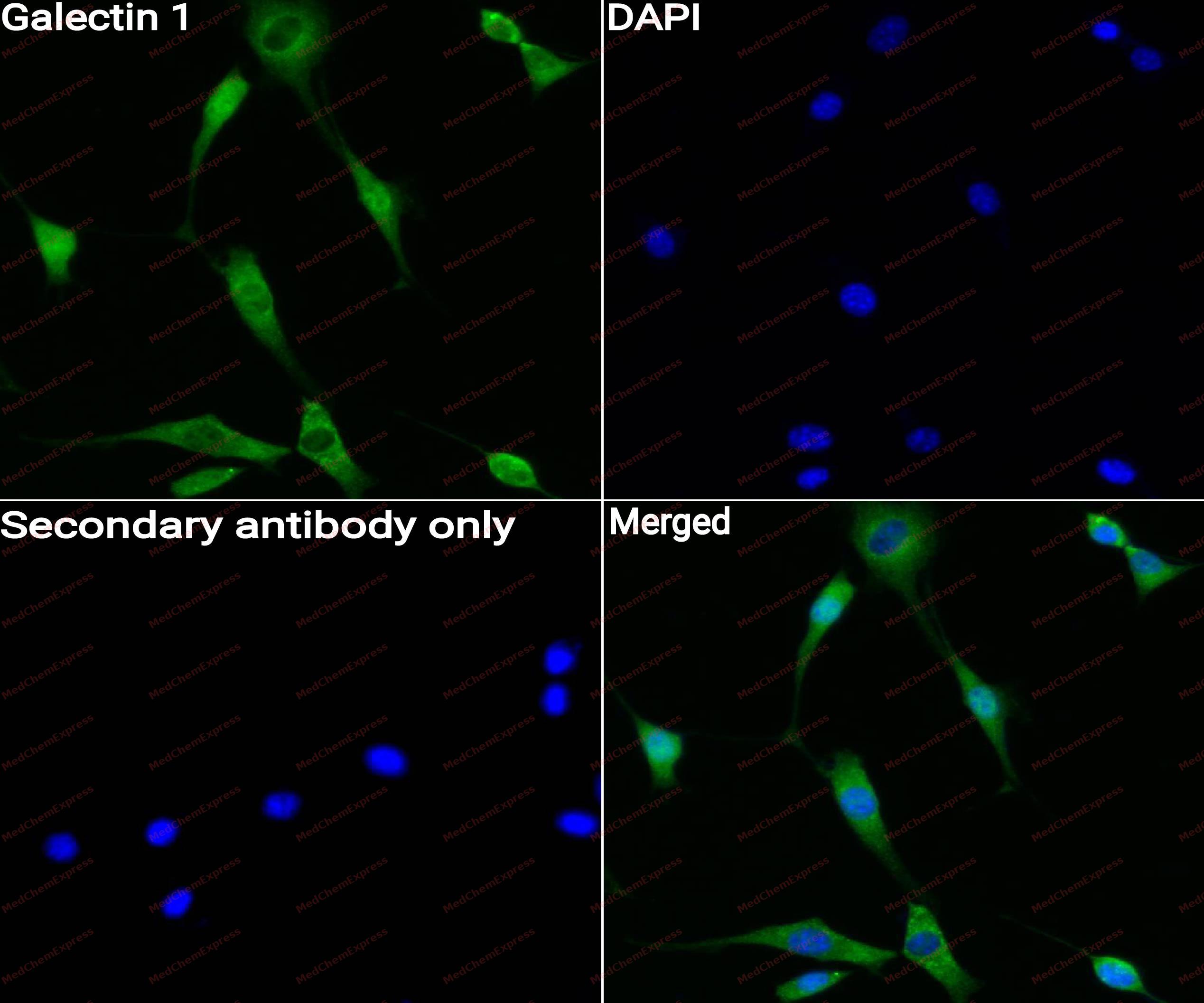 Immunocytochemistry analysis of NIH3T3 cells labeling Galectin 1 with Galectin 1 Antibody (HY-P80403)at 1/50 dilution. Cells were fixed in 4% paraformaldehyde for 15 minutes at room temperature, permeabilized with 0.1% Triton X-100 for 10 minutes at room temperature, then blocked with QuickBlock™ Blocking Buffer for Immunol Staining for 10 min at room temperature. Cells were then incubated with Galectin 1 Antibody (HY-P80403) at 1/50 dilution in QuickBlock™ Blocking Buffer for Immunol Staining at 4 ℃. Alexa Fluor® 488-conjugated AffiniPure Goat Anti-Rabbit IgG H&L(HY-P8002, Green) was used as the secondary antibody at 1/1,000 dilution. PBS instead of the primary antibody was used as the secondary antibody only control. The Nuclear counterstain was DAPI (Blue).
 -
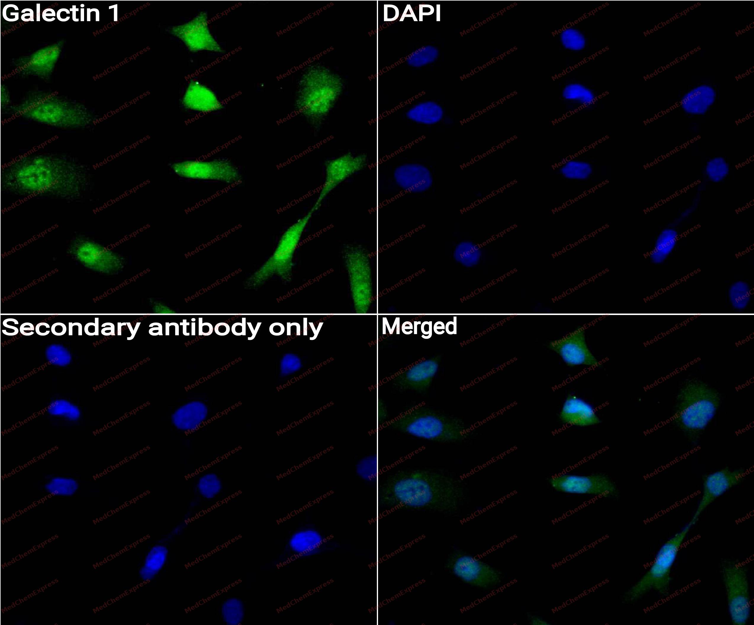 Immunocytochemistry analysis of SK-OV-3 cells labeling Galectin 1 with Galectin 1 Antibody (HY-P80403) at 1/50 dilution. Cells were fixed in 4% paraformaldehyde for 15 minutes at room temperature, permeabilized with 0.1% Triton X-100 for 10 minutes at room temperature, then blocked with QuickBlock™ Blocking Buffer for Immunol Staining for 10 min at room temperature. Cells were then incubated with Galectin 1 Antibody (HY-P80403)at 1/50 dilution in QuickBlock™ Blocking Buffer for Immunol Staining at 4 ℃. Alexa Fluor® 488-conjugated AffiniPure Goat Anti-Rabbit IgG H&L(HY-P8002, Green) was used as the secondary antibody at 1/1,000 dilution. PBS instead of the primary antibody was used as the secondary antibody only control. The Nuclear counterstain was DAPI (Blue).
 -
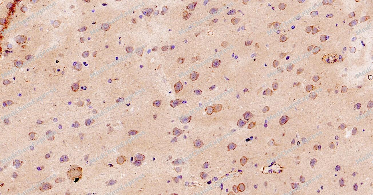 Immunohistochemical analysis of paraffin-embedded Rat brain tissue using Galectin 1 Antibody. The section was pre-treated using heat mediated antigen retrieval with Tris-EDTA buffer (pH 8.0) for 8 minutes. The tissues were blocked in QuickBlock for 20 minutes at room temperature, washed with ddH2O and PBS, and then probed with the primary antibody (HY-P80403, 1/500) in 4℃ overnight. The detection was performed using an HRP conjugated compact polymer system. DAB was used as the chromogen. Tissues were counterstained with hematoxylin and mounted with DPX.
 -
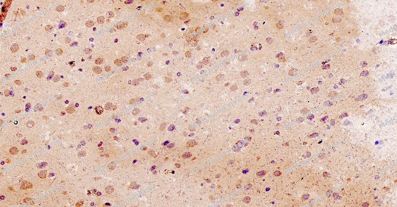 Immunohistochemical analysis of paraffin-embedded Rat brain tissue using Galectin 1 Antibody. The section was pre-treated using heat mediated antigen retrieval with Tris-EDTA buffer (pH 8.0) for 8 minutes. The tissues were blocked in QuickBlock for 20 minutes at room temperature, washed with ddH2O and PBS, and then probed with the primary antibody (HY-P80403, 1/500) in 4℃ overnight. The detection was performed using an HRP conjugated compact polymer system. DAB was used as the chromogen. Tissues were counterstained with hematoxylin and mounted with DPX.
 -
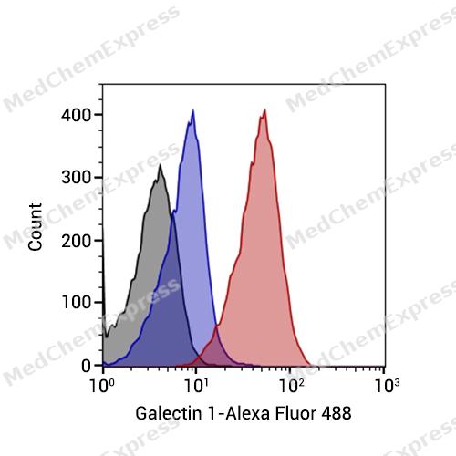 Flow cytometric analysis of 1X10^6 NIH3T3 cells labeling Galectin 1 Antibody(HY-P80403, red). Cells were fixed with 4% paraformaldehyde and permeabilised with 90% methanol. Then stained with the primary antibody at 1/1000 dilution for an hour at 4℃. Alexa Fluor® 488-conjugated AffiniPure Goat Anti-Rabbit IgG H&L (HY-P8002) was used as the secondary antibody at 1/1,000 dilution for 30 minutes at 4℃. Rabbit IgG Isotype Control (HY-P80879, blue) was used as the isotype control, cells without incubation with primary antibody were used as the unlabeled control (black).


Background
-
Function
Galectin-1 is a β-galactoside-binding protein with intracellular and extracellular activity, and evidence identifies Galectin-1 and its ligands as regulators of T-cell homeostasis, survival, immune disorders, inflammation, allergies, and host-pathogen interactions[1]. Mechanistically, Galectin-1 regulates immune and inflammatory cells through carbohydrate-dependent binding and cross-linking of cell-surface glycoproteins, including CD2, CD3, CD7, CD43, and CD45 on T cells[1]. In disease models, Galectin-1 inhibited microglial activation and ameliorated neurodegeneration in a Parkinson’s disease model through the MAPK/IκB/NFκB axis and its carbohydrate-recognition domain[2]. In collagen-induced arthritis, absence of endogenous Galectin-1 increased susceptibility and disease severity, supporting its inhibitory role in experimental inflammation[3]. Compared with related isoforms, Galectin-1 and Galectin-3 bind distinct T-cell surface glycoprotein receptors, while Galectin-9 and Galectin-1 require different glycan ligands and glycoprotein receptors to trigger T-cell death[4][5]. For experimental oncology applications, Galectin-1 overexpression was detected in hepatocellular carcinoma, and OTX008 combined with sorafenib significantly reduced tumor growth and size in tumor-cell models[6].
-
Subcellular Localization
Secreted, extracellular space, extracellular matrix; Cytoplasm; Secreted
-
Expression
Tissue_specificity:It is expressed in the placenta, maternal decidua, and fetal membranes. In the placenta, it is expressed in trophoblast cells, mesenchymal cells, villous endothelial cells, syncytiotrophoblast apical membrane, and villous mesenchyme. In the fetal membranes, it is expressed in the amnion, chorion, amniotic mesenchyme, and chorion (protein level) . It is also expressed in cardiac muscle, smooth muscle, skeletal muscle, neurons, thymus, kidneys, and hematopoietic cells. -
Subunit
Homodimer. Binds LGALS3BP. Interacts with CD2, CD3, CD4, CD6, CD7, CD43, ALCAM and CD45. Interacts with laminin (via poly-N-acetyllactosamine). Interacts with SUSD2. Interacts with cargo receptor TMED10; the interaction mediates the translocation from the cytoplasm into the ERGIC (endoplasmic reticulum-Golgi intermediate compartment) and thereby secretion (PubMed:32272059). Interacts with CD69 (PubMed:24752896)
-
SwissProt ID
-
Synonyms
Galectin-1, Gal-1, 14 kDa laminin-binding protein, 14 kDa lectin, Beta-galactoside-binding lectin L-14-I, Galaptin, HBL, HPL, Lactose-binding lectin 1, Lectin galactoside-binding soluble 1, Putative MAPK-activating protein PM12, S-Lac lectin 1, HLBP14, LGALS1
-
Research Field
Cell Biology


Documentation
-
Data Sheet (262 KB)
-
SDS (251 KB)
- English - EN (251 KB)
- Français - FR (251 KB)
- Deutsch - DE (251 KB)
- Norwegian - NO (251 KB)
- Español - ES (251 KB)
- Swedish - SV (251 KB)
- Italian - IT (251 KB)
- Korean - KR (251 KB)
- Portuguese - PT (251 KB)
-
User Guide for Antibodies (1077 KB)
References
[1]. Camby I, et al. Galectin-1: a small protein with major functions. Glycobiology. 2006 Nov;16(11):137R-157R. [Content Brief]
[2]. Li Y, et al. Galectin-1 attenuates neurodegeneration in Parkinson's disease model by modulating microglial MAPK/IκB/NFκB axis through its carbohydrate-recognition domain. Brain Behav Immun. 2020 Jan;83:214-225. [Content Brief]
[3]. Iqbal AJ, et al. Endogenous galectin-1 exerts tonic inhibition on experimental arthritis. J Immunol. 2013 Jul 1;191(1):171-7. [Content Brief]
[4]. Stillman BN, et al. Galectin-3 and galectin-1 bind distinct cell surface glycoprotein receptors to induce T cell death. J Immunol. 2006 Jan 15;176(2):778-89. [Content Brief]
[5]. Bi S, et al. Structural features of galectin-9 and galectin-1 that determine distinct T cell death pathways. J Biol Chem. 2008 May 2;283(18):12248-58. [Content Brief]
[6]. Leung Z, et al. Galectin-1 promotes hepatocellular carcinoma and the combined therapeutic effect of OTX008 galectin-1 inhibitor and sorafenib in tumor cells. J Exp Clin Cancer Res. 2019 Oct 22;38(1):423. [Content Brief]

 Powered by Bioz
