



HDAC1 Antibody (YA6183)
(Synonyms: HDAC1; RPD3L1; Histone deacetylase 1; HD1)
Based on 1 Customer Validation
HDAC1 Antibody (YA6183) is a Rabbit-derived and non-conjugated IgG monoclonal antibody, targeting to HDAC1.
-
Host:
Rabbit
-
Isotype:
IgG
-
Application:
WB, IHC-P, ICC/IF, ELISA
-
Reactivity :
Human, Mouse, Rat
-
Formulation:
Supplied in PBS, 50% glycerol, 0.05% Proclin 300, 0.05%BSA
-
Conjugation:
Non-conjugated


To place orders, for customer services and technical support, please contact: MedChemExpress USA
Tel: 609-228-6898 E-mail: [email protected] [email protected]





Applications
| Application |
IHC-P

IHC-P: Immunohistochemistry-Paraffin
|
WB
WB: Western Blot
|
ICC/IF
ICC/IF: Immunocytochemistry/
Immunofluorescence |
ELISA
ELISA: Enzyme Linked Immunosorbent Assay
|
|---|---|---|---|---|
| Dilution Ratio | 1:4000-1:10000 | 1:1000-1:5000 | 1:200-1:1000 | 1:5000-1:20000 |


Product Details
HDAC1 Antibody (YA6183) is a Rabbit-derived and non-conjugated IgG monoclonal antibody, targeting to HDAC1.
-
Host Rabbit

-
Clonality Monoclonal

-
Species ReactivityHuman, Mouse, Rat
-
Observed Molecular WeightObserved band size: 62 kDa
Note: Due to possible protein modifications or aggregation, the molecular weight should be confirmed by actual measurement, and the predicted value is for reference only.

-
Calculated Molecular Weight Predicted band size: 55 kDa

Protein A
Non-conjugated
Unmodified
IgG


Product Properties
-
Appearance
Solution
-
Formulation
Supplied in PBS, 50% glycerol, 0.05% Proclin 300, 0.05%BSA
-
Concentration
Batch-dependent, Please check the COA for the concentration of each lot. Check Lot Concentration
-
Storage & Stability
Stored at -20°C for 1 year. Avoid repeated freeze / thaw cycles.
-
Shipping
Shipping with blue ice.


Verification Images
-
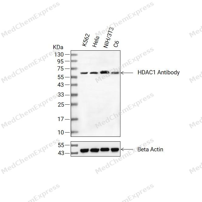 Western blot analysis of extracts from K562(lane 2(20ug) , Hela(lane 3(20ug) and NIH/3T3(lane 4(20ug),C6(lane 5(20ug) using HDAC1 Antibody (HY-P86491) Rabbit mAb. Proteins were transferred to a PVDF membrane and blocked with 5% non-fat milk in TBST for 2 hour at room temperature. The primary antibody (1/1000) and Loading control antibody (Beta Actin, HY-P83730, 1/10000) was used in 5% non-fat milk in TBST at 4°C overnight. Goat Anti-Mouse/Rabbit IgG-HRP Secondary Antibody (1/10000) was used for 1 hour at room temperature.
 -
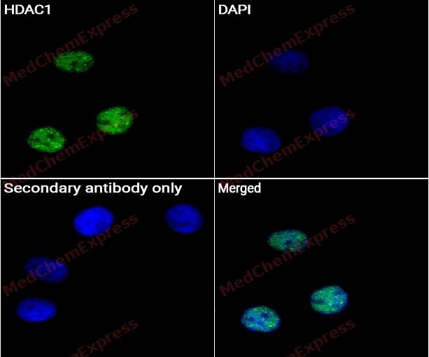 Immunocytochemistry analysis of Hela cells labeling HDAC1 Antibody (HY-P86491) at 1/50 dilution. Cells were fixed in 4% paraformaldehyde for 15 minutes at room temperature, permeabilized with 0.1% Triton X-100 for 10 minutes at room temperature, then blocked with BSA for Immunol Staining for 10 min at room temperature. Cells were then incubated with HDAC1 Antibody (HY-P86491) at 1/50 dilution in BSA for Immunol Staining at 4 ℃ overnight. AF488-conjugated AffiniPure Goat Anti-Rabbit IgG H&L(HY-P8002, Green) was used as the secondary antibody at 1/1,000 dilution. PBS instead of the primary antibody was used as the secondary antibody only control. The Nuclear counterstain was DAPI (Blue).
 -
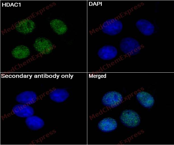 Immunocytochemistry analysis of MCF-7 cells labeling HDAC1 Antibody (HY-P86491) at 1/50 dilution. Cells were fixed in 4% paraformaldehyde for 15 minutes at room temperature, permeabilized with 0.1% Triton X-100 for 10 minutes at room temperature, then blocked with BSA for Immunol Staining for 10 min at room temperature. Cells were then incubated with HDAC1 Antibody (HY-P86491) at 1/50 dilution in BSA for Immunol Staining at 4 ℃ overnight. AF488-conjugated AffiniPure Goat Anti-Rabbit IgG H&L(HY-P8002, Green) was used as the secondary antibody at 1/1,000 dilution. PBS instead of the primary antibody was used as the secondary antibody only control. The Nuclear counterstain was DAPI (Blue).


Background
-
Function
HDAC1 is a class I histone deacetylase that supports transcriptional repression through histone deacetylase activity[1]. Mechanistically, HDAC1/2 form core catalytic components of corepressor complexes that modulate gene expression, linking chromatin regulation to proliferation and stem-cell self-renewal[2]. In human tumor cells, HDAC1 knockdown causes G1 or G2/M arrest, growth inhibition, loss of mitotic cells, and increased apoptosis[3]. In embryonic stem cells, HDAC1, but not HDAC2, is required for optimal corepressor-complex activity and cell-fate determination during differentiation[4]. Compared with HDAC2, HDAC1 shows nonredundant disease relevance in glioma stem cells, where its loss is not compensated by HDAC2 and affects the glioma stem-cell phenotype in a p53-dependent manner[5]. For experimental applications, selective HDAC1/2 inhibition suppresses colorectal cancer cells through apoptosis induction and cell-cycle regulation, supporting inhibitor use in pathway-focused cancer models[6].
-
Subcellular Localization
Nucleus
-
Expression
Tissue_specificity:It is widely present in the body, with higher concentrations in the heart, pancreas, and testes, and lower concentrations in the kidneys and brain. -
Subunit
Part of the core histone deacetylase (HDAC) complex composed of HDAC1, HDAC2, RBBP4 and RBBP7, the core complex associates with SIN3, SAP18 and SAP30 to form the SIN3 HDAC complex (PubMed:12493763, PubMed:12724404, PubMed:19061646).
-
SwissProt ID
-
Synonyms
HDAC1; RPD3L1; Histone deacetylase 1; HD1


Documentation
-
Data Sheet (262 KB)
-
SDS (251 KB)
- English - EN (251 KB)
- Français - FR (251 KB)
- Deutsch - DE (251 KB)
- Norwegian - NO (251 KB)
- Español - ES (251 KB)
- Swedish - SV (251 KB)
- Italian - IT (251 KB)
- Korean - KR (251 KB)
- Portuguese - PT (251 KB)
-
User Guide for Antibodies (1077 KB)
References
[1]. Hassig CA, et al. A role for histone deacetylase activity in HDAC1-mediated transcriptional repression. Proc Natl Acad Sci U S A. 1998 Mar 31;95(7):3519-24. [Content Brief]
[2]. Jamaladdin S, et al. Histone deacetylase (HDAC) 1 and 2 are essential for accurate cell division and the pluripotency of embryonic stem cells. Proc Natl Acad Sci U S A. 2014 Jul 8;111(27):9840-5. [Content Brief]
[3]. Senese S, et al. Role for histone deacetylase 1 in human tumor cell proliferation. Mol Cell Biol. 2007 Jul;27(13):4784-95. [Content Brief]
[4]. Dovey OM, et al. Histone deacetylase 1 (HDAC1), but not HDAC2, controls embryonic stem cell differentiation. Proc Natl Acad Sci U S A. 2010 May 4;107(18):8242-7. [Content Brief]
[5]. Lo Cascio C, et al. Nonredundant, isoform-specific roles of HDAC1 in glioma stem cells. JCI Insight. 2021 Sep 8;6(17):e149232. [Content Brief]
[6]. Lee HY, et al. A novel HDAC1/2 inhibitor suppresses colorectal cancer through apoptosis induction and cell cycle regulation. Chem Biol Interact. 2022 Jan 25;352:109778. [Content Brief]

 Powered by Bioz
