



MMP2 Antibody (YA5459)
(Synonyms: MMP2)
Based on 1 Customer Validation
MMP2 Antibody (YA5459) is a Mouse-derived and non-conjugated monoclonal antibody, targeting to MMP2.
-
Host:
Mouse
-
Application:
WB, IHC-P, ICC/IF
-
Reactivity :
Human, Rat, Mouse
-
Formulation:
Supplied in PBS containing 50% glycerol, 0.5% BSA and 0.02% sodium azide.
-
Conjugation:
Non-conjugated


To place orders, for customer services and technical support, please contact: MedChemExpress USA
Tel: 609-228-6898 E-mail: [email protected] [email protected]





Applications
| Application |
IHC-P

IHC-P: Immunohistochemistry-Paraffin
|
WB
WB: Western Blot
|
ICC/IF
ICC/IF: Immunocytochemistry/
Immunofluorescence |
|---|---|---|---|
| Dilution Ratio | 1:100-200 | 1:500-2000 | 1:50-200 |


Product Details
MMP2 Antibody (YA5459) is a Mouse-derived and non-conjugated monoclonal antibody, targeting to MMP2.
-
Host Mouse

-
Clonality Monoclonal

-
Species ReactivityHuman, Rat, Mouse
-
Observed Molecular WeightObserved band size: 64 kDa & 72 kDa
Note: Due to possible protein modifications or aggregation, the molecular weight should be confirmed by actual measurement, and the predicted value is for reference only.

Synthetic Peptide of MMP2 at AA range of INTERNAL
affinity chromatography.
Non-conjugated
Unmodified


Product Properties
-
Appearance
Solution
-
Formulation
Supplied in PBS containing 50% glycerol, 0.5% BSA and 0.02% sodium azide.
-
Concentration
Batch-dependent, Please check the COA for the concentration of each lot. Check Lot Concentration
-
Storage & Stability
Stored at -20°C for 1 year. Avoid repeated freeze / thaw cycles.
-
Shipping
Shipping with blue ice.


Verification Images
-
 Western blot analysis was performed on protein extracts (25 μg) from Mouse heart (lane 2) using MMP2 antibody. Proteins were transferred onto a 0.45 μm PVDF membrane using the Trans-Blot® Turbo™ system for 13 min. The membrane was then blocked with 5% nonfat milk in TBST (HY-K1025) for 1 h at room temperature. The primary antibody (1:1000) and loading control antibody GAPDH Antibody (HRP) (HY-P80954A) (1:5000) were diluted in 5% nonfat milk in TBST and incubated with the membrane overnight at 4°C. After washing, the membrane of primary antibody was incubated with HRP-conjugated goat anti-rabbit/mouse IgG secondary antibody (HY-P8001/HY-P8004) (1:5000) diluted in 5% nonfat milk in TBST for 1 h at room temperature. Protein bands were visualized using an Ultra High Sensitivity ECL detection kit (HY-K1005).
 -
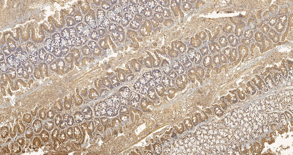 Immunohistochemical analysis of paraffin-embedded rat colon tissue using MMP2 Antibody. The section was pre-treated using heat mediated antigen retrieval with sodium citrate buffer (pH 6.0) for 8 minutes. The tissues were blocked in QuickBlock for 20 minutes at room temperature, washed with ddH2O and PBS, and then probed with the primary antibody at 1/100 dilution in 4℃ overnight. The detection was performed using an HRP conjugated compact polymer system. DAB was used as the chromogen. Tissues were counterstained with hematoxylin and mounted with DPX.
 -
Immunohistochemical analysis of paraffin-embedded rat colon tissue using MMP2 Antibody. The section was pre-treated using heat mediated antigen retrieval with sodium citrate buffer (pH 6.0) for 8 minutes. The tissues were blocked in QuickBlock for 20 minutes at room temperature, washed with ddH2O and PBS, and then probed with the primary antibody at 1/100 dilution in 4℃ overnight. The detection was performed using an HRP conjugated compact polymer system. DAB was used as the chromogen. Tissues were counterstained with hematoxylin and mounted with DPX.


Background
-
Function
MMP-2 (matrix metalloproteinase-2), also known as gelatinase A, is a zinc-dependent extracellular endopeptidase that plays a central role in extracellular matrix remodeling through the degradation of gelatin, type IV collagen, and other basement membrane components[6][7]. Mechanistically, MMP-2 is synthesized as a latent proenzyme and is activated at the cell surface through the MT1-MMP/TIMP-2/pro-MMP-2 activation complex, linking its proteolytic activity to tightly regulated pericellular signaling and matrix turnover[1][2]. Through these functions, MMP-2 contributes to cell migration, tissue remodeling, angiogenesis, and inflammatory regulation, making it an important mediator of both physiological repair processes and pathological tissue remodeling[6][7]. Dysregulated MMP-2 expression or activation has been associated with cancer progression, cardiovascular disorders, kidney disease, diabetic complications, and fibrotic conditions, where excessive extracellular matrix degradation promotes disease development and tissue dysfunction[6]. In tumor models, elevated MMP-2 activity correlates with invasive behavior, metastatic dissemination, and angiogenic remodeling, supporting its widespread use as a biomarker and mechanistic target in cancer research[3][4]. Compared with the closely related gelatinase MMP-9, MMP-2 is constitutively expressed in many tissues and is preferentially regulated through MT1-MMP- and TIMP-2-dependent activation mechanisms, whereas MMP-9 is more commonly induced by inflammatory stimuli and exhibits distinct substrate and regulatory profiles[7]. For experimental applications, broad-spectrum metalloproteinase inhibitors such as batimastat have been widely used to investigate MMP-2-dependent signaling and matrix remodeling, although selective targeting remains an active area of therapeutic development[5].
-
Subcellular Localization
Secreted, extracellular space, extracellular matrix; Membrane; Nucleus; Cytoplasm; Mitochondrion
-
Expression
Tissue_specificity:Produced by normal skin fibroblasts. PEX is expressed in a variety of tumors, including glioma, breast cancer, and prostate cancer.
Induction:Aspirin appears to inhibit expression -
Isoforms & Post-Translational Modification
P08253 has 3 isomers: P08253-1: 73882 Da (predicted); P08253-2: 65765 Da (predicted); P08253-3: 68831 Da (predicted).
Phosphorylation on multiple sites modulates enzymatic activity. Phosphorylated by PKC in vitro;The propeptide is processed by MMP14 (MT-MMP1) and MMP16 (MT-MMP3). Autocatalytic cleavage in the C-terminal produces the anti-angiogenic peptide, PEX. This processing appears to be facilitated by binding integrinv/beta3 -
Subunit
Interacts (via the C-terminal hemopexin-like domains-containing region) with the integrin alpha-V/beta-3; the interaction promotes vascular invasion in angiogenic vessels and melamoma cells. Interacts (via the C-terminal PEX domain) with TIMP2 (via the C-terminal); the interaction inhibits the degradation activity. Interacts with GSK3B
-
SwissProt ID
-
Synonyms
MMP2


Documentation
-
Data Sheet (262 KB)
-
SDS (252 KB)
- English - EN (252 KB)
- Français - FR (252 KB)
- Deutsch - DE (252 KB)
- Norwegian - NO (252 KB)
- Español - ES (252 KB)
- Swedish - SV (252 KB)
- Italian - IT (252 KB)
- Korean - KR (252 KB)
- Portuguese - PT (252 KB)
-
User Guide for Antibodies (1077 KB)
References
[1]. Khurana S, et al. MF59 adjuvant enhances diversity and affinity of antibody-mediated immune response to pandemic influenza vaccines. Sci Transl Med. 2011 Jun 1;3(85):85ra48. [Content Brief]
[2]. Heidenfelder BL, et al. Hairpin formation in Friedreich's ataxia triplet repeat expansion. J Biol Chem. 2003 Jan 24;278(4):2425-31. [Content Brief]
[3]. Forsyth PA, et al. Gelatinase-A (MMP-2), gelatinase-B (MMP-9) and membrane type matrix metalloproteinase-1 (MT1-MMP) are involved in different aspects of the pathophysiology of malignant gliomas. Br J Cancer. 1999 Apr;79(11-12):1828-35. [Content Brief]
[4]. Johnson M, et al. Multiple triangulation and collaborative research using qualitative methods to explore decision making in pre-hospital emergency care. BMC Med Res Methodol. 2017 Jan 24;17(1):11. [Content Brief]
[5]. Chang M. Matrix metalloproteinase profiling and their roles in disease. RSC Adv. 2023;13:6304-6316.

 Powered by Bioz
