



OBFC1 Antibody (YA7820)
(Synonyms: AAF-44; AAF44; bA541N10.2; RPA-32; STN1)
Based on 1 Customer Validation
OBFC1 Antibody (YA7820) is a Mouse-derived and non-conjugated IgG1 monoclonal antibody, targeting to OBFC1.
-
Host:
Mouse
-
Isotype:
IgG1
-
Application:
WB, ICC/IF, FC
-
Reactivity :
Human
-
Formulation:
Spplied in PBS (pH 7.3) containing 1% BSA, 50% glycerol and 0.02% sodium azide.
-
Conjugation:
Non-conjugated


To place orders, for customer services and technical support, please contact: MedChemExpress USA
Tel: 609-228-6898 E-mail: [email protected] [email protected]





Applications
| Application |
WB

WB: Western Blot
|
ICC/IF
ICC/IF: Immunocytochemistry/
Immunofluorescence |
FC
FC: Flow Cytometry
|
|---|---|---|---|
| Dilution Ratio | 1:2000-3000 | 1:100-250 | 1:100 |


Product Details
OBFC1 Antibody (YA7820) is a Mouse-derived and non-conjugated IgG1 monoclonal antibody, targeting to OBFC1.
-
Host Mouse

-
Clonality Monoclonal

-
Species ReactivityHuman
-
Calculated Molecular Weight Predicted band size: 41.9 kDa

Full length human recombinant protein of human OBFC1 produced in HEK293T cell.
Endogenous
Affinity purified
Non-conjugated
Unmodified
IgG1


Product Properties
-
Appearance
Solution
-
Formulation
Spplied in PBS (pH 7.3) containing 1% BSA, 50% glycerol and 0.02% sodium azide.
-
Concentration
Batch-dependent, Please check the COA for the concentration of each lot. Check Lot Concentration
-
Storage & Stability
Stored at -20°C for 1 year. Avoid repeated freeze / thaw cycles.
-
Shipping
Shipping with blue ice.


Verification Images
-
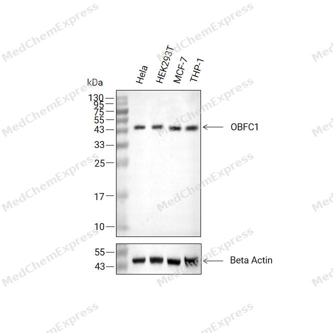 Western blot analysis of extracts from Hela (lane2(20μg), HEK293T (lane3(20μg), MCF-7 (lane4(20μg) and THP-1 (lane5(20μg) using OBFC1 Antibody (HY-P88136). Proteins were transferred to a PVDF membrane and blocked with 5% non-fat milk in TBST at 4°C overnight. The primary antibody (1/2000) and Loading control antibody (Beta Actin, HY-P80993, 1/10,000) was used in 5% non-fat milk in TBST for 2 hour at room temperature. Goat Anti-Mouse IgG-HRP Secondary Antibody (HY-P8004, 1/10,000) was used for 1 hour at room temperature.
 -
 Immunocytochemistry analysis of A431 cells labeling OBFC1 with OBFC1 Antibody (HY-P88136) at 1/100 dilution. Cells were fixed in 4% paraformaldehyde for 15 minutes at room temperature, permeabilized with 0.1% Triton X-100 in PBS for 15 minutes at room temperature, then blocked with quick block buffer for 10 minutes at room temperature. Cells were then incubated with OBFC1 Antibody (HY-P88136) at 1/100 dilution in quick block buffer overnight at 4 ℃.AF488-conjugated Goat Anti-Mouse IgG H&L(HY-P8005, Green) was used as the secondary antibody at 1/1,000 dilution. PBS instead of the primary antibody was used as the secondary antibody only control. The Nuclear counterstain was DAPI (Blue).
 -
 Immunocytochemistry analysis of HePG2 cells labeling OBFC1 with OBFC1 Antibody (HY-P88136) at 1/100 dilution . Cells were fixed in 4% paraformaldehyde for 15 minutes at room temperature, permeabilized with 0.1% Triton X-100 in PBS for 15 minutes at room temperature, then blocked with quick block buffer for 10 minutes at room temperature. Cells were then incubated with OBFC1 Antibody (HY-P88136) at 1/100 dilution in quick block buffer overnight at 4 ℃.AF488-conjugated Goat Anti-Mouse IgG H&L(HY-P8005, Green) was used as the secondary antibody at 1/1,000 dilution. PBS instead of the primary antibody was used as the secondary antibody only control. The Nuclear counterstain was DAPI (Blue).


Background
-
Function
OBFC1 component of the CST complex proposed to act as a specialized replication factor promoting DNA replication under conditions of replication stress or natural replication barriers such as the telomere duplex. The CST complex binds single-stranded DNA with high affinity in a sequence-independent manner, while isolated subunits bind DNA with low affinity by themselves. Initially the CST complex has been proposed to protect telomeres from DNA degradation. However, the CST complex has been shown to be involved in several aspects of telomere replication. The CST complex inhibits telomerase and is involved in telomere length homeostasis; it is proposed to bind to newly telomerase-synthesized 3' overhangs and to terminate telomerase action implicating the association with the ACD:POT1 complex thus interfering with its telomerase stimulation activity. The CST complex is also proposed to be involved in fill-in synthesis of the telomeric C-strand probably implicating recruitment and activation of DNA polymerase alpha. The CST complex facilitates recovery from many forms of exogenous DNA damage; seems to be involved in the re-initiation of DNA replication at repaired forks and/or dormant origins. Required for efficicient replication of the duplex region of the telomere. Promotes efficient replication of lagging-strand telomeres. Promotes general replication start following replication-fork stalling implicating new origin firing. May be in involved in C-strand fill-in during late S/G2 phase independent of its role in telomere duplex replication |Component of the CST complex, a complex that binds to single-stranded DNA and is required to protect telomeres from DNA degradation. The CST complex binds single-stranded DNA with high affinity in a sequence-independent manner, while isolated subunits bind DNA with low affinity by themselves. In addition to telomere protection, the CST complex has probably a more general role in DNA metabolism at non-telomeric sites[1][2][3][4][5][6].
-
Subcellular Localization
Nucleus,Chromosome, telomere
-
Isoforms & Post-Translational Modification
Q9H668: 368 amino acids, molecular weight 42119 Da.
-
Subunit
Component of the CST complex, composed of TEN1/C17orf106, CTC1/C17orf68 and STN1; in the complex interacts directly with TEN1 and CTC1
-
SwissProt ID
-
Synonyms
AAF-44; AAF44; bA541N10.2; RPA-32; STN1


Documentation
-
Data Sheet (231 KB)
-
SDS (251 KB)
- English - EN (251 KB)
- Français - FR (251 KB)
- Deutsch - DE (251 KB)
- Norwegian - NO (251 KB)
- Español - ES (251 KB)
- Swedish - SV (251 KB)
- Italian - IT (251 KB)
- Korean - KR (251 KB)
- Portuguese - PT (251 KB)
-
User Guide for Antibodies (1077 KB)

 Powered by Bioz
