



Phospho-MSK1 (Ser360) Antibody (YA173)
(Synonyms: RPS6KA5; MSK1; Ribosomal protein S6 kinase alpha-5; S6K-alpha-5; 90 kDa ribosomal protein S6 kinase 5; Nuclear mitogen- and stress-activated protein kinase 1; RSK-like protein kinase; RSKL)
Based on 1 Customer Validation
Phospho-MSK1 (Ser360) Antibody (YA173) is a Rabbit-derived and non-conjugated IgG monoclonal antibody, targeting to Phospho-MSK1 (Ser360).
-
Host:
Rabbit
-
Isotype:
IgG
-
Application:
WB, ICC/IF, IP
-
Reactivity :
Human, Mouse, Rat, Hamster
-
Formulation:
Supplied in 50 mM Tris-Glycine (pH 7.4), 0.15 M NaCl, 40% Glycerol and 0.05% BSA. Preservative: 0.01% Sodium azide
-
Conjugation:
Non-conjugated


To place orders, for customer services and technical support, please contact: MedChemExpress USA
Tel: 609-228-6898 E-mail: [email protected] [email protected]





Applications
| Application |
WB

WB: Western Blot
|
ICC/IF
ICC/IF: Immunocytochemistry/
Immunofluorescence |
IP
IP: Immunoprecipitation
|
|---|---|---|---|
| Dilution Ratio | 1:500-1:1000 | 1:50-1:200 | 1:20 |


Product Details
Phospho-MSK1 (Ser360) Antibody (YA173) is a Rabbit-derived and non-conjugated IgG monoclonal antibody, targeting to Phospho-MSK1 (Ser360).
-
Host Rabbit

-
Clonality Recombinant,Monoclonal

-
Species ReactivityHuman, Mouse, Rat, Hamster
-
Observed Molecular WeightObserved band size: 90 kDa
Note: Due to possible protein modifications or aggregation, the molecular weight should be confirmed by actual measurement, and the predicted value is for reference only.

-
Calculated Molecular Weight Predicted band size: 90 kDa

Entrez Gene: 9252 Human ; 73086 Mouse ; 314384 Rat
SwissProt: O75582 Human ; Q8C050 Mouse ;
OMIM: 603607 Human
Synthetic phosphopeptide corresponding to residues surrounding Ser360 of Human MSK1.The exact sequence is proprietary to MCE.
Endogenous
affinity purified
Non-conjugated
Phosphorylated
IgG


Product Properties
-
Appearance
Solution
-
Formulation
Supplied in 50 mM Tris-Glycine (pH 7.4), 0.15 M NaCl, 40% Glycerol and 0.05% BSA. Preservative: 0.01% Sodium azide
-
Concentration
Batch-dependent, Please check the COA for the concentration of each lot. Check Lot Concentration
-
Storage & Stability
Stored at -20°C for 1 year. Avoid repeated freeze / thaw cycles.
-
Shipping
Shipping with blue ice.


Verification Images
-
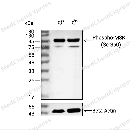 Western blot analysis of extracts from C6 (lane 2(20μg) and C6 (lane 3(40μg) using Phospho-MSK1 (Ser360) (HY-P80835) Rabbit mAb. Proteins were transferred to a PVDF membrane and blocked with 5% non-fat milk in TBST for 2 hour at room temperature. The primary antibody (1/1000) and Loading control antibody (Beta Actin, HY-P80438, 1/10000) was used in 5% non-fat milk in TBST at 4°C overnight. Goat Anti-Mouse/Rabbit IgG-HRP Secondary Antibody (1/10000) was used for 1 hour at room temperature.
 -
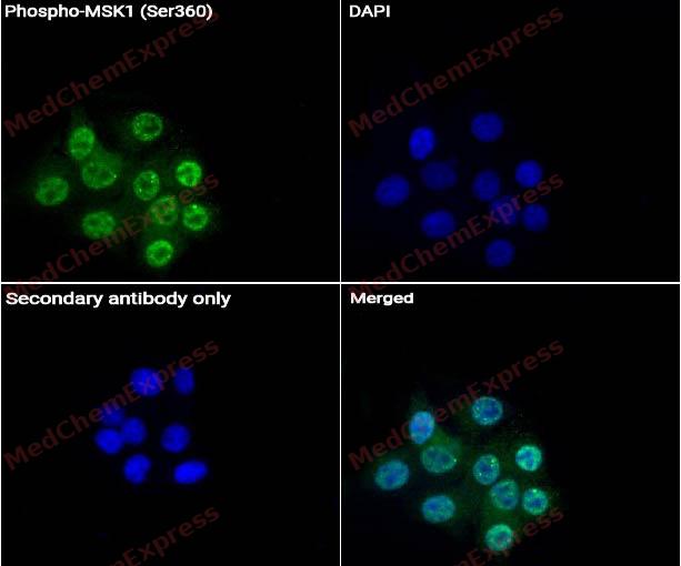 Immunocytochemistry analysis of MCF-7 cells labeling Phospho-MSK1 (Ser360) Antibody (HY-P80835) at 1/50 dilution. Cells were fixed in 4% paraformaldehyde for 15 minutes at room temperature, permeabilized with 0.1% Triton X-100 for 10 minutes at room temperature, then blocked with QuickBlock™ Blocking Buffer for Immunol Staining for 10 min at room temperature. Cells were then incubated with Phospho-MSK1 (Ser360) Antibody (HY-P80835) at 1/50 dilution in QuickBlock™ Blocking Buffer for Immunol Staining at 4 ℃. Alexa Fluor® 488-conjugated AffiniPure Goat Anti-Rabbit IgG H&L(HY-P8002, Green) was used as the secondary antibody at 1/1,000 dilution. PBS instead of the primary antibody was used as the secondary antibody only control. The Nuclear counterstain was DAPI (Blue).
 -
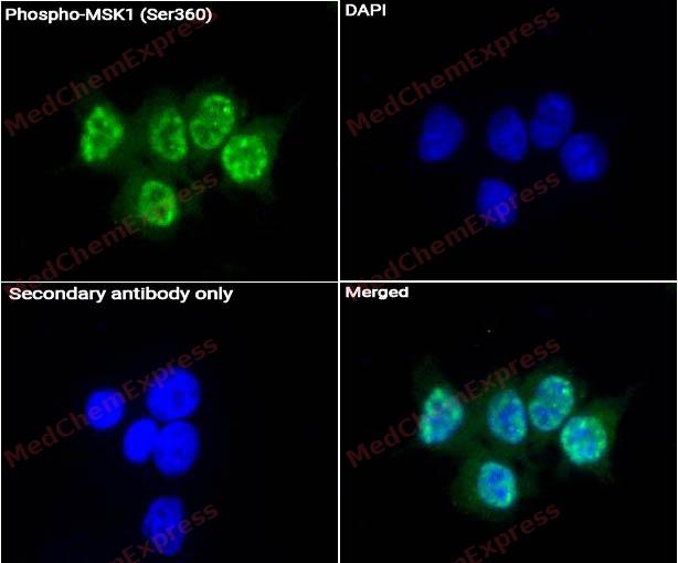 Immunocytochemistry analysis of MCF-7 cells labeling Phospho-MSK1 (Ser360) Antibody (HY-P80835) at 1/200 dilution. Cells were fixed in 4% paraformaldehyde for 15 minutes at room temperature, permeabilized with 0.1% Triton X-100 for 10 minutes at room temperature, then blocked with QuickBlock™ Blocking Buffer for Immunol Staining for 10 min at room temperature. Cells were then incubated with Phospho-MSK1 (Ser360) Antibody (HY-P80835) at 1/200 dilution in QuickBlock™ Blocking Buffer for Immunol Staining at 4 ℃. Alexa Fluor® 488-conjugated AffiniPure Goat Anti-Rabbit IgG H&L(HY-P8002, Green) was used as the secondary antibody at 1/1,000 dilution. PBS instead of the primary antibody was used as the secondary antibody only control. The Nuclear counterstain was DAPI (Blue).


Background
-
Function
MSK1 (mitogen- and stress-activated protein kinase 1) is a predominantly nuclear serine/threonine kinase that functions downstream of the ERK1/2 and p38 MAPK pathways, integrating mitogenic and stress signals into transcriptional responses through phosphorylation-dependent activation mechanisms[1][2]. MSK1 regulates gene expression by phosphorylating key nuclear substrates, including CREB, ATF1, and histone H3, thereby controlling immediate-early gene induction and chromatin-associated transcriptional programs[2][3]. Mechanistically, activation of upstream MAPKs promotes sequential phosphorylation and autophosphorylation events within MSK1, enabling its N-terminal kinase domain to phosphorylate downstream targets involved in stimulus-responsive gene regulation[1][2]. In disease-relevant systems, MSK1 contributes to anti-inflammatory transcriptional networks, regulates cytokine expression, and participates in neuronal proliferation, synaptic plasticity, and neurogenesis-related processes[2]. MSK1 also functions as a chromatin-associated transcriptional coactivator, directly interacting with p53 and promoting p21 gene expression through p53-dependent histone H3 phosphorylation at target promoters[3]. Compared with the closely related isoform MSK2, MSK1 shares substantial functional overlap and is often considered functionally redundant in many MAPK-dependent signaling contexts, although isoform-specific expression patterns and experimental models continue to support investigation of distinct biological contributions[2]. For experimental applications, pharmacological inhibitors such as H89 and dimethylfumarate have been used to suppress MSK1/MSK2 signaling and evaluate downstream transcriptional and inflammatory responses in cellular and disease models[2].
-
Subcellular Localization
Nucleus; Cytoplasm
-
Expression
Tissue_specificity:It is widely expressed in the heart, brain, and placenta, at high levels. It is present in lower levels in the lungs, kidneys, and liver. -
Isoforms & Post-Translational Modification
O75582 has 3 isomers: O75582-1: 89865 Da (predicted); O75582-2: 61769 Da (predicted); O75582-3: 81780 Da (predicted).
Ser-376 and Thr-581 phosphorylation is required for kinase activity. Ser-376 and Ser-212 are autophosphorylated by the C-terminal kinase domain, and their phosphorylation is essential for the catalytic activity of the N-terminal kinase domain. Phosphorylated at Ser-360, Thr-581 and Thr-700 by MAPK1/ERK2, MAPK3/ERK1 and MAPK14/p38-alpha. Autophosphorylated at Ser-750, Ser-752 and Ser-758 by the N-terminal kinase domain;Ubiquitinated -
Subunit
Forms a complex with either MAPK1/ERK2 or MAPK3/ERK1 in quiescent cells which transiently dissociates following mitogenic stimulation. Also associates with MAPK14/p38-alpha. Activated RPS6KA5 associates with and phosphorylates the NF-kappa-B p65 subunit RELA. Interacts with CREBBP and EP300
-
SwissProt ID
-
Synonyms
RPS6KA5; MSK1; Ribosomal protein S6 kinase alpha-5; S6K-alpha-5; 90 kDa ribosomal protein S6 kinase 5; Nuclear mitogen- and stress-activated protein kinase 1; RSK-like protein kinase; RSKL
-
Research Field
Signal Transduction


Documentation
-
Data Sheet (260 KB)
-
SDS (252 KB)
- English - EN (252 KB)
- Français - FR (252 KB)
- Deutsch - DE (252 KB)
- Norwegian - NO (252 KB)
- Español - ES (252 KB)
- Swedish - SV (252 KB)
- Italian - IT (252 KB)
- Korean - KR (252 KB)
- Portuguese - PT (252 KB)
-
User Guide for Antibodies (1077 KB)
References
[1]. McCoy CE, et al. MSK1 activity is controlled by multiple phosphorylation sites. Biochem J. 2005 Apr 15;387(Pt 2):507-17. [Content Brief]
[2]. Reyskens KM, et al. Emerging Roles of the Mitogen and Stress Activated Kinases MSK1 and MSK2. Front Cell Dev Biol. 2016 Jun 10;4:56. [Content Brief]
[3]. Ahn J, et al. MSK1 functions as a transcriptional coactivator of p53 in the regulation of p21 gene expression. Exp Mol Med. 2018 Oct 10;50(10):1-12. [Content Brief]

 Powered by Bioz
