



PKA C-alpha Antibody (YA6154)
(Synonyms: PRKACA; PKACA; cAMP-dependent protein kinase catalytic subunit alpha; PKA C-alpha)
Based on 1 Customer Validation
PKA C-alpha Antibody (YA6154) is a Rabbit-derived and non-conjugated IgG monoclonal antibody, targeting to PKA C-alpha.
-
Host:
Rabbit
-
Isotype:
IgG
-
Application:
WB, IHC-P, ICC/IF, IP, ELISA
-
Reactivity :
Human, Mouse, Rat
-
Formulation:
Supplied in PBS, 50% glycerol, 0.05% Proclin 300, 0.05%BSA
-
Conjugation:
Non-conjugated


To place orders, for customer services and technical support, please contact: MedChemExpress USA
Tel: 609-228-6898 E-mail: [email protected] [email protected]





Applications
| Application |
IHC-P

IHC-P: Immunohistochemistry-Paraffin
|
WB
WB: Western Blot
|
ICC/IF
ICC/IF: Immunocytochemistry/
Immunofluorescence |
ELISA
ELISA: Enzyme Linked Immunosorbent Assay
|
IP
IP: Immunoprecipitation
|
|---|---|---|---|---|---|
| Dilution Ratio | 1:2000-1:10000 | 1:1000-1:5000 | 1:200-1:1000 | 1:5000-1:20000 | 1:50-1:200 |


Product Details
PKA C-alpha Antibody (YA6154) is a Rabbit-derived and non-conjugated IgG monoclonal antibody, targeting to PKA C-alpha.
-
Host Rabbit

-
Clonality Monoclonal

-
Species ReactivityHuman, Mouse, Rat
-
Observed Molecular WeightObserved band size: 40 kDa
Note: Due to possible protein modifications or aggregation, the molecular weight should be confirmed by actual measurement, and the predicted value is for reference only.

-
Calculated Molecular Weight Predicted band size: 40 kDa

Protein A
Non-conjugated
Unmodified
IgG


Product Properties
-
Appearance
Liquid
-
Formulation
Supplied in PBS, 50% glycerol, 0.05% Proclin 300, 0.05%BSA
-
Concentration
Batch-dependent, Please check the COA for the concentration of each lot. Check Lot Concentration
-
Storage & Stability
Stored at -20°C for 1 year. Avoid repeated freeze / thaw cycles.
-
Shipping
Shipping with blue ice.


Verification Images
-
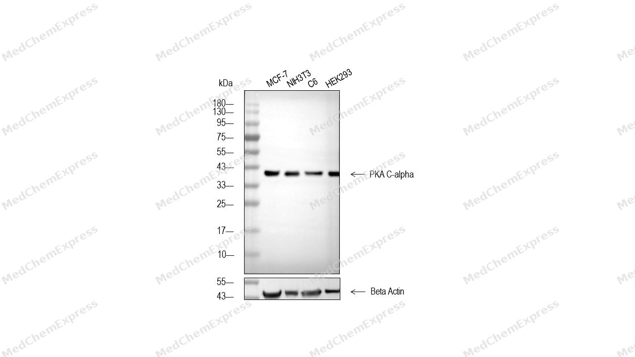 Western blot analysis of extracts from MCF-7 (lane2(20μg), NIH3T3 (lane3(20μg), C6 (lane4(20μg) and HEK293 (lane5(20μg) using PKA C-alpha Antibody (HY-P86462). Proteins were transferred to a PVDF membrane and blocked with 5% non-fat milk in TBST for 2 hour at room temperature. The primary antibody (1/3000) and Loading control antibody (Beta Actin, HY-P80993, 1/10000) was used in 5% non-fat milk in TBST at 4°C overnight. Goat Anti-Rabbit IgG-HRP Secondary Antibody (HY-P8001 ,1/10,000) was used for 1 hour at room temperature.
 -
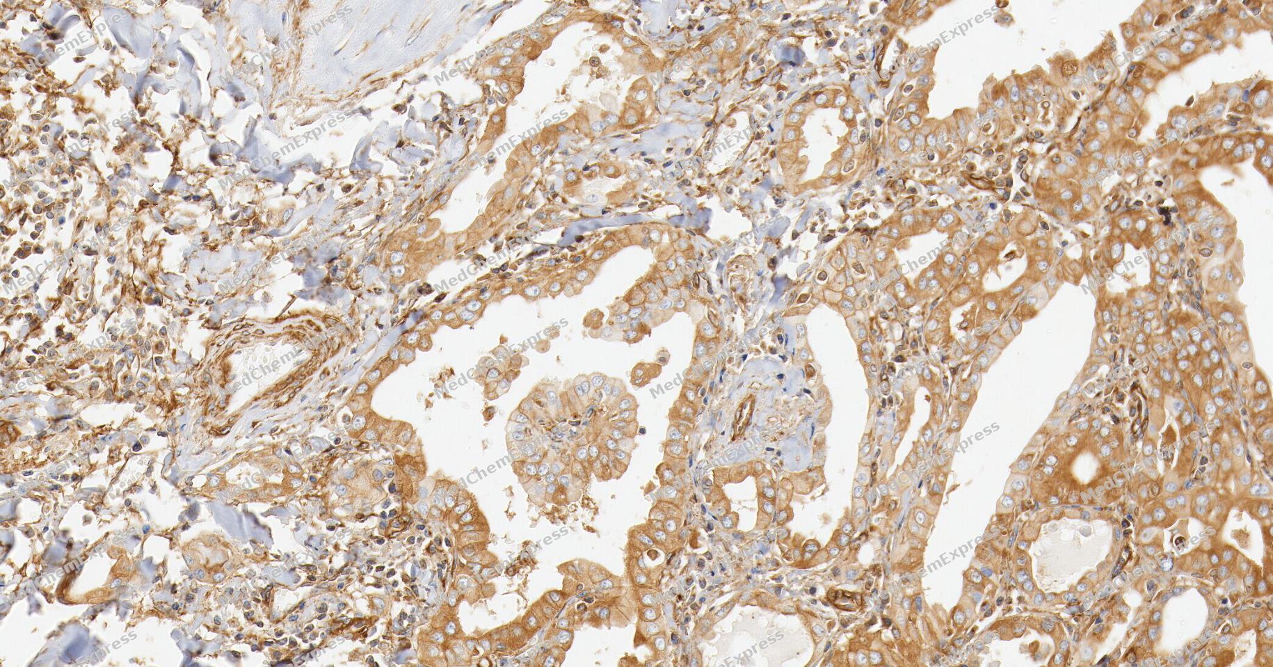 Immunohistochemical analysis of paraffin-embedded human thyroid cancer tissue using PKA C-alpha Antibody (YA6154). The section was pre-treated using heat mediated antigen retrieval with Tris/EDTA buffer (pH 9.0) for 20 minutes. The tissues were probed with the primary antibody (HY-P86462, 1/300) overnight at 4℃. The detection was performed using an HRP conjugated compact polymer system. DAB was used as the chromogen. Tissues were counterstained with hematoxylin and mounted with neutral balsam.
 -
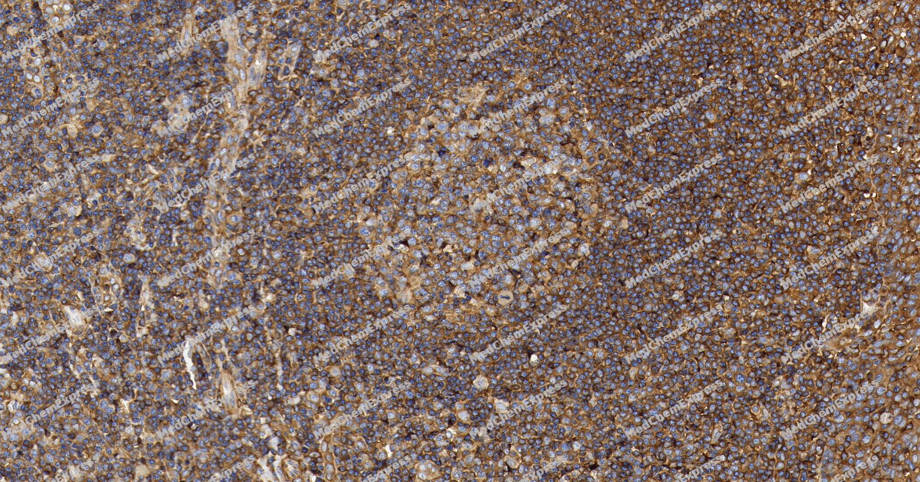 Immunohistochemical analysis of paraffin-embedded human tonsil tissue using PKA C-alpha Antibody (YA6154). The section was pre-treated using heat mediated antigen retrieval with Tris/EDTA buffer (pH 9.0) for 20 minutes. The tissues were probed with the primary antibody (HY-P86462, 1/300) overnight at 4℃. The detection was performed using an HRP conjugated compact polymer system. DAB was used as the chromogen. Tissues were counterstained with hematoxylin and mounted with neutral balsam.
 -
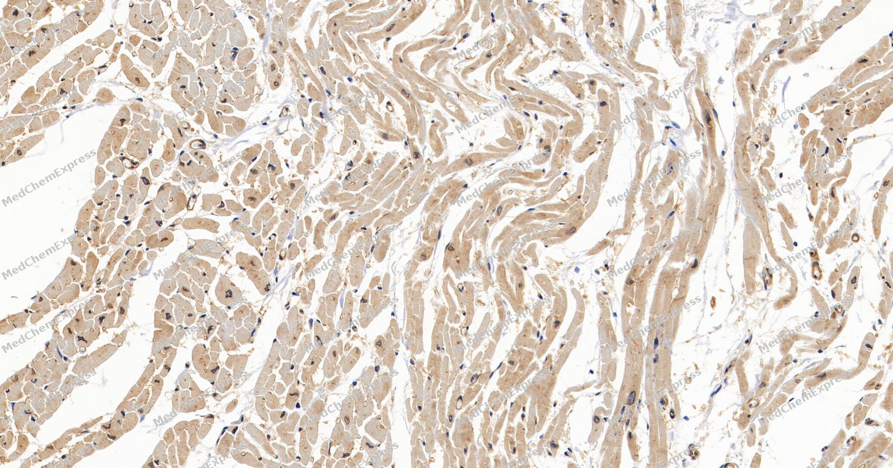 Immunohistochemical analysis of paraffin-embedded human heart tissue using PKA C-alpha Antibody (YA6154). The section was pre-treated using heat mediated antigen retrieval with Tris/EDTA buffer (pH 9.0) for 20 minutes. The tissues were probed with the primary antibody (HY-P86462, 1/300) overnight at 4℃. The detection was performed using an HRP conjugated compact polymer system. DAB was used as the chromogen. Tissues were counterstained with hematoxylin and mounted with neutral balsam.
 -
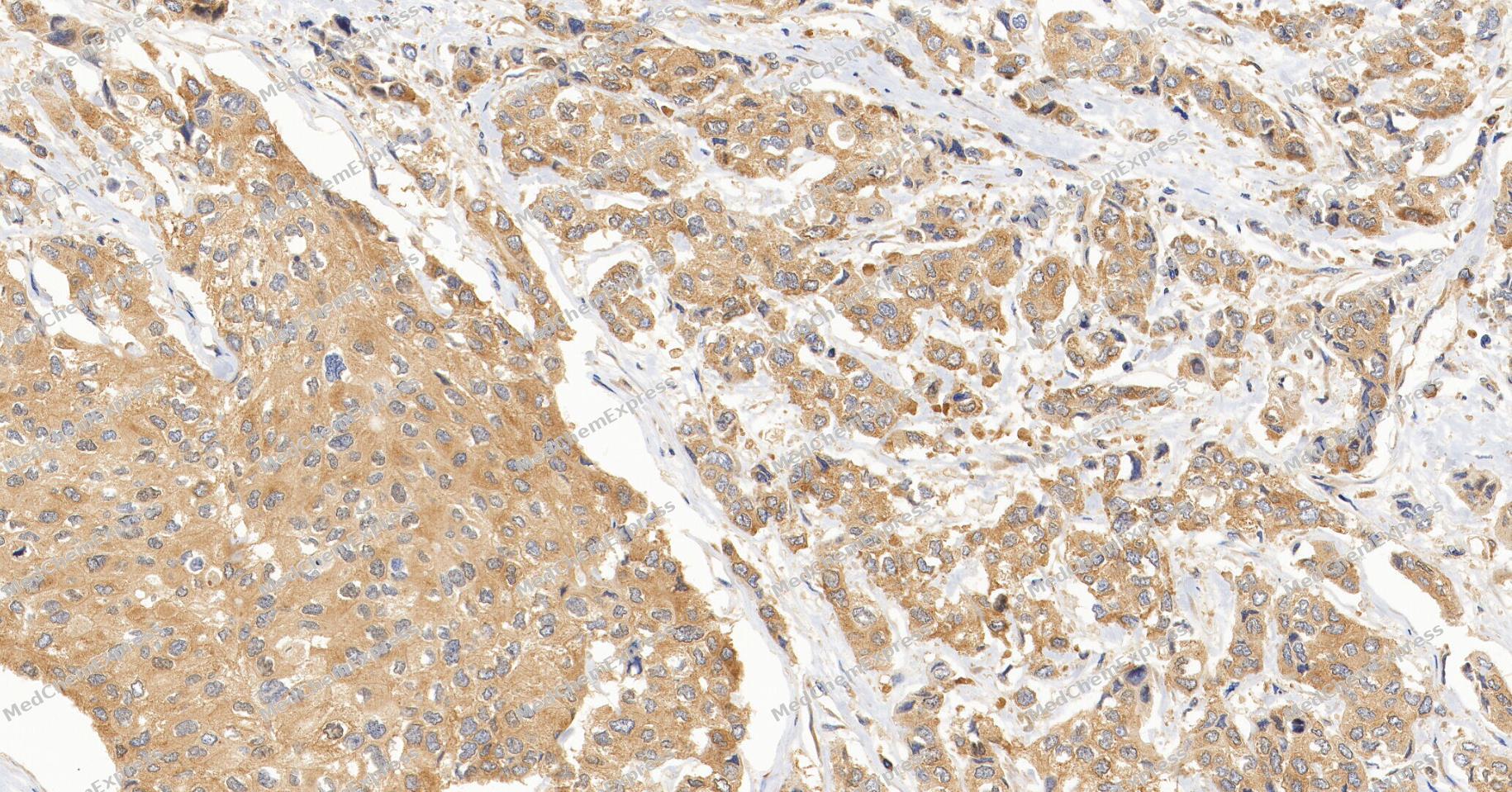 Immunohistochemical analysis of paraffin-embedded human breast cancer tissue using PKA C-alpha Antibody (YA6154). The section was pre-treated using heat mediated antigen retrieval with Tris/EDTA buffer (pH 9.0) for 20 minutes. The tissues were probed with the primary antibody (HY-P86462, 1/300) overnight at 4℃. The detection was performed using an HRP conjugated compact polymer system. DAB was used as the chromogen. Tissues were counterstained with hematoxylin and mounted with neutral balsam.
 -
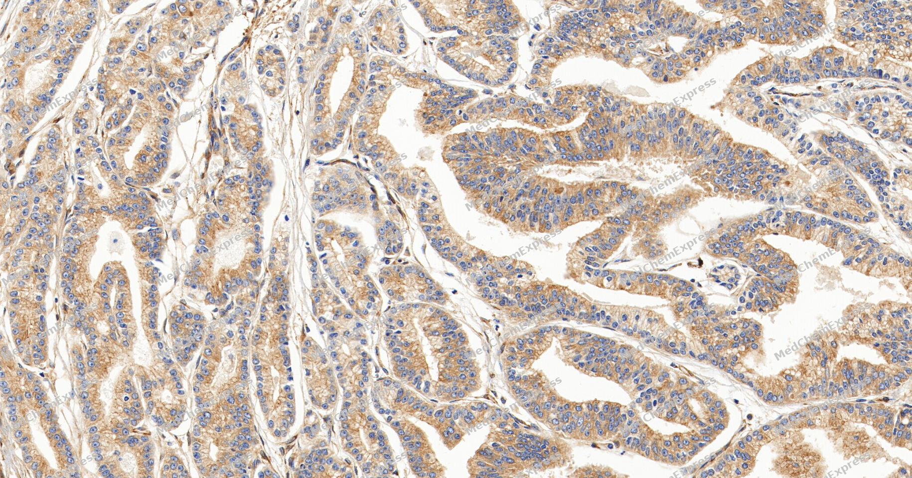 Immunohistochemical analysis of paraffin-embedded human prostate cancer tissue using PKA C-alpha Antibody (YA6154). The section was pre-treated using heat mediated antigen retrieval with Tris/EDTA buffer (pH 9.0) for 20 minutes. The tissues were probed with the primary antibody (HY-P86462, 1/300) overnight at 4℃. The detection was performed using an HRP conjugated compact polymer system. DAB was used as the chromogen. Tissues were counterstained with hematoxylin and mounted with neutral balsam.
 -
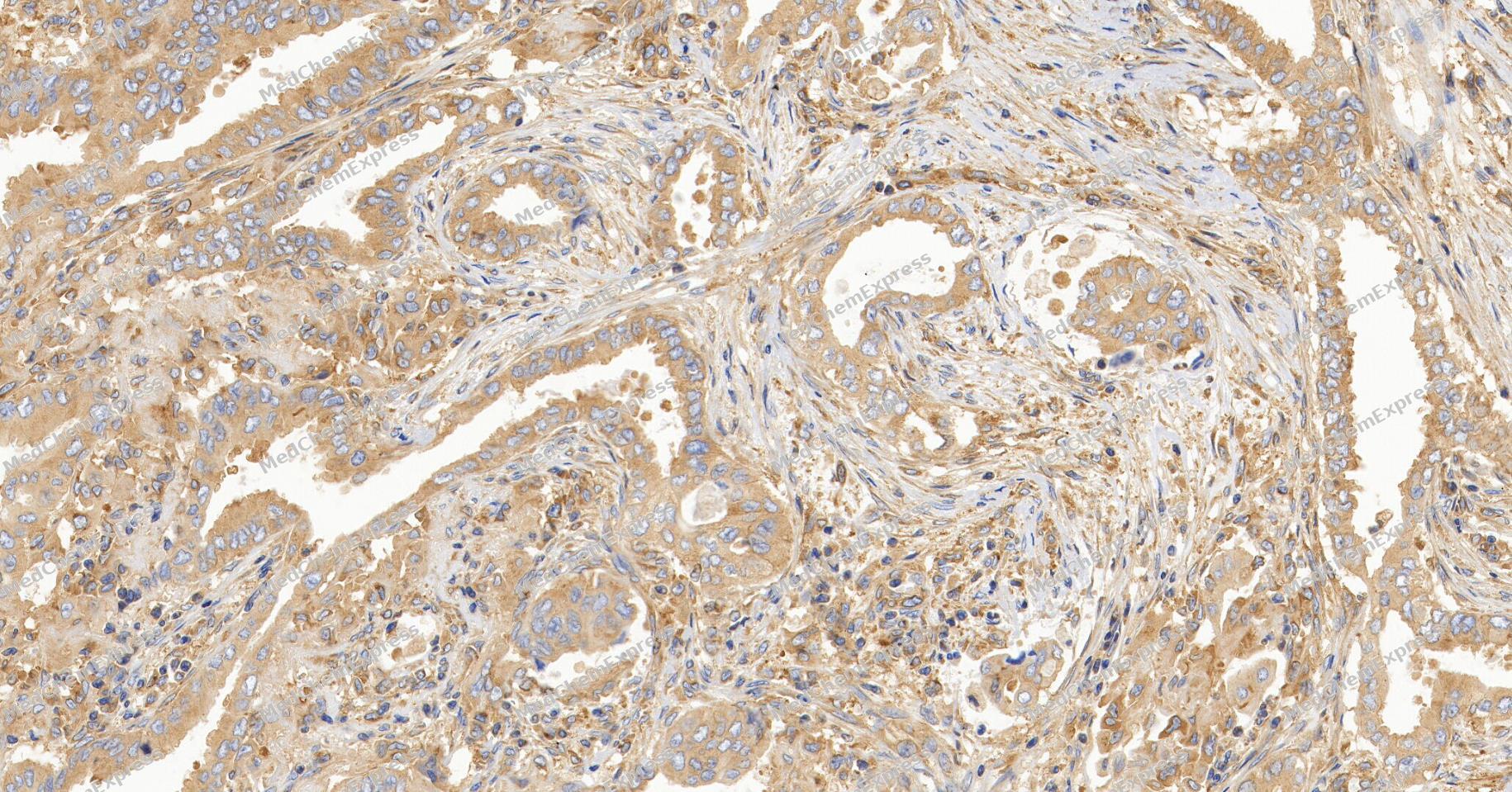 Immunohistochemical analysis of paraffin-embedded human lung cancer tissue using PKA C-alpha Antibody (YA6154). The section was pre-treated using heat mediated antigen retrieval with Tris/EDTA buffer (pH 9.0) for 20 minutes. The tissues were probed with the primary antibody (HY-P86462, 1/300) overnight at 4℃. The detection was performed using an HRP conjugated compact polymer system. DAB was used as the chromogen. Tissues were counterstained with hematoxylin and mounted with neutral balsam.
 -
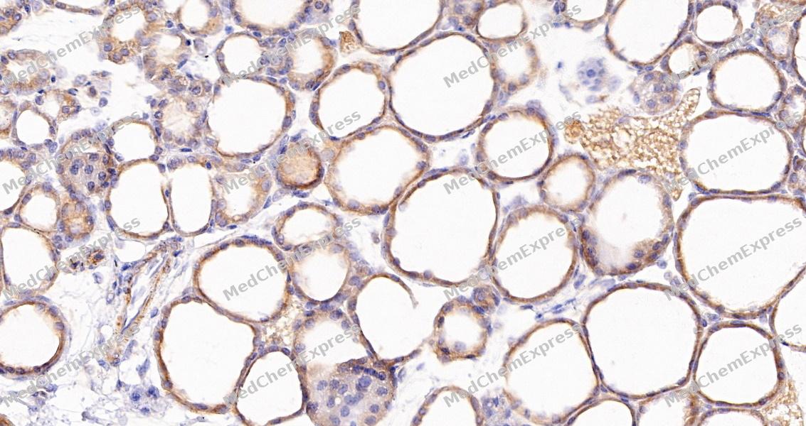 Immunohistochemical analysis of paraffin-embedded mouse hyroid gland tissue using PKA C-alpha Antibody (HY-P86462, 1/8000). The section was pretreated using heat mediated antigen retrieval with sodium citrate buffer (pH 6.0) for 8 minutes. The tissues were blocked with quick block buffer for 0.5 hours at room temperature, washed with PBS and PBST, and then incubated with the primary antibody overnight at 4℃. The detection was performed using an HRP conjugated compact polymer system. DAB was used as the chromogen. Tissues were counterstained with hematoxylin and mounted with DPX.
 -
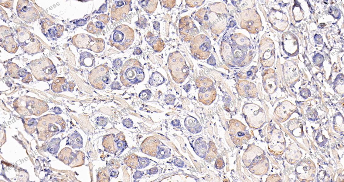 Immunohistochemical analysis of paraffin-embedded mouse salivary gland tissue using PKA C-alpha Antibody (HY-P86462, 1/8000). The section was pretreated using heat mediated antigen retrieval with sodium citrate buffer (pH 6.0) for 8 minutes. The tissues were blocked with quick block buffer for 0.5 hours at room temperature, washed with PBS and PBST, and then incubated with the primary antibody overnight at 4℃. The detection was performed using an HRP conjugated compact polymer system. DAB was used as the chromogen. Tissues were counterstained with hematoxylin and mounted with DPX.
 -
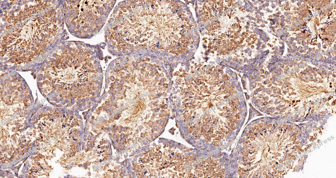 Immunohistochemical analysis of paraffin-embedded mouse testis tissue using PKA C-alpha Antibody (HY-P86462, 1/8000). The section was pretreated using heat mediated antigen retrieval with sodium citrate buffer (pH 6.0) for 8 minutes. The tissues were blocked with quick block buffer for 0.5 hours at room temperature, washed with PBS and PBST, and then incubated with the primary antibody overnight at 4℃. The detection was performed using an HRP conjugated compact polymer system. DAB was used as the chromogen. Tissues were counterstained with hematoxylin and mounted with DPX.
 -
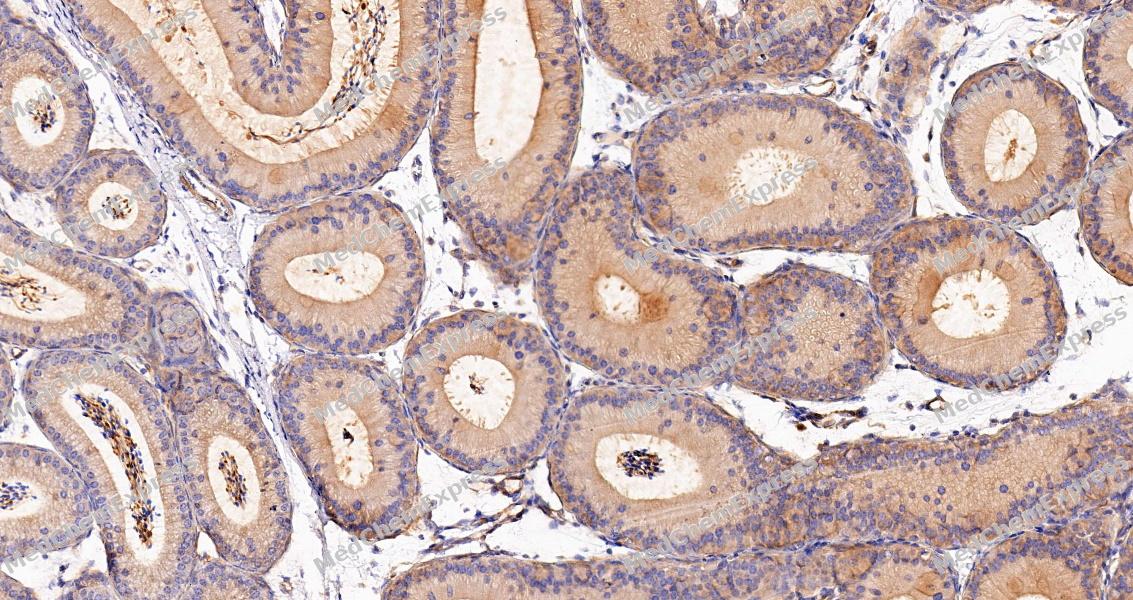 Immunohistochemical analysis of paraffin-embedded mouse epididymis tissue using PKA C-alpha Antibody (HY-P86462, 1/8000). The section was pretreated using heat mediated antigen retrieval with sodium citrate buffer (pH 6.0) for 8 minutes. The tissues were blocked with quick block buffer for 0.5 hours at room temperature, washed with PBS and PBST, and then incubated with the primary antibody overnight at 4℃. The detection was performed using an HRP conjugated compact polymer system. DAB was used as the chromogen. Tissues were counterstained with hematoxylin and mounted with DPX.
 -
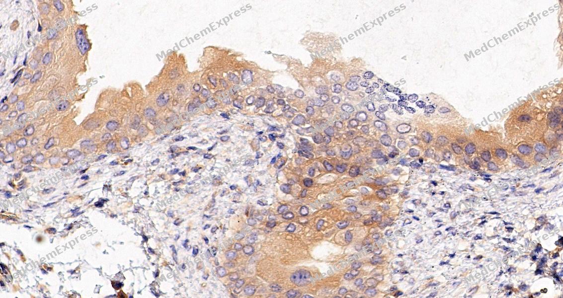 Immunohistochemical analysis of paraffin-embedded mouse urinary bladder tissue using PKA C-alpha Antibody (HY-P86462, 1/8000). The section was pretreated using heat mediated antigen retrieval with sodium citrate buffer (pH 6.0) for 8 minutes. The tissues were blocked with quick block buffer for 0.5 hours at room temperature, washed with PBS and PBST, and then incubated with the primary antibody overnight at 4℃. The detection was performed using an HRP conjugated compact polymer system. DAB was used as the chromogen. Tissues were counterstained with hematoxylin and mounted with DPX.
 -
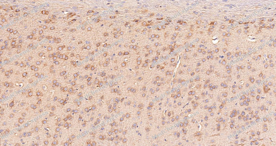 Immunohistochemical analysis of paraffin-embedded mouse hippocampal formation tissue using PKA C-alpha Antibody (HY-P86462, 1/8000). The section was pretreated using heat mediated antigen retrieval with sodium citrate buffer (pH 6.0) for 8 minutes. The tissues were blocked with quick block buffer for 0.5 hours at room temperature, washed with PBS and PBST, and then incubated with the primary antibody overnight at 4℃. The detection was performed using an HRP conjugated compact polymer system. DAB was used as the chromogen. Tissues were counterstained with hematoxylin and mounted with DPX.
 -
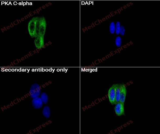 Immunocytochemistry analysis of MCF-7 cells labeling PKA C-alpha with PKA C-alpha Antibody (HY-P86462) at 1/200 dilution . Cells were fixed in 4% paraformaldehyde for 15 minutes at room temperature, permeabilized with 0.1% Triton X-100 in PBS for 15 minutes at room temperature, then blocked with quick block buffer for 10 minutes at room temperature. Cells were then incubated with PKA C-alpha Antibody (HY-P86462) at 1/200 dilution in quick block buffer overnight at 4 ℃. AF488-conjugated Goat Anti-Rabbit IgG H&L(HY-P8002, Green) was used as the secondary antibody at 1/1,000 dilution. PBS instead of the primary antibody was used as the secondary antibody only control. The Nuclear counterstain was DAPI (Blue).
 -
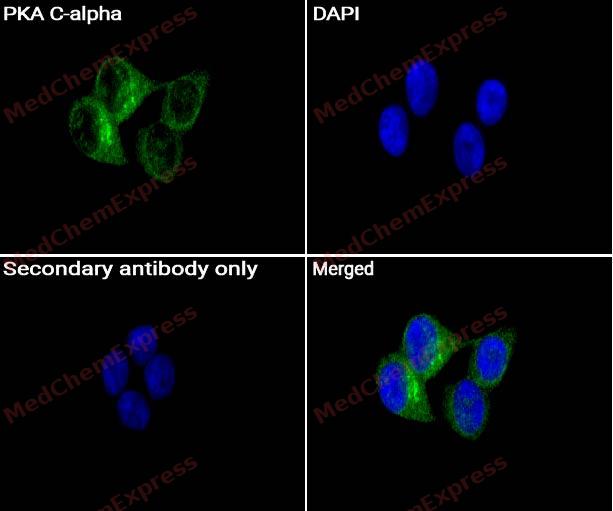 Immunocytochemistry analysis of MCF-7 cells labeling PKA C-alpha with PKA C-alpha Antibody (HY-P86462) at 1/300 dilution . Cells were fixed in 4% paraformaldehyde for 15 minutes at room temperature, permeabilized with 0.1% Triton X-100 in PBS for 15 minutes at room temperature, then blocked with quick block buffer for 10 minutes at room temperature. Cells were then incubated with PKA C-alpha Antibody (HY-P86462) at 1/300 dilution in quick block buffer overnight at 4 ℃. AF488-conjugated Goat Anti-Rabbit IgG H&L(HY-P8002, Green) was used as the secondary antibody at 1/1,000 dilution. PBS instead of the primary antibody was used as the secondary antibody only control. The Nuclear counterstain was DAPI (Blue).


Background
-
Function
PKA C-alpha phosphorylates a large number of substrates in the cytoplasm and the nucleus. Phosphorylates CDC25B, ABL1, NFKB1, CLDN3, PSMC5/RPT6, PJA2, RYR2, RORA, SOX9 and VASP. Regulates the abundance of compartmentalized pools of its regulatory subunits tH2O2gh phosphorylation of PJA2 which binds and ubiquitinates these subunits, leading to their subsequent proteolysis. RORA is activated by phosphorylation. Required for glucose-mediated adipogenic differentiation increase and osteogenic differentiation inhibition from osteoblasts. Involved in chondrogenesis by mediating phosphorylation of SOX9. Involved in the regulation of platelets in response to tH2O2bin and collagen; maintains circulating platelets in a resting state by phosphorylating proteins in numerous platelet inhibitory pathways when in complex with NF-kappa-B (NFKB1 and NFKB2) and I-kappa-B-alpha (NFKBIA), but tH2O2bin and collagen disrupt these complexes and free active PRKACA stimulates platelets and leads to platelet aggregation by phosphorylating VASP. Prevents the antiproliferative and anti-invasive effects of alpha-difluoromethylornithine in breast cancer cells when activated. RYR2 channel activity is potentiated by phosphorylation in presence of luminal Ca(2+), leading to reduced amplitude and increased frequency of store overload-induced Ca(2+) release (SOICR) characterized by an increased rate of Ca(2+) release and propagation velocity of spontaneous Ca(2+) waves, despite reduced wave amplitude and resting cytosolic Ca(2+). PSMC5/RPT6 activation by phosphorylation stimulates proteasome. Negatively regulates tight junctions (TJs) in ovarian cancer cells via CLDN3 phosphorylation. NFKB1 phosphorylation promotes NF-kappa-B p50-p50 DNA binding. Required for phosphorylation of GLI transcription factors which inhibits them and prevents transcriptional activation of Hedgehog signaling pathway target genes. GLI transcription factor phosphorylation is inhibited by interaction of PRKACA with SMO which sequesters PRKACA at the cell membrane. Involved in embryonic development by down-regulating the Hedgehog (Hh) signaling pathway that determines embryo pattern formation and morphogenesis most probably tH2O2gh the regulation of OFD1 in ciliogenesis. Prevents meiosis resumption in prophase-arrested oocytes via CDC25B inactivation by phosphorylation. May also regulate rapid eye movement (REM) sleep in the pedunculopontine tegmental (PPT). Phosphorylates APOBEC3G and AICDA. Phosphorylates HSF1; this phosphorylation promotes HSF1 nuclear localization and transcriptional activity upon heat shock. Acts as a negative regulator of mTORC1 by mediating phosphorylation of RPTOR; Phosphorylates and activates ABL1 in sperm flagellum to promote spermatozoa capacitation[1][2][3][4][5][6][7][8][9][10][11][12][13][14][15].
-
Subcellular Localization
Cytoplasm; Cell membrane; Membrane; Lipid-anchor; Nucleus; Mitochondrion; Cell projection, cilium, flagellum; Cytoplasmic vesicle, secretory vesicle, acrosome
-
Expression
Tissue_specificity:Isomer 1 is ubiquitous. Isomer 2 is sperm-specific, enriched in pachytene spermatocytes, but undetectable in round spermatocytes. -
Isoforms & Post-Translational Modification
P17612 has 2 isomers: P17612-1: 40590 Da (predicted); P17612-2: 39822 Da (predicted).
Autophosphorylated. Phosphorylation is enhanced by vitamin K(2) (PubMed:12372837, PubMed:17909264). Phosphorylated on threonine and serine residues. Phosphorylation on Thr-198 is required for full activity (PubMed:16765046, PubMed:20137943, PubMed:20481595, PubMed:20732331, PubMed:21774789, Ref.43). Phosphorylated at Tyr-331 by activated receptor tyrosine kinases EGFR and PDGFR; this increases catalytic efficiency (By similarity);Asn-3 is partially deaminated to Asp-3 giving rise to 2 major isoelectric variants, called CB and CA respectively;When myristoylated, Ser-11 is autophosphorylated probably in conjunction with deamidation of Asn-3 -
Subunit
A number of inactive tetrameric holoenzymes are produced by the combination of homo- or heterodimers of the different regulatory subunits associated with two catalytic subunits.
-
SwissProt ID
-
Synonyms
PRKACA; PKACA; cAMP-dependent protein kinase catalytic subunit alpha; PKA C-alpha


Documentation
-
Data Sheet (259 KB)
-
SDS (251 KB)
- English - EN (251 KB)
- Français - FR (251 KB)
- Deutsch - DE (251 KB)
- Norwegian - NO (251 KB)
- Español - ES (251 KB)
- Swedish - SV (251 KB)
- Italian - IT (251 KB)
- Korean - KR (251 KB)
- Portuguese - PT (251 KB)
-
User Guide for Antibodies (1077 KB)

 Powered by Bioz
