



TCF3 / E2A Antibody (YA3815)
(Synonyms: E2A; ITF1; VDIR; bHLHb21; MGC129647; MGC129648; TCF3)Based on 1 Customer Validation
TCF3 / E2A Antibody (YA3815) is a Mouse-derived and non-conjugated IgG1 monoclonal antibody, targeting to TCF3 / E2A.
-
Host:
Mouse
-
Isotype:
IgG1
-
Application:
WB, FC, ELISA
-
Reactivity :
Human
-
Formulation:
Supplied in PBS with 0.05% sodium azide.
-
Conjugation:
Non-conjugated


To place orders, for customer services and technical support, please contact: MedChemExpress USA
Tel: 609-228-6898 E-mail: [email protected] [email protected]





Applications
| Application |
WB

WB: Western Blot
|
FC
FC: Flow Cytometry
|
ELISA
ELISA: Enzyme Linked Immunosorbent Assay
|
|---|---|---|---|
| Dilution Ratio | 1:500-1:2000 | 1:200-1:400 | 1:10000 |


Product Details
TCF3 / E2A Antibody (YA3815) is a Mouse-derived and non-conjugated IgG1 monoclonal antibody, targeting to TCF3 / E2A.
-
Host Mouse

-
Clonality Monoclonal

-
Species ReactivityHuman
-
Observed Molecular WeightObserved band size: 68 kDa
Note: Due to possible protein modifications or aggregation, the molecular weight should be confirmed by actual measurement, and the predicted value is for reference only.

-
Calculated Molecular Weight Predicted band size: 67.6 kDa

Purified recombinant fragment of human TCF3 aa 1-200.
affinity purified.
Non-conjugated
Unmodified
IgG1


Product Properties
-
Appearance
Solution
-
Formulation
Supplied in PBS with 0.05% sodium azide.
-
Concentration
Batch-dependent, Please check the COA for the concentration of each lot. Check Lot Concentration
-
Storage & Stability
Stored at -20°C for 1 year. Avoid repeated freeze / thaw cycles.
-
Shipping
Shipping with blue ice.


Verification Images
-
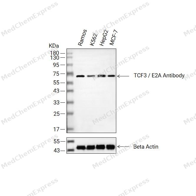 Western blot analysis of extracts from Ramos (lane 2(20μg), K562 (lane 3(20μg), HepG2 (lane 4(20μg), MCF-7 (lane 5(20μg) using TCF3 / E2A Antibody. Proteins were transferred to a PVDF membrane and blocked with 5% BSA in TBST for 2 hour at room temperature. The primary antibody (1/1000)and Loading control antibody (Beta Actin, HY-P80438, 1/3000) was used in 5% BSA in TBST at 4°C overnight. Goat Anti-Mouse/Rabbit IgG-HRP Secondary Antibody (HY-P8004/HY-P8001, 1/10,000) was used for 1 hour at room temperature.
 -
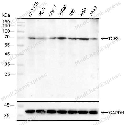 Western blot analysis of extracts from HCT116(lane 1(40μg) ),PC-3(lane 2(40μg) ),COS-7(lane 3(40μg) ),Jurkat(lane 4(40μg) ),Raji(lane 5(40μg) ),Hela(lane 6(40μg) )and A549(lane 7(40μg) ) using TCF3 antibody. Proteins were transferred to a NC membrane and blocked with 5% Skim milk in TBST for 2 hour at room temperature. The primary antibody ( 1/1000) and Loading control antibody (GAPDH, 1/1000) was used in 5% Skim milk in TBST at 4°C overnight. Goat Anti-Mouse IgG-HRP Secondary Antibody (1/10,000) was used for 1 hour at room temperature.
 -
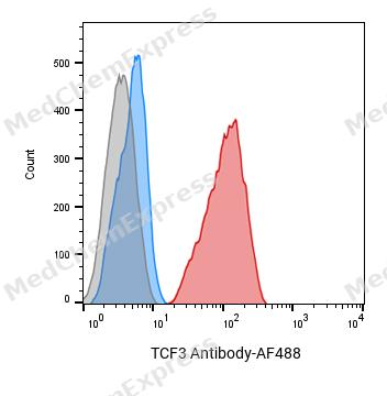 Flow cytometric analysis of 1X10^6 Ramos cells labeling TCF3 Antibody(red). Cells were fixed with 4% paraformaldehyde and permeabilised with 0.2% Triton X-100. Then stained with the primary antibody at 1/400 dilution overnight at 4℃. AF 488 Goat Anti-mouse IgG H&L was used as the secondary antibody at 1/1,000 dilution for 45 minutes at room temperature. Mouse IgG Isotype Control (blue) was used as the isotype control, cells without incubation with primary antibody were used as the unlabeled control (black).


Background
-
Function
TCF3 / E2A transcriptional regulator involved in the initiation of neuronal differentiation and mesenchymal to epithelial transition. Heterodimers between TCF3 and tissue-specific basic helix-loop-helix (bHLH) proteins play major roles in determining tissue-specific cell fate during embryogenesis, like muscle or early B-cell differentiation. Together with TCF15, required for the mesenchymal to epithelial transition. Dimers bind DNA on E-box motifs: 5'-CANNTG-3'. Binds to the kappa-E2 site in the kappa immunoglobulin gene enhancer. Binds to IEB1 and IEB2, which are short DNA sequences in the insulin gene transcription control region; Facilitates ATOH7 binding to DNA at the consensus sequence 5'-CAGGTG-3', and positively regulates transcriptional activity[1].
-
Subcellular Localization
Nucleus
-
Isoforms & Post-Translational Modification
P15923 has 3 isomers: P15923-1: 67600 Da (predicted); P15923-2: 67265 Da (predicted); P15923-3: 68510 Da (predicted).
Phosphorylated following NGF stimulation;Undergoes Notch-induced ubiquitination and subsequent proteasomal degradation which is mediated by ASB1 or ASB2, the substrate-recognition components of probable ECS E3 ubiquitin-protein ligase complexes -
Subunit
Homodimer (PubMed:14752053, PubMed:2112746). Heterodimer; efficient DNA binding requires dimerization with another bHLH protein (By similarity). Forms a heterodimer with ASH1, TWIST1 and TWIST2 (By similarity). Forms a heterodimer with MYOG; heterodimerization enhances MYOG DNA-binding and transcriptional activities (By similarity). Forms a heterodimer with NEUROD1; the heterodimer is inhibited in presence of ID2, but not NR0B2, to E-box element (PubMed:14752053). Forms a heterodimer with TCF15; the heterodimer binds E-box element (By similarity). Forms a heterodimer with ATOH8; repress transcription of TCF3 and TCF3/NEUROG3 dimer-induced transactivation of E box-dependent promoters (By similarity). Component of a nuclear TAL-1 complex composed at least of CBFA2T3, LDB1, TAL1 and TCF3 (By similarity). Interacts with NEUROD2, PTF1A and TGFB1I1 (By similarity). Interacts with EP300 and UBE2I (PubMed:14752053, PubMed:9409784). Interacts with BHLHA9 (PubMed:25466284). Interacts with ASB2; the interaction is mediated by SKP2 and targets TCF3 for Notch-induced proteasomal degradation (PubMed:21119685)
-
SwissProt ID
-
Synonyms
E2A; ITF1; VDIR; bHLHb21; MGC129647; MGC129648; TCF3


Documentation
-
Data Sheet (234 KB)
-
SDS (252 KB)
- English - EN (252 KB)
- Français - FR (252 KB)
- Deutsch - DE (252 KB)
- Norwegian - NO (252 KB)
- Español - ES (252 KB)
- Swedish - SV (252 KB)
- Italian - IT (252 KB)
- Korean - KR (252 KB)
- Portuguese - PT (252 KB)
-
User Guide for Antibodies (1077 KB)

 Powered by Bioz
