



PMS2 Antibody (YA3328)
(Synonyms: TGT; tRNA guanine transglycosylase 60 kD subunit; Ubiquitin carboxyl terminal hydrolase 14; Ubiquitin specific peptidase 14; USP14)
Based on 1 Customer Validation
PMS2 Antibody (YA3328) is a Rabbit-derived and non-conjugated IgG monoclonal antibody, targeting to PMS2.
-
Host:
Rabbit
-
Isotype:
IgG
-
Application:
WB, ICC/IF, FC
-
Reactivity :
Human, Mouse, Rat
-
Formulation:
Supplied in Rabbit IgG in 10mM phosphate buffered saline , pH 7.4, 150mM sodium chloride, 0.05% BSA, 0.02% sodium azide and 50% glycerol.
-
Conjugation:
Non-conjugated


To place orders, for customer services and technical support, please contact: MedChemExpress USA
Tel: 609-228-6898 E-mail: [email protected] [email protected]





Applications
| Application |
WB

WB: Western Blot
|
ICC/IF
ICC/IF: Immunocytochemistry/
Immunofluorescence |
FC
FC: Flow Cytometry
|
|---|---|---|---|
| Dilution Ratio | 1:500-1:1000 | 1:50-1:200 | 1:50-1:100 |


Product Details
PMS2 Antibody (YA3328) is a Rabbit-derived and non-conjugated IgG monoclonal antibody, targeting to PMS2.
-
Host Rabbit

-
Clonality Recombinant,Monoclonal

-
Species ReactivityHuman, Mouse, Rat
-
Observed Molecular WeightObserved band size: 56 kDa
Note: Due to possible protein modifications or aggregation, the molecular weight should be confirmed by actual measurement, and the predicted value is for reference only.

-
Calculated Molecular Weight Predicted band size: 56 kDa

Entrez Gene: 9097 Human ; 59025 Mouse ; 291796 Rat
SwissProt: P54578 Human ; Q9JMA1 Mouse ;
OMIM: 607274 Human
A synthesized peptide derived from human USP14 aa350-494/494.
Endogenous
Affinity Chromatography
Non-conjugated
Unmodified
IgG


Product Properties
-
Appearance
Solution
-
Formulation
Supplied in Rabbit IgG in 10mM phosphate buffered saline , pH 7.4, 150mM sodium chloride, 0.05% BSA, 0.02% sodium azide and 50% glycerol.
-
Concentration
Batch-dependent, Please check the COA for the concentration of each lot. Check Lot Concentration
-
Storage & Stability
Stored at -20°C for 1 year. Avoid repeated freeze / thaw cycles.
-
Shipping
Shipping with blue ice.


Verification Images
-
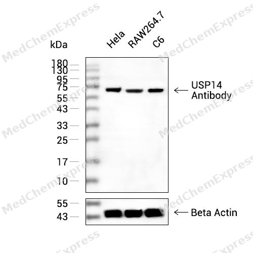 Western blot analysis of extracts from Hela(lane 2(20ug) , RAW264.7(lane 3(20ug) and C6(lane 4(20ug) using USP14 Antibody (HY-P83583) Rabbit mAb. Proteins were transferred to a PVDF membrane and blocked with 5% non-fat milk in TBST for 2 hour at room temperature. The primary antibody (1/1000) and Loading control antibody (Beta Actin, HY-P80438, 1/10000) was used in 5% non-fat milk in TBST at 4°C overnight. Goat Anti-Mouse/Rabbit IgG-HRP Secondary Antibody (1/10000) was used for 1 hour at room temperature.
 -
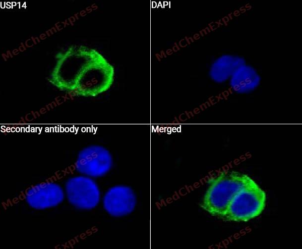 Immunocytochemistry analysis of HepG2 cells labeling USP14 with USP14 Antibody (HY-P83583)at 1/50 dilution. Cells were fixed in 4% paraformaldehyde for 15 minutes at room temperature, permeabilized with 0.1% Triton X-100 for 10 minutes at room temperature, then blocked with QuickBlock™ Blocking Buffer for Immunol Staining for 10 min at room temperature. Cells were then incubated with USP14 Antibody (HY-P83583) at 1/50 dilution in QuickBlock™ Blocking Buffer for Immunol Staining at 4 ℃. Alexa Fluor® 488-conjugated AffiniPure Goat Anti-Rabbit IgG H&L(HY-P8002, Green) was used as the secondary antibody at 1/1,000 dilution. PBS instead of the primary antibody was used as the secondary antibody only control. The Nuclear counterstain was DAPI (Blue).
 -
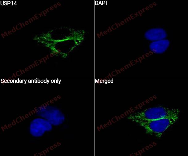 Immunocytochemistry analysis of HepG2 cells labeling USP14 with USP14 Antibody (HY-P83583) at 1/100 dilution. Cells were fixed in 4% paraformaldehyde for 15 minutes at room temperature, permeabilized with 0.1% Triton X-100 for 10 minutes at room temperature, then blocked with QuickBlock™ Blocking Buffer for Immunol Staining for 10 min at room temperature. Cells were then incubated with USP14 Antibody (HY-P83583) at 1/100 dilution in QuickBlock™ Blocking Buffer for Immunol Staining at 4 ℃. Alexa Fluor® 488-conjugated AffiniPure Goat Anti-Rabbit IgG H&L(HY-P8002, Green) was used as the secondary antibody at 1/1,000 dilution. PBS instead of the primary antibody was used as the secondary antibody only control. The Nuclear counterstain was DAPI (Blue).
 -
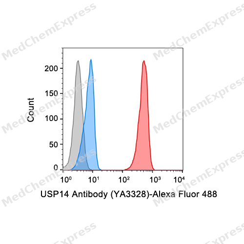 Flow cytometric analysis of 1X10^6 Jurkat cells labeling USP14 Antibody (YA3328) (HY-P83583, red). Cells were fixed with 4% paraformaldehyde and permeabilised with 90% methanol. Then stained with the primary antibody at 1/50 dilution for an hour at 4℃. Alexa Fluor® 488-conjugated AffiniPure Goat Anti-Rabbit IgG H&L (HY-P8002) was used as the secondary antibody at 1/1,000 dilution for 30 minutes at 4℃. Rabbit IgG Isotype Control (HY-P80879, blue) was used as the isotype control, cells without incubation with primary antibody were used as the unlabeled control (black).


Background
-
Function
PMS2 is a Proteasome-associated deubiquitinase which releases ubiquitin from the proteasome targeted ubiquitinated proteins. Ensures the regeneration of ubiquitin at the proteasome. Is a reversibly associated subunit of the proteasome and a large fraction of proteasome-free protein exists within the cell. Required for the degradation of the chemokine receptor CXCR4 which is critical for CXCL12-induced cell chemotaxis. Also serves as a physiological inhibitor of endoplasmic reticulum-associated degradation (ERAD) under the non-stressed condition by inhibiting the degradation of unfolded endoplasmic reticulum proteins via interaction with ERN1. Indispensable for synaptic development and function at neuromuscular junctions (NMJs). Plays a role in the innate immune defense against viruses by stabilizing the viral DNA sensor CGAS and thus inhibiting its autophagic degradation. Inhibits OPTN-mediated selective autophagic degradation of KDM4D and thereby negatively regulates H3K9me2 and H3K9me3[1][2][3][4][5][6].
-
Subcellular Localization
Cytoplasm; Cell membrane; Peripheral membrane protein
-
Isoforms & Post-Translational Modification
P54578 has 3 isomers: P54578-1: 56069 Da (predicted); P54578-2: 52386 Da (predicted); P54578-3: 54770 Da (predicted).
-
Subunit
Homodimer (Potential). Associates with the 26S proteasome. Interacts with FANCC, CXCR4 and ERN1. Interacts with TRIM14; this interaction recruits USP14 to cleave ubiquitin chains of CGAS and KDM4D (PubMed:27666593, PubMed:35145029)
-
SwissProt ID
-
Synonyms
TGT; tRNA guanine transglycosylase 60 kD subunit; Ubiquitin carboxyl terminal hydrolase 14; Ubiquitin specific peptidase 14; USP14
-
Research Field
Cell Biology


Documentation
-
Data Sheet (261 KB)
-
SDS (252 KB)
- English - EN (252 KB)
- Français - FR (252 KB)
- Deutsch - DE (252 KB)
- Norwegian - NO (252 KB)
- Español - ES (252 KB)
- Swedish - SV (252 KB)
- Italian - IT (252 KB)
- Korean - KR (252 KB)
- Portuguese - PT (252 KB)
-
User Guide for Antibodies (1077 KB)
References
[1]. Liu D, et al. TRIM14 inhibits OPTN-mediated autophagic degradation of KDM4D to epigenetically regulate inflammation. Proc Natl Acad Sci U S A. 2022 Feb 15;119(7):. [Content Brief]
[2]. Koulich E, et al. Relative structural and functional roles of multiple deubiquitylating proteins associated with mammalian 26S proteasome. Mol Biol Cell. 2008 Mar;19(3):1072-82. [Content Brief]
[3]. Kuo CL, et al. Ubiquitinated proteins promote the association of proteasomes with the deubiquitinating enzyme Usp14 and the ubiquitin ligase Ube3c. Proc Natl Acad Sci U S A. 2017 Apr 25;114(17):E3404-E3413. [Content Brief]
[4]. Mines MA, et al. Deubiquitination of CXCR4 by USP14 is critical for both CXCL12-induced CXCR4 degradation and chemotaxis but not ERK ativation. J Biol Chem. 2009 Feb 27;284(9):5742-52. [Content Brief]
[5]. Nagai A, et al. USP14 inhibits ER-associated degradation via interaction with IRE1alpha. Biochem Biophys Res Commun. 2009 Feb 20;379(4):995-1000. [Content Brief]
[6]. Chen M, et al. TRIM14 Inhibits cGAS Degradation Mediated by Selective Autophagy Receptor p62 to Promote Innate Immune Responses. Mol Cell. 2016 Oct 6;64(1):105-119. [Content Brief]

 Powered by Bioz
