



Wnt-1 Antibody (YA3644)
(Synonyms: INT1)Based on 1 publication(s) in Google Scholar
Wnt-1 Antibody (YA3644) is a Mouse-derived and non-conjugated IgG1 monoclonal antibody, targeting to Wnt-1.
-
Host:
Mouse
-
Isotype:
IgG
-
Application:
WB, IHC-P, ICC/IF, FC, ELISA
-
Reactivity :
Human, Mouse
-
Formulation:
Supplied in PBS with 0.05% sodium azide.
-
Conjugation:
Non-conjugated


To place orders, for customer services and technical support, please contact: MedChemExpress USA
Tel: 609-228-6898 E-mail: [email protected] [email protected]



Publications Citing Use of MedChemExpress (MCE) Wnt-1 Antibody (YA3644)
More

Applications
| Application |
WB

WB: Western Blot
|
IHC-P
IHC-P: Immunohistochemistry-Paraffin
|
ICC/IF
ICC/IF: Immunocytochemistry/
Immunofluorescence |
FC
FC: Flow Cytometry
|
ELISA
ELISA: Enzyme Linked Immunosorbent Assay
|
|---|---|---|---|---|---|
| Dilution Ratio | 1:500-1:2000 | 1:200-1:1000 | 1:200-1:1000 | 1:200-1:400 | 1:10000 |


Product Details
Wnt-1 Antibody (YA3644) is a Mouse-derived and non-conjugated IgG1 monoclonal antibody, targeting to Wnt-1.
-
Host Mouse

-
Clonality Monoclonal

-
Species ReactivityHuman, Mouse
-
Observed Molecular WeightObserved band size: 45 kDa
Note: Due to possible protein modifications or aggregation, the molecular weight should be confirmed by actual measurement, and the predicted value is for reference only.

-
Calculated Molecular Weight Predicted band size: 41 kDa

Purified recombinant fragment of WNT1 aa 28-370.
affinity purified.
Non-conjugated
Unmodified
IgG


Product Properties
-
Appearance
Solution
-
Formulation
Supplied in PBS with 0.05% sodium azide.
-
Concentration
Batch-dependent, Please check the COA for the concentration of each lot. Check Lot Concentration
-
Storage & Stability
Stored at -20°C for 1 year. Avoid repeated freeze / thaw cycles.
-
Shipping
Shipping with blue ice.


Publications (1)
-
Journal Impact Factor
-
Most Recent
-
Mol Immunol
Indole-3-propionic acid exerts radio-protective effects by modulating Wnt1/STAT3 pathway to alleviate oxidative stress and neuroinflammation in microglia. [Abstract]2025 Dec 22:189:192-205. PMID: 41435624


Verification Images
-
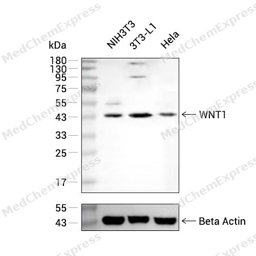 Western blot analysis of extracts from NIH/3T3(lane 1(40μg) ) and 3T3-L1(lane 2(40μg) ) using WNT1 antibody. Proteins were transferred to a NC membrane and blocked with 5% Skim milk in TBST for 2 hour at room temperature. The primary antibody ( 1/1000) and Loading control antibody (GAPDH,1/1000) was used in 5% Skim milk in TBST at 4°C overnight. Goat Anti-Mouse IgG-HRP Secondary Antibody (1/10
 -
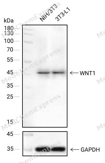 Western blot analysis of extracts from NIH/3T3(lane 1(40μg) ) and 3T3-L1(lane 2(40μg) ) using WNT1 antibody. Proteins were transferred to a NC membrane and blocked with 5% Skim milk in TBST for 2 hour at room temperature. The primary antibody ( 1/1000) and Loading control antibody (GAPDH, 1/1000) was used in 5% Skim milk in TBST at 4°C overnight. Goat Anti-Mouse IgG-HRP Secondary Antibody (1/10,000) was used for 1 hour at room temperature.
 -
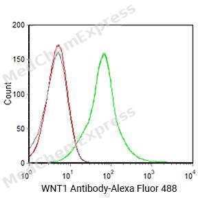 Flow cytometric analysis of 1X10^6 Hela cells labeling WNT1 Antibody(HY-P83947
 -
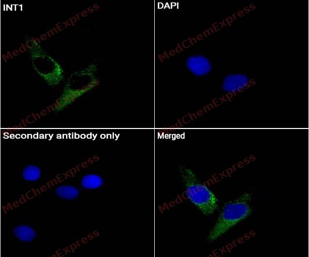 Immunocytochemistry analysis of HeLa cells labeling INT1 Antibody (HY-P83947) at 1/50 dilution. Cells were fixed in 4% paraformaldehyde for 15 minutes at room temperature, permeabilized with 0.1% Triton X-100 for 10 minutes at room temperature, then blocked with BSA for Immunol Staining for 10 min at room temperature. Cells were then incubated with INT1 Antibody (HY-P83947) at 1/50 dilution in BSA for Immunol Staining at 4 ℃overnight. AF488-conjugated AffiniPure Goat Anti-Mouse IgG H&L(HY-P8005, Green) was used as the secondary antibody at 1/1,000 dilution. PBS instead of the primary antibody was used as the secondary antibody only control. The Nuclear counterstain was DAPI (Blue).
 -
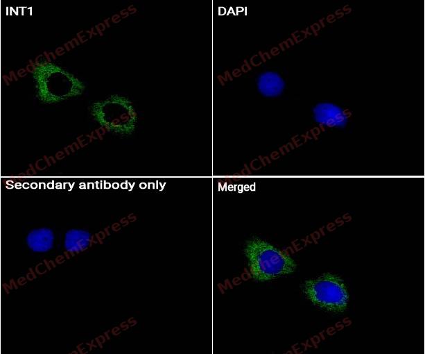 Immunocytochemistry analysis of mouse 3T3 cells labeling INT1 Antibody (HY-P83947) at 1/50 dilution. Cells were fixed in 4% paraformaldehyde for 15 minutes at room temperature, permeabilized with 0.1% Triton X-100 for 10 minutes at room temperature, then blocked with BSA for Immunol Staining for 10 min at room temperature. Cells were then incubated with INT1 Antibody (HY-P83947) at 1/50 dilution in BSA for Immunol Staining at 4 ℃overnight. AF488-conjugated AffiniPure Goat Anti-Mouse IgG H&L(HY-P8005, Green) was used as the secondary antibody at 1/1,000 dilution. PBS instead of the primary antibody was used as the secondary antibody only control. The Nuclear counterstain was DAPI (Blue).


Background
-
Function
Wnt1 is a secreted Wnt ligand that mediates signaling through Frizzled/LRP5/6 and the β-catenin-dependent canonical Wnt pathway, a pathway linked to cell proliferation and differentiation[1]. Mechanistically, Wnt1 has essential developmental relevance because Wnt1- mice lose the midbrain and adjacent cerebellar component, showing its requirement for midbrain-hindbrain development[2][3]. In disease-focused skeletal research, WNT1 mutations cause early-onset osteoporosis and osteogenesis imperfecta, identifying WNT1 as a key ligand regulating human bone mass[4][5]. Wnt1 mutant mouse models reproduce bone fragility and support experimental analysis of osteogenesis imperfecta mechanisms[6]. Compared with Wnt5a, which can antagonize canonical Wnt signaling by promoting β-catenin degradation, Wnt1 is best positioned as a canonical Wnt ligand for β-catenin-centered experimental design[1][7]. For applied studies, Wnt1-Cre models remain useful for neural crest and midbrain research, but developmental phenotypes from ectopic Wnt signaling require careful controls[8]. Wnt-pathway inhibitors such as PORCN inhibitors can block Wnt ligand processing and secretion, providing tools to test Wnt-ligand-dependent signaling in Wnt-driven models[9].
-
Subcellular Localization
Secreted, extracellular space, extracellular matrix; Secreted
-
Subunit
Forms a soluble 1:1 complex with AFM; this prevents oligomerization and is required for prolonged biological activity (PubMed:26902720). The complex with AFM may represent the physiological form in body fluids (PubMed:26902720). Interacts with PORCN. Interacts with RSPO1, RSPO2 and RSPO3 (By similarity). Interacts with WLS (By similarity)
-
SwissProt ID
-
Synonyms
INT1
Documentation
-
Data Sheet (262 KB)
-
SDS (251 KB)
- English - EN (251 KB)
- Français - FR (251 KB)
- Deutsch - DE (251 KB)
- Norwegian - NO (251 KB)
- Español - ES (251 KB)
- Swedish - SV (251 KB)
- Italian - IT (251 KB)
- Korean - KR (251 KB)
- Portuguese - PT (251 KB)
-
User Guide for Antibodies (1077 KB)
References
[1]. Qin K, et al. Canonical and noncanonical Wnt signaling: Multilayered mediators, signaling mechanisms and major signaling crosstalk. Genes Dis. 2023 Mar 24;11(1):103-134. [Content Brief]
[2]. McMahon AP, et al. The Wnt-1 (int-1) proto-oncogene is required for development of a large region of the mouse brain. Cell. 1990 Sep 21;62(6):1073-85. [Content Brief]
[3]. McMahon AP, et al. The midbrain-hindbrain phenotype of Wnt-1-/Wnt-1- mice results from stepwise deletion of engrailed-expressing cells by 9.5 days postcoitum. Cell. 1992 May 15;69(4):581-95. [Content Brief]
[4]. Laine CM, et al. WNT1 mutations in early-onset osteoporosis and osteogenesis imperfecta. N Engl J Med. 2013 May 9;368(19):1809-16. [Content Brief]
[5]. Keupp K, et al. Mutations in WNT1 cause different forms of bone fragility. Am J Hum Genet. 2013 Apr 4;92(4):565-74. [Content Brief]
[6]. Vollersen N, et al. The WNT1G177C mutation specifically affects skeletal integrity in a mouse model of osteogenesis imperfecta type XV. Bone Res. 2021 Nov 10;9(1):48. [Content Brief]
[7]. Topol L, et al. Wnt-5a inhibits the canonical Wnt pathway by promoting GSK-3-independent beta-catenin degradation. J Cell Biol. 2003 Sep 1;162(5):899-908. [Content Brief]
[8]. Lewis AE, et al. The widely used Wnt1-Cre transgene causes developmental phenotypes by ectopic activation of Wnt signaling. Dev Biol. 2013 Jul 15;379(2):229-34. [Content Brief]
[9]. Liu J, et al. Targeting Wnt-driven cancer through the inhibition of Porcupine by LGK974. Proc Natl Acad Sci U S A. 2013 Dec 10;110(50):20224-9. [Content Brief]

 Powered by Bioz
