



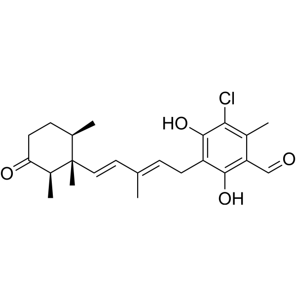
Ascochlorin
Ascochlorin (Ilicicolin D), an isoprenoid antibiotic, mediates its anti-tumor effects predominantly through the suppression of STAT3 signaling cascade. Ascochlorin induces apoptosis. Anti-inflammatory activity.
For research use only. We do not sell to patients.
- CAS No.: 26166-39-2
- Formula: C23H29ClO4
- Molecular Weight:404.93
-
Storage:
Please store the product under the recommended conditions in the Certificate of Analysis.

To place orders, for customer services and technical support, please contact: MedChemExpress USA
Tel: 609-228-6898 E-mail: [email protected] [email protected]


-
 Biological Activity
-
Chemical Information
- Protocols
- Purity & Documentation
- References
-
 Help & FAQs



All Antibiotic Isoforms
More

Biological Activity
Description
IC50 & Target
[3]|
STAT3 |
Apoptosis |
Cellular Effect
|
Cell Line

|
Type | Value | Description | References |
|---|---|---|---|---|
| A549 | IC50 |
0.9 μM
Compound: 7
|
Cytotoxicity against human A549 cells assessed as reduction in cell viability incubated for 48 hrs by CCK8 assay
Cytotoxicity against human A549 cells assessed as reduction in cell viability incubated for 48 hrs by CCK8 assay
|
[PMID: 34781681] |
| C3H 10T1/2 | IC50 |
1 μM
Compound: 1
|
Concentration required for inhibition of C3H10T1/2 progenitor cell growth
Concentration required for inhibition of C3H10T1/2 progenitor cell growth
|
[PMID: 12954063] |
| HepG2 | IC50 |
0.9 μM
Compound: 7
|
Cytotoxicity against human HepG2 cells assessed as reduction in cell viability incubated for 48 hrs by CCK8 assay
Cytotoxicity against human HepG2 cells assessed as reduction in cell viability incubated for 48 hrs by CCK8 assay
|
[PMID: 34781681] |
| MDA-MB-231 | IC50 |
2.2 μM
Compound: 12
|
Cytotoxicity against human MDA-MB-231 cells assessed as reduction in cell viability incubated for 24 hrs by CellTiter-Glo luminescent assay
Cytotoxicity against human MDA-MB-231 cells assessed as reduction in cell viability incubated for 24 hrs by CellTiter-Glo luminescent assay
|
[PMID: 34516133] |
| MDA-MB-468 | IC50 |
1.1 μM
Compound: 12
|
Cytotoxicity against human MDA-MB-468 cells assessed as reduction in cell viability incubated for 24 hrs by CellTiter-Glo luminescent assay
Cytotoxicity against human MDA-MB-468 cells assessed as reduction in cell viability incubated for 24 hrs by CellTiter-Glo luminescent assay
|
[PMID: 34516133] |
In Vitro
Ascochlorin (Ilicicolin D) (10-50 μM; 24-72 hours) inhibits the viability of HepG2, HCCLM3 and Huh7 cells in a time and dose dependent manner[3].
Ascochlorin (50 μM; 48 hours) induces apoptosis in HCC cells[3].
Ascochlorin (1-50 μM) significantly suppresses the production of nitric oxide (NO) and prostaglandin E2 (PGE2) and decreases the gene expression of inducible NO synthase (iNOS) and cyclooxygenase-2 (COX-2) in a dose-dependent manner. Ascochlorin inhibits the mRNA expression and the protein secretion of interleukin (IL)-1β and IL-6 but not tumor necrosis factor (TNF)-α in LPS-stimulated RAW 264.7 macrophage cells. Ascochlorin suppresses nuclear translocation and DNA binding affinity of nuclear factor-κB (NF-κB). Ascochlorin down-regulates phospho-extracellular signal-regulated kinase 1/2 (p-ERK1/2) and p-p38[2].
MedChemExpress (MCE) has not independently confirmed the accuracy of these methods. They are for reference only.
-
Cell Line:HepG2, HCCLM3, Huh7 cells
-
Concentration:10, 25, 50 μM
-
Incubation Time:24, 48, 72 hours
-
Result:Inhibit the viability of three different HCC cell lines tested (HepG2, HCCLM3 and Huh7) in a time and dose dependent manner.
-
Cell Line:HepG2 cells
-
Concentration:50 μM
-
Incubation Time:48 hours
-
Result:Inhibited expression of the cell cycle regulator protein cyclin D1, the anti-apoptotic proteins Bcl-2, Mcl-1, survivin and XIAP, and the invasive gene product MMP-9.
In Vivo
MedChemExpress (MCE) has not independently confirmed the accuracy of these methods. They are for reference only.
-
Animal Model:Eight week-old athymic balb/c nude female mice (HCCLM3-Luc2 tumors)[3]
-
Dosage:2.5 mg/kg, 5 mg/kg
-
Administration:I.p.; day 0, 1, 2, 3, 13, 15, 17, 20, 22, 24, 27, 29 and 31
-
Result:Induced significant inhibition of tumor growth.


Chemical Information
-
CAS No. 26166-39-2
-
Molecular Weight 404.93
-
Formula C23H29ClO4
-
SMILES
O=CC1=C(O)C(C/C=C(C)/C=C/[C@]2(C)[C@@H](C)C(CC[C@H]2C)=O)=C(O)C(Cl)=C1C
-
Synonyms
Ilicicolin D
-
Initial Source
-
Shipping
Room temperature in continental US; may vary elsewhere.
-
Storage
Please store the product under the recommended conditions in the Certificate of Analysis.


Purity & Documentation
References
[1]. Min-Wen JC, et al. Molecular Targets of Ascochlorin and Its Derivatives for Cancer Therapy. Adv Protein Chem Struct Biol. 2017;108:199-225. [Content Brief]
[2]. Lee SH, et al. Anti-Inflammatory Effect of Ascochlorin in LPS-Stimulated RAW 264.7 Macrophage Cells Is Accompanied With the Down-Regulation of iNOS, COX-2 and Proinflammatory Cytokines Through NF-κB, ERK1/2, and p38 Signaling Pathway. J Cell Biochem. 2016 Apr;117(4):978-87. [Content Brief]
[3]. Dai X, et al. Ascochlorin, an isoprenoid antibiotic inhibits growth and invasion of hepatocellular carcinoma by targeting STAT3 signaling cascade through the induction of PIAS3. Mol Oncol. 2015 Apr;9(4):818-33. [Content Brief]

Calculators
Concentration (start) × Volume (start) = Concentration (final) × Volume (final)


Ascochlorin Related Classifications
HY-101021 Related Classifications
 Powered by Bioz