



GXF-111
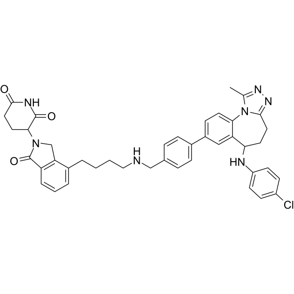
GXF-111 is a BRD3 and BRD4-L PROTAC degrader. Its BD1 and BD2 Ki values against human BRD3 are 11.97 nM and 2.45 nM, respectively. The degradation activity of GXF-111 depends on its binding to BET proteins and Cereblon, as well as the involvement of a functional proteasome. The degradation selectivity of GXF-111 is mainly determined by differences in degradation kinetics and cell types. GXF-111 induces G1 phase cell cycle arrest, downregulates c-Myc expression, upregulates p21 expression, and exhibits antiproliferative activity against a variety of cancer cell lines. GXF-111 can serve as a research tool for cancer-related studies.
(Pink: BRD3 and BRD4-L Target protein ligand; Blue: Cereblon ligand (HY-138793); Black: linker).
Nos produits utilisent uniquement pour la recherche. Nous ne vendons pas aux patients.
- Formule: C42H42ClN7O3
- Masse moléculaire:728.28
-
Stockage:
Please store the product under the recommended conditions in the Certificate of Analysis.

To place orders, for customer services and technical support, please contact: MedChemExpress USA
Tel: 609-228-6898 E-mail: [email protected] [email protected]


-
 Activité biologique
-
Chemical Information
- Protocole
- Pureté et documentation
- Références
-
 Help & FAQs


Voir tous les produits spécifiques à Isoform PROTACs
More

Activité biologique
Description
IC50 & Target
|
BRD3 BD2 2.45 nM (Ki) |
BRD3 BD1 11.97 nM (Ki) |
BRD4-L |
Cellular Effect
|
Cell Line

|
Type | Value | Description | References |
|---|---|---|---|---|
| A549 | IC50 |
14.26 μM
Compound: 24; GXF111
|
Antiproliferative activity against human A549 cells assessed as cell growth inhibition measured after 4 days by CCK-8 assay
Antiproliferative activity against human A549 cells assessed as cell growth inhibition measured after 4 days by CCK-8 assay
|
[PMID: 37084596] |
| HeLa | IC50 |
6.96 μM
Compound: 24; GXF111
|
Antiproliferative activity against human HeLa cells assessed as cell growth inhibition measured after 4 days by CCK-8 assay
Antiproliferative activity against human HeLa cells assessed as cell growth inhibition measured after 4 days by CCK-8 assay
|
[PMID: 37084596] |
| HepG2 | IC50 |
17.25 μM
Compound: 24; GXF111
|
Antiproliferative activity against human HepG2 cells assessed as cell growth inhibition measured after 4 days by CCK-8 assay
Antiproliferative activity against human HepG2 cells assessed as cell growth inhibition measured after 4 days by CCK-8 assay
|
[PMID: 37084596] |
| HGC-27 | IC50 |
2.35 μM
Compound: 24; GXF111
|
Antiproliferative activity against human HGC-27 cells assessed as cell growth inhibition measured after 4 days by CCK-8 assay
Antiproliferative activity against human HGC-27 cells assessed as cell growth inhibition measured after 4 days by CCK-8 assay
|
[PMID: 37084596] |
| MCF7 | IC50 |
0.23 μM
Compound: 24; GXF111
|
Antiproliferative activity against human MCF7 cells assessed as cell growth inhibition measured after 4 days by CCK-8 assay
Antiproliferative activity against human MCF7 cells assessed as cell growth inhibition measured after 4 days by CCK-8 assay
|
[PMID: 37084596] |
| MM1.S | IC50 |
95.2 nM
Compound: 24; GXF111
|
Antiproliferative activity against human MM1.S cells assessed as cell growth inhibition measured after 4 days by CellTitre-Glo assay
Antiproliferative activity against human MM1.S cells assessed as cell growth inhibition measured after 4 days by CellTitre-Glo assay
|
[PMID: 37084596] |
| MV4-11 | IC50 |
6.31 nM
Compound: 24; GXF111
|
Antiproliferative activity against human MV4-11 cells assessed as cell growth inhibition measured after 3 days by CellTitre-Glo assay
Antiproliferative activity against human MV4-11 cells assessed as cell growth inhibition measured after 3 days by CellTitre-Glo assay
|
[PMID: 37084596] |
In Vitro
GXF-111 (compound 24) binds to BRD3 BD2 with high affinity (Ki = 2.45 nM) and shows lower affinity for BRD3 BD1 (Ki = 11.97 nM), exhibiting approximately 5-fold selectivity for the BD2 domain of BRD3[1].
GXF-111 (3-4) potently inhibits the growth of MV4-11 and MM.1S cells, with IC50 values of 6.31 nM and 95.2 nM after 3-day and 4-day treatment, respectively[1].
GXF-111 (for 4 days) inhibits the growth of MCF-7 (IC50 = 0.23 μM), HGC-27 (IC50 = 2.35 μM), A549 (IC50 = 14.26 μM), HeLa (IC50 = 6.96 μM) and HepG2 (IC50 = 17.25 μM) cells[1].
GXF-111 (0.1-30 nM; 24 h) selectively degrades BRD3 (1-3 nM) and BRD4-L in MM.1S cells without affecting BRD4-S, and only exerts a partial effect on BRD2 at higher doses; meanwhile, this compound regulates the expression of c-Myc and p21 without inducing significant apoptosis[1].
GXF-111 (30 nM; 1-24 h) induces time-dependent selective degradation of BRD3 and BRD4-L in MM.1S, HGC-27 and MCF-7 cells. Compared with BRD2, the degradation of BRD3/BRD4-L initiates earlier, and GXF-111 exerts minimal effects on BRD4-S within 24 hours[1].
GXF-111 (0.1-1000 nM; 24 h) selectively degrades BRD3 and BRD4-L (but not BRD2 or BRD4-S) in HGC-27, MCF-7, A549, HeLa and HepG2 cells, while it selectively degrades BRD4-L and BRD4-S in MV4-11 cells; its degradation efficacy varies among different cell lines[1].
The degradation of BRD3 and BRD4-L in MM.1S cells induced by GXF-111 (30 nM; 4 h) depends on its binding to BET proteins, binding to Cereblon, and functional proteasome activity[1].
GXF-111 (1-300 nM; 24 h) induces G1-phase cell cycle arrest in MM.1S cells in a dose-dependent manner[1].
MedChemExpress (MCE) has not independently confirmed the accuracy of these methods. They are for reference only. Further protocols information, click here.
-
Cell Line:MM.1S
-
Concentration:0.1, 0.3, 1, 3, 10 and 30 nM
-
Incubation Time:24 h
-
Result:Dose-dependently depleted BRD3 and BRD4-L; BRD3 was depleted at 1-3 nM, while BRD2 was only partially depleted at 10 nM, and BRD4-S remained intact.
Decreased c-Myc expression and increased p21 expression, with minimal PARP cleavage detected.
-
Cell Line:MM.1S, HGC-27, MCF-7
-
Concentration:30 nM
-
Incubation Time:1, 3, 6, 8 and 24 h
-
Result:In MM.1S and MCF-7 cells, BRD3 and BRD4-L degradation began as early as 3-6 h, while BRD2 degradation onset was delayed until ~24 h.
In HGC-27 cells, BRD3 and BRD4-L degradation onset occurred after 8 h.
BRD4-S remained minimally degraded across all time points in all three cell lines.
-
Cell Line:HGC-27, MCF-7, A549, HeLa, HepG2, MV4-11
-
Concentration:0.1, 0.3, 1, 3, 10 and 30 nM (HGC-27, MV4-11); 1, 3, 10, 30, 100 and 300 nM (MCF-7); 3, 10, 30, 100, 300 and 1000 nM (A549); 1, 3, 10, 30, 100 and 300 (HeLa); 10, 30, 100, 300, 1000 and 3000 (HepG2)
-
Incubation Time:24 h
-
Result:In HGC-27, MCF-7, A549, HeLa, and HepG2 cells, BRD3 and BRD4-L were depleted dose-dependently, while BRD2 and BRD4-S remained intact.
In MV4-11 cells, BRD4-L and BRD4-S were selectively degraded over BRD2 and BRD3.
Degradation potency varied by cell line: 3-10 nM was sufficient for BRD3 degradation in HGC-27 and MCF-7, while 30-100 nM was required in A549, HeLa, and HepG2.
-
Cell Line:MM.1S
-
Concentration:30 nM
-
Incubation Time:4 h
-
Result:Degradation of BRD3 and BRD4-L by this compound was rescued by co-treatment with BY27 (HY-126325) (a BET binder) or Lenalidomide (HY-A0003) (a Cereblon binder).
The proteasome inhibitor MG132 (HY-13259) dose-dependently abolished this compound-induced degradation of BRD3 and BRD4-L.
-
Cell Line:MM.1S
-
Concentration:1-300 nM
-
Incubation Time:24 h
-
Result:Dose-dependently induced cell cycle arrest at the G1 phase of the cell cycle.


Chemical Information
-
Masse moléculaire 728.28
-
Formule C42H42ClN7O3
-
SMILES
O=C1N(C2C(NC(CC2)=O)=O)CC3=C1C=CC=C3CCCCNCC4=CC=C(C5=CC=C6C(C(NC7=CC=C(Cl)C=C7)CCC8=NN=C(C)N86)=C5)C=C4
-
Livraison
Room temperature in continental US; may vary elsewhere.
-
Stockage
Please store the product under the recommended conditions in the Certificate of Analysis.

Protocole
-
RNA extraction experimental
By lysing cells, releasing RNA, and removing impurities such as proteins and DNA, high-purity RNA products are finally obtained. The commonly used traditional method is the guanidine isothiocyanate/phenol/chloroform method (Trizol), which is suitable for a variety of animal materials including animal tissues, microorganisms, cultured cells, etc., and most plant materials.
-
Flow cytometric DNA-content cell-cycle staining
Flow cytometric DNA-content cell-cycle staining measures the fluorescence intensity of DNA-bound fluorochromes in single cells or nuclei to estimate DNA content distributions, allowing assignment of populations to G0/G1, S, and G2/M phases by DNA histogram deconvolution. Propidium iodide (PI) intercalates into DNA, and PI fluorescence is proportional to cellular DNA content when staining is performed under conditions that make DNA accessible and minimize non-DNA signal. Cells with G2/M DNA content are expected to show approximately twice the fluorescence intensity of G0/G1 cells, while S-phase cells occupy intermediate fluorescence values. PI-based DNA-content analysis can also detect cells with fractional DNA content, often reported as sub-G1, when DNA fragmentation and extraction during staining reduce retained DNA signal in apoptotic cells. DAPI is an alternative DNA fluorochrome for univariate DNA-content analysis, while bivariate approaches combining DNA content with proliferation
-
BrdU Incorporation Assay
Bromodeoxyuridine (BrdU) incorporation assay is based on the principle that BrdU, a thymidine analog, is incorporated into newly synthesized DNA during the S phase of the cell cycle, thereby serving as a marker of DNA replication and cellular proliferation. Incorporated BrdU can be detected using anti-BrdU antibodies following DNA denaturation, enabling visualization or quantification of proliferating cells through immunochemical detection methods such as immunofluorescence or immunohistochemistry.
-
Protocol for Cell Cycle
Cell-cycle analysis by flow cytometry measures DNA content in single cells to estimate the fraction of cells in G0/G1, S, and G2/M phases. Propidium iodide intercalates into DNA, and after RNA removal with RNase, fluorescence intensity reflects cellular DNA content: 2N cells are assigned to G0/G1, cells between 2N and 4N to S phase, and 4N cells to G2/M. DNA-content analysis alone cannot reliably separate G0 from G1 or G2 from M. Ki-67 can distinguish quiescent G0 cells from cycling cells, EdU or BrdU incorporation marks active DNA synthesis in S phase, and phospho-histone H3 staining identifies mitotic cells within the 4N population.
-
Cell Viability Determination by MTT Colorimetric Assay
The following protocol uses the MTT colorimetric assay as a classic literature-established method for assessing cell viability/metabolic activity in cultured mammalian cells. MTT[3-(4,5-dimethylthiazol-2-yl)-2,5-diphenyltetrazolium bromide] is reduced by metabolically active cells to a colored formazan product; the amount of formazan is quantified spectrophotometrically and provides an indirect measure of metabolically active viable cells. Importantly, MTT reduction reflects cellular oxidoreductase/metabolic activity rather than an absolute direct count of living cells, so changes in cellular metabolism can alter the signal independently of cell number.


Pureté et documentation
Références

Calculators
Concentration (start) × Volume (start) = Concentration (final) × Volume (final)


GXF-111 Related Classifications
HY-153414 Related Classifications
 Powered by Bioz