



HDAC-IN-57
Based on 1 publication(s) in Google Scholar
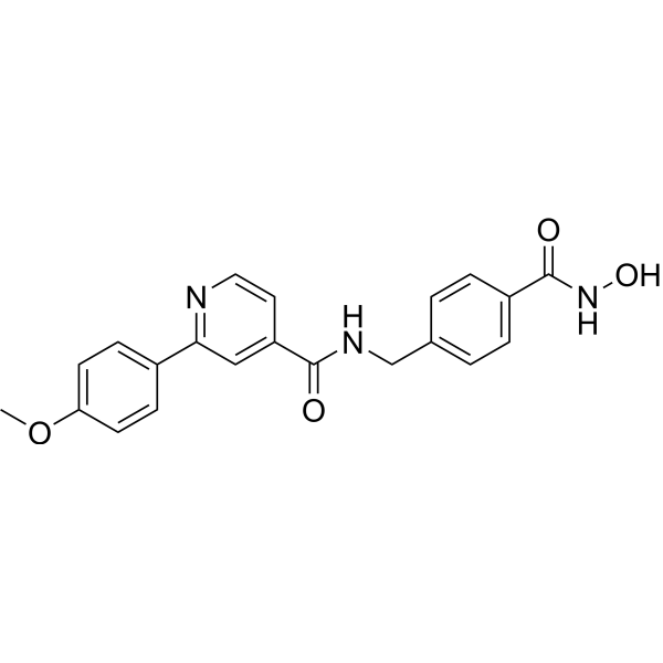
HDAC-IN-57 is an orally active inhibitor of histone deacetylases (HDAC), with IC50s of 2.07 nM, 4.71 nM, 2.4 nM and 107 nM for HDAC1, HDAC2, HDAC6, HDAC8, respectively. HDAC-IN-57 can inhibits LSD1, with IC50 of 1.34 μΜ. HDAC-IN-57 induces apoptosis, and has anti-tumor activity.
For research use only. We do not sell to patients.
- Purity : 99.61%
- CAS No.: 2716217-79-5
- Formula: C21H19N3O4
- Molecular Weight:377.39
-
Storage:
4°C, sealed storage, away from moisture
* In solvent : -80°C, 6 months; -20°C, 1 month (sealed storage, away from moisture)

To place orders, for customer services and technical support, please contact: MedChemExpress USA
Tel: 609-228-6898 E-mail: [email protected] [email protected]


-
 Biological Activity
-
Chemical Information
-
Solvent & Solubility
- Protocols
- Purity & Documentation
- References
-
 Help & FAQs


Publications Citing Use of MedChemExpress (MCE) HDAC-IN-57
More

Biological Activity
Description
IC50 & Target
[1]|
HDAC1 2.07 nM (IC50) |
HDAC2 4.71 nM (IC50) |
HDAC6 2.4 nM (IC50) |
HDAC8 107 nM (IC50) |
Cellular Effect
|
Cell Line

|
Type | Value | Description | References |
|---|---|---|---|---|
| A549 | IC50 |
1.48 μM
Compound: 5e
|
Antiproliferative activity against human A549 cells assessed as inhibition of cell growth incubated for 72 hrs by celltiter glo assay
Antiproliferative activity against human A549 cells assessed as inhibition of cell growth incubated for 72 hrs by celltiter glo assay
|
[PMID: 37086699] |
| GES1 | IC50 |
8.66 μM
Compound: 5e
|
Cytotoxicity against human GES1 cells harboring low LSD1 expression assessed as inhibition of cell growth incubated for 72 hrs by celltiter glo assay
Cytotoxicity against human GES1 cells harboring low LSD1 expression assessed as inhibition of cell growth incubated for 72 hrs by celltiter glo assay
|
[PMID: 37086699] |
| HCT-116 | IC50 |
0.57 μM
Compound: 5e
|
Antiproliferative activity against human HCT-116 cells assessed as inhibition of cell growth incubated for 72 hrs by celltiter glo assay
Antiproliferative activity against human HCT-116 cells assessed as inhibition of cell growth incubated for 72 hrs by celltiter glo assay
|
[PMID: 37086699] |
| MGC-803 | IC50 |
0.45 μM
Compound: 5e
|
Antiproliferative activity against human MGC-803 cells assessed as inhibition of cell growth incubated for 72 hrs by celltiter glo assay
Antiproliferative activity against human MGC-803 cells assessed as inhibition of cell growth incubated for 72 hrs by celltiter glo assay
|
[PMID: 37086699] |
| MOLT-4 | IC50 |
0.25 μM
Compound: 5e
|
Antiproliferative activity against human MOLT-4 cells assessed as inhibition of cell growth incubated for 72 hrs by celltiter glo assay
Antiproliferative activity against human MOLT-4 cells assessed as inhibition of cell growth incubated for 72 hrs by celltiter glo assay
|
[PMID: 37086699] |
| MV4-11 | IC50 |
0.14 μM
Compound: 5e
|
Antiproliferative activity against human MV4-11 cells assessed as inhibition of cell growth incubated for 72 hrs by celltiter glo assay
Antiproliferative activity against human MV4-11 cells assessed as inhibition of cell growth incubated for 72 hrs by celltiter glo assay
|
[PMID: 37086699] |
| THP-1 | IC50 |
0.47 μM
Compound: 5e
|
Antiproliferative activity against human THP-1 cells assessed as inhibition of cell growth incubated for 72 hrs by celltiter glo assay
Antiproliferative activity against human THP-1 cells assessed as inhibition of cell growth incubated for 72 hrs by celltiter glo assay
|
[PMID: 37086699] |
In Vitro
HDAC-IN-57 (Compound 5e) (1.0 μM, 2.5 μM, 5.0 μΜ;48 hour) inhibits migration and invasion activity of MGC-803 and HCT-116 cells[1].
HDAC-IN-57 (1.0 μM, 2.5 μM, 5.0 μΜ;48 hour) significantly inhibits the growth of solid tumor cell lines MGC-803, A549, and HCT-116, with IC50s of 0.45 μM. 1.48 μM and 0.57 μM, respectively[1].
HDAC-IN-57 (1.0 μM. 2.5 μM, 5.0 μΜ; 48 hours) triggers apoptosis of MGC-803 and HCT -116 cells in a dose-dependent manner[1].
HDAC-IN-57 (1.0 μM. 2.5 μM, 5.0 μΜ; 48 hours) inhibits LSD1 and HDACs of MGC-803 and HCT -116 cells[1].
HDAC-IN-57 (1.0 μM. 2.5 μM, 5.0 μΜ; 48 hours) induces G2/M cycle arrest in MGC-803 and HCT-116 cells[1].
HDAC-IN-57 (Compound 5e) showes excellent metabolic stability in human liver (HLM) and rat liver microsomes (RLM), maintaining 86.1% and 87.4%, respectively, of the parent compound after incubation for 1 h, with T1/2 values over 120 min[1].
MedChemExpress (MCE) has not independently confirmed the accuracy of these methods. They are for reference only.
-
Cell Line:MGC-803 cells, HCT-116 cells
-
Concentration:1.5 μM
-
Incubation Time:48 hours
-
Result:Inhibited cellular LSD1 and HDACs.
Upregulated the expression of apoptotic markers, including cytochrome C, Bax, cleaved caspase-3/7/9, and cleaved PARP, while downregulating the expression of anti-apoptotic protein Bcl-2.
-
Cell Line:MGC-803 cells, HCT-116 cells
-
Concentration:1.0 μM, 2.5 μM, 5.0 μM
-
Incubation Time:48 hours
-
Result:Triggered MGC-803 and HCT116 cells apoptosis in a dose-dependent manner.
Induced about 55.4% and 51.5% MGC-803 cell apoptosis at a concentration of 5 μM.
-
Cell Line:MGC-803 cells, HCT-116 cells
-
Concentration:1.0 μM, 2.0 μM, 4 μM
-
Incubation Time:48 hours
-
Result:Reduced the number of migrated of MGC-803 and HCT-116 cells. Inhibited the migration and invasion of cancer cells.
-
Cell Line:MGC-803 cells, HCT-116 cells
-
Concentration:1.0 μM, 2.5 μM, 5.0 μM
-
Incubation Time:48 hours
-
Result:Induced G2/M cycle arrest in MGC-803 and HCT-116 cells.
Parmacokinetics
In Vivo
HDAC-IN-57 (25 or 50 mg/kg, oral gavage once daily for 21 consecutive days) achieves a dose-dependent inhibition for tumor growth in an MGC-803 xenograft model with NOD-SCID mice[1].
.
MedChemExpress (MCE) has not independently confirmed the accuracy of these methods. They are for reference only.
-
Animal Model:NOD-SCID mouse MGC-803 xenograft model[1]
-
Dosage:25 mg/kg; 50 mg/kg
-
Administration:p.o.; once daily; for 21 days
-
Result:Inhibited tumor growth in a dose-dependent manner.


Chemical Information
-
CAS No. 2716217-79-5
-
Appearance Solid
-
Molecular Weight 377.39
-
Formula C21H19N3O4
-
Color White to off-white
-
SMILES
O=C(C1=CC=C(CNC(C2=CC(C3=CC=C(OC)C=C3)=NC=C2)=O)C=C1)NO
-
Shipping
Room temperature in continental US; may vary elsewhere.
-
Storage
4°C, sealed storage, away from moisture
* In solvent : -80°C, 6 months; -20°C, 1 month (sealed storage, away from moisture)


Publications (1)
-
Journal Impact Factor
-
Most Recent


Solvent & Solubility
In Vitro:
DMSO : 50 mg/mL (132.49 mM; Need ultrasonic; Hygroscopic DMSO has a significant impact on the solubility of product, please use newly opened DMSO)
Please refer to the solubility information to select the appropriate solvent. Once prepared, please aliquot and store the solution to prevent product inactivation from repeated freeze-thaw cycles.
Storage method and period of stock solution: -80°C, 6 months; -20°C, 1 month (sealed storage, away from moisture). When stored at -80°C, please use it within 6 months. When stored at -20°C, please use it within 1 month.
Please refer to the solubility information to select the appropriate solvent. Once prepared, please aliquot and store the solution to prevent product inactivation from repeated freeze-thaw cycles.
Storage method and period of stock solution: -80°C, 6 months; -20°C, 1 month (sealed storage, away from moisture). When stored at -80°C, please use it within 6 months. When stored at -20°C, please use it within 1 month.
Concentration (start) × Volume (start) = Concentration (final) × Volume (final)


Purity & Documentation
-
Data Sheet (275 KB)
-
SDS (252 KB)
- English - EN (252 KB)
- Français - FR (252 KB)
- Deutsch - DE (252 KB)
- Norwegian - NO (252 KB)
- Español - ES (252 KB)
- Swedish - SV (252 KB)
- Italian - IT (252 KB)
- Korean - KR (252 KB)
- Portuguese - PT (252 KB)
-
Handling Instructions (2659 KB)
References


Complete Stock Solution Preparation Table
Please refer to the solubility information to select the appropriate solvent. Once prepared, please aliquot and store the solution to prevent product inactivation from repeated freeze-thaw cycles.
Storage method and period of stock solution: -80°C, 6 months; -20°C, 1 month (sealed storage, away from moisture). When stored at -80°C, please use it within 6 months. When stored at -20°C, please use it within 1 month.
| Optional Solvent | Concentration Solvent Mass | 1 mg | 5 mg | 10 mg | 25 mg |
|---|---|---|---|---|---|
| DMSO | 1 mM | 2.6498 mL | 13.2489 mL | 26.4978 mL | 66.2445 mL |
| 5 mM | 0.5300 mL | 2.6498 mL | 5.2996 mL | 13.2489 mL | |
| 10 mM | 0.2650 mL | 1.3249 mL | 2.6498 mL | 6.6244 mL | |
| 15 mM | 0.1767 mL | 0.8833 mL | 1.7665 mL | 4.4163 mL | |
| 20 mM | 0.1325 mL | 0.6624 mL | 1.3249 mL | 3.3122 mL | |
| 25 mM | 0.1060 mL | 0.5300 mL | 1.0599 mL | 2.6498 mL | |
| 30 mM | 0.0883 mL | 0.4416 mL | 0.8833 mL | 2.2081 mL | |
| 40 mM | 0.0662 mL | 0.3312 mL | 0.6624 mL | 1.6561 mL | |
| 50 mM | 0.0530 mL | 0.2650 mL | 0.5300 mL | 1.3249 mL | |
| 60 mM | 0.0442 mL | 0.2208 mL | 0.4416 mL | 1.1041 mL | |
| 80 mM | 0.0331 mL | 0.1656 mL | 0.3312 mL | 0.8281 mL | |
| 100 mM | 0.0265 mL | 0.1325 mL | 0.2650 mL | 0.6624 mL |


HDAC-IN-57 Related Classifications
HY-149946 Related Classifications
 Powered by Bioz