



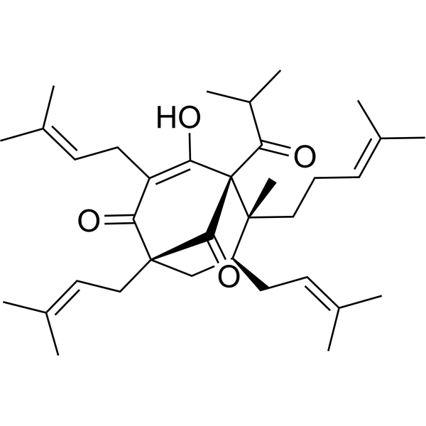
Hyperforin
Based on 3 publication(s) in Google Scholar
Hyperforin is a transient receptor canonical 6 (TRPC6) channels activator. Hyperforin modulates Ca2+ levels by activating Ca2+-conducting non-selective canonical TRPC6 channels and triggers adipose tissue thermogenesis via the Dlat-AMPK signaling axis to suppress obesity. Hyperforin also shows diverse pharmacological activities including anti-depression, anti-tumor, anti-dementia, anti-diabetes. Hyperforin modulates γδ T cells to secret IL-17α, improves Imiquimod (HY-B0180)-induced psoriasis-like mice model.
Nur für Forschungszwecke. Wir verkaufen nicht an Patienten.
- CAS. Nr.: 11079-53-1
- Formel: C35H52O4
- Molecular Weight:536.78
-
Speicherung:
Please store the product under the recommended conditions in the Certificate of Analysis.

To place orders, for customer services and technical support, please contact: MedChemExpress USA
Tel: 609-228-6898 E-mail: [email protected] [email protected]


-
 Biologische Aktivität
-
Chemical Information
- Protokoll
- Reinheit & Dokumentation
- Verweise
-
 Help & FAQs


Publications Citing Use of MedChemExpress (MCE) Hyperforin
More Customer Validation & Images

-
Bio/Physico-chemical Assay

-
WB

-
WB

Alle Calcium Channel Isoform-spezifische Produkte anzeigen
More

Biologische Aktivität
Beschreibung
IC50 & Target
TRPC6[1]
Cellular Effect
|
Cell Line

|
Type | Value | Description | References |
|---|---|---|---|---|
| A549 | IC50 |
>40 μM
Compound: 1
|
Cytotoxicity against human A549 cells assessed as reduction in cell viability after 48 hrs by MTT assay
Cytotoxicity against human A549 cells assessed as reduction in cell viability after 48 hrs by MTT assay
|
[PMID: 25871261] |
| HL-60 | IC50 |
>40 μM
Compound: 1
|
Cytotoxicity against human HL60 cells assessed as reduction in cell viability after 48 hrs by MTT assay
Cytotoxicity against human HL60 cells assessed as reduction in cell viability after 48 hrs by MTT assay
|
[PMID: 25871261] |
| MCF7 | IC50 |
>40 μM
Compound: 1
|
Cytotoxicity against human MCF7 cells assessed as reduction in cell viability after 48 hrs by MTT assay
Cytotoxicity against human MCF7 cells assessed as reduction in cell viability after 48 hrs by MTT assay
|
[PMID: 25871261] |
| SMMC-7721 | IC50 |
>40 μM
Compound: 1
|
Cytotoxicity against human SMMC7721 cells assessed as reduction in cell viability after 48 hrs by MTT assay
Cytotoxicity against human SMMC7721 cells assessed as reduction in cell viability after 48 hrs by MTT assay
|
[PMID: 25871261] |
| SW480 | IC50 |
>40 μM
Compound: 1
|
Cytotoxicity against human SW480 cells assessed as reduction in cell viability after 48 hrs by MTT assay
Cytotoxicity against human SW480 cells assessed as reduction in cell viability after 48 hrs by MTT assay
|
[PMID: 25871261] |
In Vitro
Hyperforin has a multi-directional mechanism of action. It blocks conductance of ligand-gated (GABA, NMDA, and AMPA receptors) and voltage-gated channels (Ca2+, K+, and Na+)[2].
Hyperforin (0.1, 1, 10 μM; 2 h) reduces the expression and secretion of IL-17A in γδ T cell in vitro cultured murine splenic γδ T cells[3].
Hyperforin (0.1, 1, 10 μM; 2 h) suppresses phosphorylation of MAPK and STAT3 pathways in TNF-α stimulated HaCaT cells[3].
Hyperforin (IC50=3.7 μmol/L) inhibits the microvascular tube formation and proliferation of HDMEC in a dose-dependent manner without toxic effects[4].
MedChemExpress (MCE) has not independently confirmed the accuracy of these methods. They are for reference only. Further protocols information, click here.
-
Cell Line:HaCaT cells
-
Concentration:0.1, 1, 10 μM; with or without 10, 20 ng/mL TNF-α
-
Incubation Time:2 hours
-
Result:Reduced the expressions of p-p38, p-ERK, p-JNK, and p-STAT3, especially at the dosage of 10 μM.
In Vivo
MedChemExpress (MCE) has not independently confirmed the accuracy of these methods. They are for reference only.
-
Animal Model:IMQ-induced psoriasis-like mice model[3]
-
Dosage:5 mg/kg
-
Administration:Intraperitoneal injection; once daily for 7 days
-
Result:Significantly ameliorated skin lesion throughout the treatment period, demonstrated by the reduced severity score of skin inflammation.
Suppressed infiltration of CD3+ T cells and downregulated expression of Il1, Il6, Il23, Il17a, Il22, antimicrobial peptides (AMPs) in the skin lesion.


Chemical Information
-
CAS. Nr. 11079-53-1
-
Molecular Weight 536.78
-
Formel C35H52O4
-
SMILES
CC(C)C([C@@]12[C@@](C)([C@H](C[C@](C/C=C(C)/C)(C(C(C/C=C(C)/C)=C2O)=O)C1=O)C/C=C(C)/C)CC/C=C(C)/C)=O
-
Structure Classification
-
Initial Source
-
Versand
Room temperature in continental US; may vary elsewhere.
-
Speicherung
Please store the product under the recommended conditions in the Certificate of Analysis.


Publications (3)
-
Journal Impact Factor
-
Most Recent
-
Mol Metab
2026 Apr:106:102341. PMID: 41747880
Hyperforin purchased from MedChemExpress. Usage Cited in: Mol Metab. 2026 Apr:106:102341. [Abstract]
Primary brown adipocytes loaded with Fluo-4 AM. Baseline [Ca2+]i was acquired. Primary brown adipocytes, incubated in Ca2+-containing or Ca2+-free bath solutions, were exposed to HPF (Hyperforin dicyclohexylammonium salt, 5 μM) at 150 s.
Hyperforin purchased from MedChemExpress. Usage Cited in: Mol Metab. 2026 Apr:106:102341. [Abstract]
Primary brown adipocytes were treated with vehicle (Ctrl) or HPF (Hyperforin dicyclohexylammonium salt, 5 μM) for 48 h. The protein levels of UCP1, PGC-1α, β-Actin, and p38 MAPK (n = 3).
Hyperforin purchased from MedChemExpress. Usage Cited in: Mol Metab. 2026 Apr:106:102341. [Abstract]
Trpc6−/− adipocytes were treated with vehicle or HPF (Hyperforin dicyclohexylammonium salt, 5 μM) for 48 h.
-
Drug Discov Ther
2025 Oct 16. PMID: 41093589 -
Transfus Med
2023 Jun;33(3):232-243. PMID: 36746770

Protokoll
-
Imiquimod-Induced Psoriasiform Dermatitis
Imiquimod (IMQ)-induced psoriasiform dermatitis is a widely used murine model in which topical application of IMQ, a Toll-like receptor 7 (TLR7) agonist, triggers innate immune activation in the skin and induces a psoriasis-like inflammatory cascade characterized by epidermal hyperplasia, immune cell infiltration, and cytokine production dominated by the IL-23/IL-17 axis. This inflammatory response is mediated through activation of dendritic cells and downstream induction of IL-23, IL-17A, IL-22, and related pro-inflammatory mediators, recapitulating key features of human plaque psoriasis and enabling mechanistic and therapeutic studies. The model is commonly induced using Aldara (5% IMQ cream) applied topically to murine skin, resulting in rapid onset of erythema, scaling, and thickening that can be quantified as disease severity indices and validated histologically.
-
Large-size fat particle sorting
Large-size fat particle sorting is widely used to isolate cells up to 200 μm in diameter. Single-cell flow sorting will allow greater insight into adipocyte heterogeneity by identifying gene expression, protein composition, and metabolic signatures at the single-cell level.
-
Ca2+ Staining Technique
Ca2+ staining is an experimental technique that utilizes specific fluorescent probes (such as Fluo-4 AM, Fura-2, etc.) to qualitatively or quantitatively detect dynamic changes in intracellular Ca2+ concentrations; this is achieved by monitoring the changes in fluorescent signals generated when these probes bind to free intracellular calcium ions. The underlying principle relies primarily on the presence of chelating groups within the probe's molecular structure that possess high affinity for calcium ions.
-
Research Protocol for Metabolic Diseases
AMP-activated protein kinase, AMPK, is a conserved cellular energy sensor that responds to reduced cellular energy status and coordinates metabolism by increasing ATP-generating catabolic pathways while suppressing ATP-consuming anabolic processes. In metabolic disease research, the AMPK pathway is experimentally relevant because it regulates hepatic lipid synthesis, fatty acid oxidation, glucose production, skeletal-muscle glucose disposal, mTORC1-linked biosynthesis, autophagy, mitochondrial homeostasis, and whole-body energy balance. The central pathway logic is that energy stress, metformin, exercise-like stimulation, or direct AMPK activators increase AMPKα Thr172 phosphorylation and downstream substrate phosphorylation, including ACC and RAPTOR. Phosphorylation of ACC suppresses lipogenesis and supports fatty acid oxidation, whereas phosphorylation of RAPTOR suppresses mTORC1 signaling and links cellular energy status to growth and protein synthesis control. The pathway is linked
-
Cuproptosis Solutions
Cuproptosis is a copper-dependent regulated cell-death pathway in which intracellular copper binds lipoylated tricarboxylic acid cycle proteins, especially DLAT-containing pyruvate dehydrogenase complex components, causing lipoylated protein aggregation, iron-sulfur cluster protein loss, proteotoxic stress, and cell death. The pathway is functionally linked to mitochondrial respiration because copper-ionophore sensitivity is higher in cells dependent on oxidative phosphorylation, and FDX1 and protein lipoylation machinery are required for copper-ionophore-induced death. Elesclomol-Cu and related copper-loading strategies are widely used experimental tools to induce cuproptosis, whereas copper chelation with tetrathiomolybdate or genetic suppression of FDX1, LIAS, LIPT1, or DLAT can test pathway dependence. The major unresolved questions are how disease context determines cuproptosis sensitivity, how copper transporters such as SLC31A1/CTR1 and ATP7A/ATP7B regulate the pathway, and whic


Reinheit & Dokumentation
Verweise
[1]. Heiser JH, et al. TRPC6 channel-mediated neurite outgrowth in PC12 cells and hippocampal neurons involves activation of RAS/MEK/ERK, PI3K, and CAMKIV signaling. J Neurochem. 2013 Nov;127(3):303-13. [Content Brief]
[2]. Pochwat B, et al. Hyperforin Potentiates Antidepressant-Like Activity of Lanicemine in Mice. Front Mol Neurosci. 2018 Dec 12;11:456. [Content Brief]
[3]. Zhang S, et al. Hyperforin Ameliorates Imiquimod-Induced Psoriasis-Like Murine Skin Inflammation by Modulating IL-17A-Producing γδ T Cells. Front Immunol. 2021 May 5;12:635076. [Content Brief]
[4]. Adam P, et al. Biosynthesis of hyperforin in Hypericum perforatum. J Med Chem. 2002 Oct 10;45(21):4786-93. [Content Brief]
[5]. Li XX, et al. Hyperforin: A natural lead compound with multiple pharmacological activities. Phytochemistry. 2023 Feb;206:113526. [Content Brief]
[6]. Suzhen Chen, et al. "The phytochemical hyperforin triggers thermogenesis in adipose tissue via a Dlat-AMPK signaling axis to curb obesity." Cell Metabolism 33.3 (2021): 565-580. [Content Brief]

Calculators
Konzentration (Stammlösung) × Volumen (Stammlösung) = Konzentration (Ziellösung) × Volumen (Ziellösung)


Hyperforin Related Classifications
HY-116330 Related Classifications
 Powered by Bioz