



Frozen sectioning is a cryostat-based tissue preparation method in which fresh, fixed, or cryoprotected tissue is rapidly frozen, embedded in a support medium such as OCT, cut into thin cryosections, and mounted on slides for rapid morphology, histochemistry, immunofluorescence, in situ hybridization, enzyme activity, lipid, mucin, or downstream molecular analysis. The method preserves antigenicity and avoids dehydration/clearing steps used in paraffin processing, but frozen sections are physically less stable and generally show more freezing or cutting artifact than FFPE sections; therefore, frozen sectioning is useful for rapid diagnosis, spatial molecular assays, and preservation of labile targets, but not always optimal for fine morphology alone.
Frozen sectioning technique
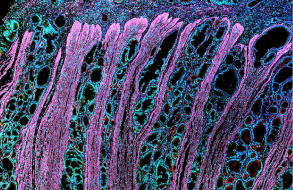
Frozen sectioning is a method used to prepare sections for the study of cells and tissues. First, the tissue sample is encapsulated in cryogen by freezing to form a frozen block. Then, use a freezing microtome or microtome to cut thin sections at very low temperatures. This technology retains the natural structure and biological activity of cells and tissues, and is suitable for studies such as immunohistochemistry, fluorescence microscopy, and in situ hybridization. Frozen sectioning technology is important for preserving the activity of biomolecules and providing high-resolution images.
Related Experimental Schemes


