



PLK1 Protein, Human (Active, sf9, His)
Based on 3 publication(s) in Google Scholar
PLK1 is a serine/threonine protein kinase that centrally coordinates key functions during the M phase of the cell cycle. It regulates centrosome maturation, spindle assembly, cohesin removal, inactivates APC/C inhibitors, and controls mitotic exit and cytokinesis. PLK1 Protein, Human (sf9, His) is the recombinant human-derived PLK1 protein, expressed by Sf9 insect cells , with N-10*His labeled tag.
- Species: Human
- Source: Sf9 insect cells
-
Storage:Stored at -80°C for 1 year from date of receipt. It is stable at -20°C for 3 months after opening. It is recommended to freeze aliquots at -80°C for extended storage. Avoid repeated freeze-thaw cycles.


To place orders, for customer services and technical support, please contact: MedChemExpress USA
Tel: 609-228-6898 E-mail: [email protected] [email protected]


-
 Biological Activity
-
Technical Parameters
-
Product Properties
- Documentation
- Help & FAQs




Biological Activity
Description
PLK1 is a serine/threonine protein kinase that centrally coordinates key functions during the M phase of the cell cycle. It regulates centrosome maturation, spindle assembly, cohesin removal, inactivates APC/C inhibitors, and controls mitotic exit and cytokinesis. PLK1 Protein, Human (sf9, His) is the recombinant human-derived PLK1 protein, expressed by Sf9 insect cells , with N-10*His labeled tag.
Background
PLK1, a serine/threonine-protein kinase, plays a pivotal role in orchestrating various essential functions throughout the M phase of the cell cycle. It regulates centrosome maturation and spindle assembly, facilitates the removal of cohesins from chromosome arms, inactivates anaphase-promoting complex/cyclosome (APC/C) inhibitors, and governs mitotic exit and cytokinesis. Operating by binding and phosphorylating proteins that are already phosphorylated on specific motifs recognized by the POLO box domains, PLK1 phosphorylates an extensive array of substrates, including BORA, BUB1B/BUBR1, CCNB1, CDC25C, CEP55, ECT2, ERCC6L, FBXO5/EMI1, FOXM1, KIF20A/MKLP2, CENPU, NEDD1, NINL, NPM1, NUDC, PKMYT1/MYT1, PRC1, RACGAP1/CYK4, SGO1, STAG2/SA2, TEX14, TOPORS, p73/TP73, TPT1, WEE1, HNRNPU, and others. PLK1's crucial roles include promoting centrosome functions and bipolar spindle assembly through the phosphorylation of KIZ, NEDD1, and NINL. Additionally, it governs mitotic exit and cytokinesis by phosphorylating CEP55, ECT2, KIF20A/MKLP2, CENPU, PRC1, and RACGAP1. PLK1's involvement extends to kinetochore functions, sister chromatid cohesion, and the regulation of various checkpoint proteins, positioning it as a key player in the intricate network governing cell cycle progression.
Verified Bioactivity
Measured by its ability to catalyze 0.2 mg/mL PLKtide peptide substrate and 25 μM ATP that incubate at room temperature for 40 mins. The specific activity is >40 nmo/min/mg.
MCE Validation Data
-
Purity - SDS-PAGE
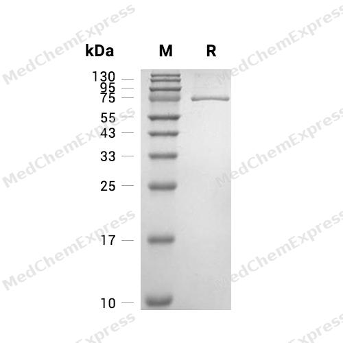
Purity - SDS-PAGE

Assay Procedure
Materials
Assay buffer (5x): 200 mM Tris-HCl, pH 7.4, 100 mM MgCl2, and 0.5 mg/mL BSA. Add fresh DTT prior to use to a final concentration of 250 μM.
PLK1 Protein, Human (sf9, His) (HY-P76550)
Substrate: PLKtide peptide substrate
Assay Kit: ADP-GloTM Kinase Assay
Standard: ATP/ADP
Procedure
Preparation of Standard Curve
1. Dilute the 5x assay buffer to 1x with ddH2O.
2. Dilute ADP and ATP solutions using assay buffer (1X) to prepare 1 mL of 100 μM ADP and ATP solutions.
Well No. A1 A2 A2 A4 A5 A6 A7 A8 A9 A10 A11 A12
ADP(μL) 100 80 60 40 20 10 5 4 3 2 1 0
ATP(μL) 0 20 40 60 80 90 95 96 97 98 99 100
Add 100, 80, 60, 40, 20, 10, 5, 4, 3, 2, 1, 0 μL of 100 μM ADP and 0, 20, 40, 60, 80, 90, 95, 96, 97, 98, 99, 100 μL of 100 μM ATP solution into wells A1-A12 respectively, and mix as shown in the table above to simulate the ATP and ADP concentrations corresponding to each conversion percentage. This series constitutes the 100 μM concentration series.
3. Add 5 μL of the mixed solution to each well, followed by 5 μL of ADP-GloTM Reagent.
4. Incubate at room temperature for 40 min.
5. Add 10 μL of Kinase Detection Reagent to the 384-well plate.
6. Incubate at room temperature for 30 min.
7. Measure RLU values under Luminescence using a microplate reader.
8. Plot the standard curve equation with measured RLU values on the y-axis and ATP molar quantity on the x-axis.
Protein Activity Assay
1. Dilute PLK1 protein to 20 and 100 μg/mL using 1x assay buffer.
2. Prepare the mixture according to the following system:
10 mM ATP solution: 1 μL
Assay buffer (5x): 79 μL
1 mg/mL Substrate: 80 μL
3. Add 3 μL of 20 or 100 μg/mL protein solution to a 384-well plate, followed by 2 μL of the mixture. For blank wells, add 3 μL of assay buffer followed by 2 μL of the mixture.
4. Incubate at room temperature for 40 min.
5. After incubation, add 5 μL ADP-GloTM Reagent to each well.
6. Incubate at room temperature for 40 min.
7. After incubation, add 10 μL Kinase Detection Reagent to each well.
8. Incubate at room temperature for 30 min.
9. Measure the RLU value under Luminescence using a microplate reader.
10. CalcμLate specific activity:
Specific Activity (pmol/min/μg) = | Corrected RLU from reaction* |
| SA of ADP** (RFU/pmol) x Reaction time (min) x amount of enzyme (mg) |
*Corrected for Substrate Blank
**Derived using calibration standard


Publications (3)
-
Journal Impact Factor
-
Most Recent
-
Cell Death Dis
USP10 promotes the progression and attenuates gemcitabine chemotherapy sensitivity via stabilizing PLK1 in PDAC. [Abstract]2025 Jun 14;16(1):449. PMID: 40517136 -
J Cell Sci
In vitro reconstitution of a minimal human centrosome scaffold capable of forming and clustering microtubule asters. [Abstract]2025 Jun 15;138(12):jcs264121. PMID: 40454523 -
bioRxiv
2025 Feb 20:2025.02.20.639226. PMID: 40027679


Technical Parameters
-
Species Human
-
Source Sf9 insect cells
-
Tag N-10*His
-
Accession
P53350 (M1-S603)
-
Molecular Construction
-
N-term
-
10*His
-
PLK1 (M1-S603)
Accession # P53350 -
C-term
-
-
Protein Length
Full Length
-
Synonyms
PLK1; STPK13; Prev. PLK; Cell Cycle Regulated Protein Kinase; Serine/Threonine-Protein Kinase PLK1; Polo-Like Kinase (Drosophila); Polo-Like Kinase 1; Polo (Drosophia)-Like Kinase; Serine/Threonine-Protein Kinase 13; Polo Like Kinase 1; PLK-1
-
AA Sequence
MSAAVTAGKLARAPADPGKAGVPGVAAPGAPAAAPPAKEIPEVLVDPRSRRRYVRGRFLGKGGFAKCFEISDADTKEVFAGKIVPKSLLLKPHQREKMSMEISIHRSLAHQHVVGFHGFFEDNDFVFVVLELCRRRSLLELHKRRKALTEPEARYYLRQIVLGCQYLHRNRVIHRDLKLGNLFLNEDLEVKIGDFGLATKVEYDGERKKTLCGTPNYIAPEVLSKKGHSFEVDVWSIGCIMYTLLVGKPPFETSCLKETYLRIKKNEYSIPKHINPVAASLIQKMLQTDPTARPTINELLNDEFFTSGYIPARLPITCLTIPPRFSIAPSSLDPSNRKPLTVLNKGLENPLPERPREKEEPVVRETGEVVDCHLSDMLQQLHSVNASKPSERGLVRQEEAEDPACIPIFWVSKWVDYSDKYGLGYQLCDNSVGVLFNDSTRLILYNDGDSLQYIERDGTESYLTVSSHPNSLMKKITLLKYFRNYMSEHLLKAGANITPREGDELARLPYLRTWFRTRSAIILHLSNGSVQINFFQDHTKLILCPLMAAVTYIDEKRDFRTYRLSLLEEYGCCKELASRLRYARTMVDKLLSSRSASNRLKAS
-
Molecular Weight
Approximately 66 kDa, based on SDS-PAGE under reducing conditions.
-
Glycosylation
Yes
-
Purity
≥ 90%, as determined by reducing SDS-PAGE.


Product Properties
Solution.
Supplied as a 0.22 μm filtered solution of 50 mM Tris, 100 mM NaCl, pH 7.4, 0.5 mM EDTA, 0.5 mM EGTA, 0.5 mM PMSF, 25% glycerol or 50 mM PB, 300 mM NaCl, pH 7.0, 25% Glycerol, 0.1 mM EDTA, 0.5 mM PMSF, 3 mM DTT.
Note: For SPR assay, please replace the buffer. Primary amine components (e.g., Tris, imidazole) can affect protein-coupled chips.
<1 EU/μg, determined by LAL method.
Please use rapid thawing with running water to thaw the protein.
Stored at -80°C for 1 year from date of receipt. It is stable at -20°C for 3 months after opening. It is recommended to freeze aliquots at -80°C for extended storage. Avoid repeated freeze-thaw cycles.
Shipping with dry ice.


Documentation
-
Data Sheet (239 KB)
-
SDS (252 KB)
- English - EN (252 KB)
- Français - FR (252 KB)
- Deutsch - DE (252 KB)
- Norwegian - NO (252 KB)
- Español - ES (252 KB)
- Swedish - SV (252 KB)
- Italian - IT (252 KB)
- Korean - KR (252 KB)
- Portuguese - PT (252 KB)
-
Handling Instructions (2659 KB)

Calculators
Concentration (start) × Volume (start) = Concentration (final) × Volume (final)


 Powered by Bioz

