



Trimethylamine N-oxide
Based on 12 publication(s) in Google Scholar
Trimethylamine N-oxide is a gut microbe-dependent metabolite of dietary choline and other trimethylamine-containing nutrients. Trimethylamine N-oxide induces inflammation by activating the ROS/NLRP3 inflammasome. Trimethylamine N-oxide also accelerates fibroblast-myofibroblast differentiation and induces cardiac fibrosis by activating the TGF-β/smad2 signaling pathway.
For research use only. We do not sell to patients.
- Purity : 98.0%
- CAS No.: 1184-78-7
- Formula: C3H9NO
- Molecular Weight:75.11
-
Storage:Powder -20°C, 3 years , 4°C, 2 years ; In solvent -80°C, 6 months , -20°C, 1 month


To place orders, for customer services and technical support, please contact: MedChemExpress USA
Tel: 609-228-6898 E-mail: [email protected] [email protected]


-
 Biological Activity
-
Chemical Information
-
Solvent & Solubility
- Protocols
- Purity & Documentation
- References
-
 Help & FAQs


-
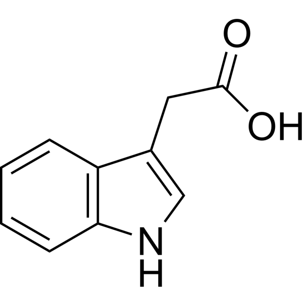
HY-18569
3-Indoleacetic acidPlant Growth Regulator -
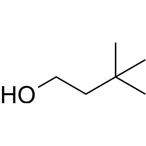
HY-W012977
3,3-Dimethyl-1-butanolTMA /TMAO Inhinbitor -
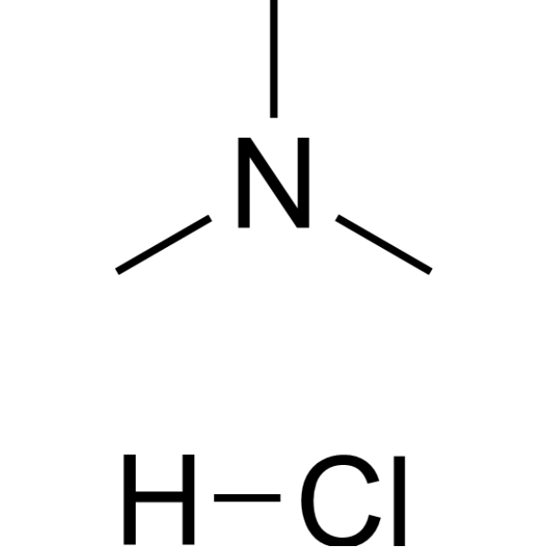
HY-Y0504
Trimethylammonium chlorideAcetylcholinesterase Inhibitor -

HY-B0215
AcetylcysteineMucolytic Agent -
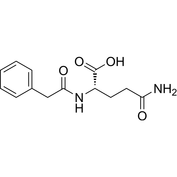
HY-W050026
Phenylacetylglutamine -
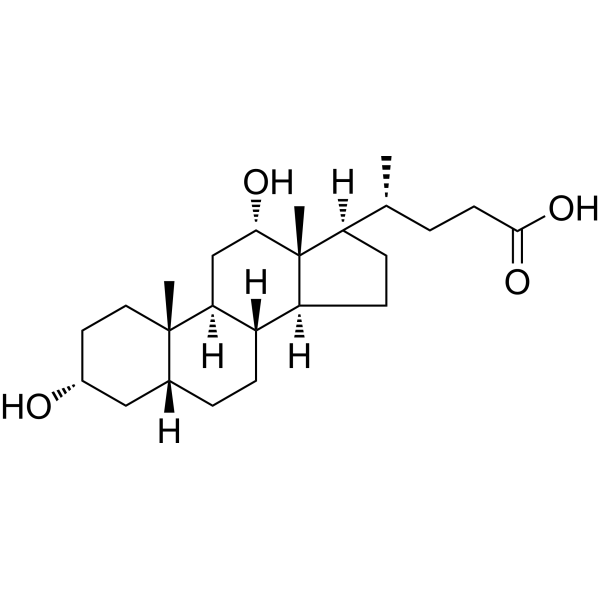
HY-N0593
Deoxycholic acidBile Acid Receptor Activator -
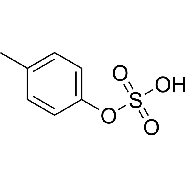
HY-111431
p-Cresyl sulfateUremic Toxin -
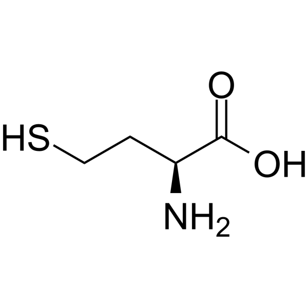
HY-W010347
L-HomocysteineAmino Acid -
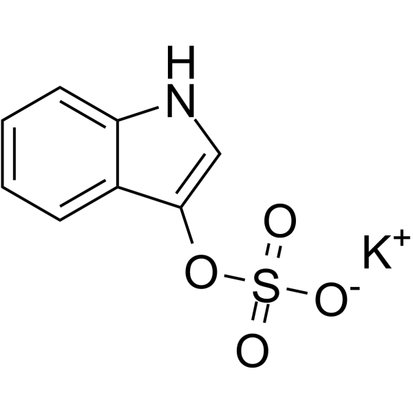
HY-W011910
Potassium 1H-indol-3-yl sulfateMetabolite of Tryptophan -
HY-B0351
TaurineAutophagy Inducer -

HY-13259
MG-132Proteasome Inhibitor -

HY-K0301
Cell Counting Kit-8
Publications Citing Use of MedChemExpress (MCE) Trimethylamine N-oxide
More- Gut Microbes. 2026 Dec 31;18(1):2605768. [Abstract]
- Phytomedicine. 2022 Mar 21;100:154067. [Abstract]
- Free Radic Biol Med. 2025 May:232:214-230. [Abstract]
- Chin Med. 2026 Mar 9;21(1):81. [Abstract]
- Eur Polym J. 2026 Feb 5.
- Ecotoxicol Environ Saf. 2025 Sep 1:302:118691. [Abstract]
- FASEB J. 2025 Dec 15;39(23):e71269. [Abstract]
- Biol Trace Elem Res. 2025 Apr;203(4):2295-2309. [Abstract]
- Clin Immunol. 2026 Jun:285:110706. [Abstract]
- Clin Chim Acta. 2024 Jan 15:553:117726. [Abstract]
- J Chromatogr B Analyt Technol Biomed Life Sci. 2024 Aug 1:1243:124210. [Abstract]
- Mol Cell Toxicol. 2025 Dec 10.
 Customer Validation & Images

-
Cell Proliferation/Viability Assay
-
In Vivo Efficacy Study
-
Cell Proliferation/Viability Assay
-
WB
-
IF
All Endogenous Metabolite Isoforms
More

Biological Activity
Description
IC50 & Target
|
NLRP3 |
Microbial Metabolite |
Human Endogenous Metabolite |
In Vitro
The size and migration of fibroblasts are increased after Trimethylamine N-oxide (TMAO) treatment compared with non-treated fibroblasts in vitro. Trimethylamine N-oxide increases TGF-β receptor I expression, which promotes the phosphorylation of Smad2 and up-regulates the expression of α-SMA and collagen I. The ubiquitination of TGF-βRI is decreased in neonatal mouse fibroblasts after Trimethylamine N-oxide treatment. Trimethylamine N-oxide also inhibits the expression of smurf2[2].
Trimethylamine N-oxide is frequently found in the tissues of a variety of marine organisms that protects against the adverse effects of temperature, salinity, high urea and hydrostatic pressure[3].
MedChemExpress (MCE) has not independently confirmed the accuracy of these methods. They are for reference only. Further protocols information, click here.
Induced Disease Models
Please do not refer to only one article to determine the experimental conditions. It is recommended to determine the optimal experimental conditions (animal strain, age, dosage, frequency and cycle, detection time and indicators, etc.) through preliminary experiments before the formal experiment.
Trimethylamine N-oxide can be used in animal modeling to create cardiovascular and metabolic disease models[1].
Administration: 100 μM and 1 mM• perfusion or incubation in TMAO-containing buffer solution• incubated for 1 h[1]
Mice: CD-1• male• weighing 25-30 g• 6-8 weeks of age[1]
Administration: 120 mg/kg• mixed with drinking water• a single dose or daily for 7 days[1]
(2) All experimental animals were housed under standard conditions (21-23°C, 12-hour light/dark cycle, relative humidity 45-65%) with unlimited access to food (R70 diet) and water[1].
(3) The mice from the first experimental group received Isoproterenol (HY-B1670A) at a dose of 10 μg/mouse, but the animals from the second group received Isoproterenol (HY-B1670A) and Trimethylamine N-oxide at doses of 10 μg/mouse and 120 mg/kg, respectively. After 30 min, the experimental animals were anesthetized with isoflurane once more to record the cardiac response to acute cardiac stress and the impact of Trimethylamine N-oxide on the inotropic and chronotropic effects. For the next seven days, the mice in the second group received Trimethylamine N-oxide together with drinking water at a dose of 120 mg/kg, while the animals from the first group received pure drinking water[1].
Pathology change: Trimethylamine N-oxide had no influence on Isoproterenol (HY-B1670A)-induced increase on left ventricular ejection fraction, fractional shortening and heart rate[1].
Histological analysis: Promote myocardial hypertrophy, fibrosis, and inflammation in a model of cardiovascular diseases (CVDs)[3].
MedChemExpress (MCE) has not independently confirmed the accuracy of these methods. They are for reference only.


Chemical Information
-
CAS No. 1184-78-7
-
Appearance Solid
-
Molecular Weight 75.11
-
Formula C3H9NO
-
Color White to off-white
-
SMILES
CN(C)(C)=O
-
Structure Classification
-
Initial Source
Host intestinal bacteria
-
Shipping
Room temperature in continental US; may vary elsewhere.
-
Storage
Powder -20°C 3 years 4°C 2 years In solvent -80°C 6 months -20°C 1 month


Publications (12)
-
Journal Impact Factor
-
Most Recent
-
Gut Microbes
Blautia coccoides-derived metabolite trimethylamine-N-oxide exacerbates Alzheimer's disease progression via targeting HIF1α signaling. [Abstract]2026 Dec 31;18(1):2605768. PMID: 41459734
Trimethylamine N-oxide purchased from MedChemExpress. Usage Cited in: Gut Microbes. 2026 Dec 31;18(1):2605768. [Abstract]
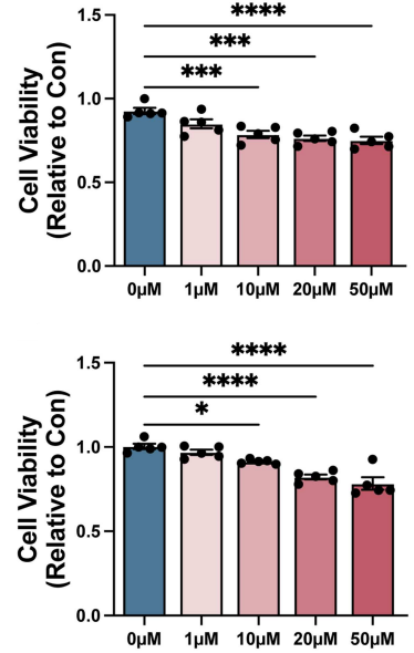
P301L-N2a and P301L-293T cells were treated with 1, 10, 20 and 50 μM Trimethylamine N-oxide (TMAO, 24 h), and cell viability was assessed using the CCK-8 method (n = 5).
-
Phytomedicine
2022 Mar 21;100:154067. PMID: 35349832 -
Free Radic Biol Med
Trimethylamine N-oxide (TMAO) treatment triggers premature ovarian insufficiency (POI) via the activation of mitochondrial pathway apoptosis in granulosa cells. [Abstract]2025 May:232:214-230. PMID: 40054636
Trimethylamine N-oxide purchased from MedChemExpress. Usage Cited in: Free Radic Biol Med. 2025 May:232:214-230. [Abstract]
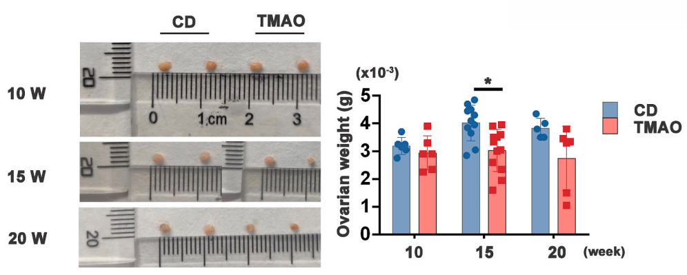
Representative ovarian size and ovarian weight quantification in the two groups across developmental stages. The results showed that at 15 weeks of age, mice in the Trimethylamine N-oxide (TMAO, 0.12% in drinking water for 11 weeks)-treated group exhibited significant reductions in both ovarian volume and ovarian weight.
-
Chin Med
Based on the gut-heart axis: Polygonum capitatum improves atherosclerosis by modulating gut microbiota and TMAO, supporting MCPIP1/p53-associated endothelial protection. [Abstract]2026 Mar 9;21(1):81. PMID: 41803870 -
-
Ecotoxicol Environ Saf
Involvement of TMAO in exacerbating arsenic-induced neurotoxicity by activating the PERK/eIF2α/ATF4 pathway. [Abstract]2025 Sep 1:302:118691. PMID: 40684638
Trimethylamine N-oxide purchased from MedChemExpress. Usage Cited in: Ecotoxicol Environ Saf. 2025 Sep 1:302:118691. [Abstract]
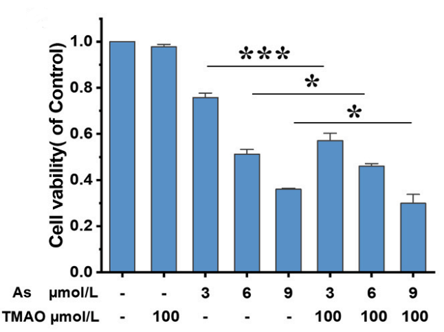
Cell viability after treatment with As (3–9 µmol/L) and/or Trimethylamine N-oxide (TMAO) (100 µmol/L) for 24 h.
Trimethylamine N-oxide purchased from MedChemExpress. Usage Cited in: Ecotoxicol Environ Saf. 2025 Sep 1:302:118691. [Abstract]
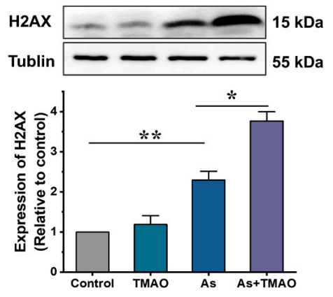
Representative western blots and quantification of H2AX in ASTs. Values represent the mean ± standard deviation from three separate experiments. Western blot analysis confirmed that Trimethylamine N-oxide (TMAO, 100 μM; 24 h) enhanced As-induced H2AX upregulation.
Trimethylamine N-oxide purchased from MedChemExpress. Usage Cited in: Ecotoxicol Environ Saf. 2025 Sep 1:302:118691. [Abstract]
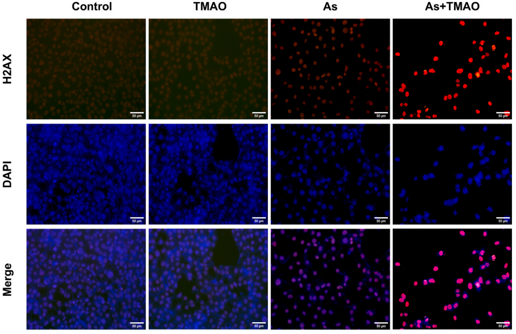
Representative immunofluorescence images of H2AX staining in ASTs. Immunofluorescence analysis revealed significantly increased γH2AX expression in ASTs after As (3 μM; 24 h) treatment This effect was further amplified by Trimethylamine N-oxide (TMAO, 100 µM; 24 h).
-
FASEB J
POSTN Enhances the Proliferation of Goat Skin Fibroblasts Through the TGFβ Pathway Activation. [Abstract]2025 Dec 15;39(23):e71269. PMID: 41324916 -
Biol Trace Elem Res
TGF-β/SMAD Pathway Mediates Cadmium Poisoning-Induced Chicken Liver Fibrosis and Epithelial-Mesenchymal Transition. [Abstract]2025 Apr;203(4):2295-2309. PMID: 38958867 -
Clin Immunol
Short-chain fatty acid deficiency drives aberrant B cell differentiation in systemic lupus erythematosus. [Abstract]2026 Jun:285:110706. PMID: 41974394 -
Clin Chim Acta
SAH and SAM/SAH ratio associate with acute kidney injury in critically ill patients: A case-control study. [Abstract]2024 Jan 15:553:117726. PMID: 38110027 -
J Chromatogr B Analyt Technol Biomed Life Sci
Simultaneous determination of choline, L-carnitine, betaine, trimethylamine, trimethylamine N-oxide, and creatinine in plasma, liver, and feces of hyperlipidemic rats by UHPLC-MS/MS. [Abstract]2024 Aug 1:1243:124210. PMID: 38936270 -


Solvent & Solubility
In Vitro:
H2O : 100 mg/mL (1331.38 mM; Need ultrasonic)
DMSO : 100 mg/mL (1331.38 mM; Need ultrasonic; Hygroscopic DMSO has a significant impact on the solubility of product, please use newly opened DMSO)
Please refer to the solubility information to select the appropriate solvent. Once prepared, please aliquot and store the solution to prevent product inactivation from repeated freeze-thaw cycles.
Storage method and period of stock solution: -80°C, 6 months; -20°C, 1 month. When stored at -80°C, please use it within 6 months. When stored at -20°C, please use it within 1 month.
* Note: If you choose water as the stock solution, please dilute it to the working solution, then filter and sterilize it with a 0.22 μm filter before use.
Please refer to the solubility information to select the appropriate solvent. Once prepared, please aliquot and store the solution to prevent product inactivation from repeated freeze-thaw cycles.
Storage method and period of stock solution: -80°C, 6 months; -20°C, 1 month. When stored at -80°C, please use it within 6 months. When stored at -20°C, please use it within 1 month.
* Note: If you choose water as the stock solution, please dilute it to the working solution, then filter and sterilize it with a 0.22 μm filter before use.
Concentration (start) × Volume (start) = Concentration (final) × Volume (final)
In Vivo:
Select the appropriate dissolution method based on your experimental animal and administration route.
- For the following dissolution methods, please ensure to first prepare a clear stock solution using an In Vitro approach and then sequentially add co-solvents:
- To ensure reliable experimental results, the clarified stock solution can be appropriately stored based on storage conditions. As for the working solution for In Vivo experiments, it is recommended to prepare freshly and use it on the same day.
- The percentages shown for the solvents indicate their volumetric ratio in the final prepared solution. If precipitation or phase separation occurs during preparation, heat and/or sonication can be used to aid dissolution.
Add each solvent one by one: 10% DMSO 40% PEG300 5% Tween-80 45% Saline
Solubility: ≥ 2.5 mg/mL (33.28 mM); Clear solution
This protocol yields a clear solution of ≥ 2.5 mg/mL (saturation unknown).
Taking 1 mL working solution as an example, add 100 μL DMSO stock solution (25.0 mg/mL) to 400 μL PEG300, and mix evenly; then add 50 μL Tween-80 and mix evenly; then add 450 μL Saline to adjust the volume to 1 mL.
Preparation of Saline: Dissolve 0.9 g sodium chloride in ddH₂O and dilute to 100 mL to obtain a clear Saline solution.
Add each solvent one by one: 10% DMSO 90% (20% SBE-β-CD in Saline)
Solubility: ≥ 2.5 mg/mL (33.28 mM); Clear solution
This protocol yields a clear solution of ≥ 2.5 mg/mL (saturation unknown).
Taking 1 mL working solution as an example, add 100 μL DMSO stock solution (25.0 mg/mL) to 900 μL 20% SBE-β-CD in Saline, and mix evenly.
Preparation of 20% SBE-β-CD in Saline (4°C, storage for one week): 2 g SBE-β-CD powder is dissolved in 10 mL Saline, completely dissolve until clear.
For the following dissolution methods, please prepare the working solution directly:
It is recommended to prepare fresh solutions and use them promptly within a short period of time.
The percentages shown for the solvents indicate their volumetric ratio in the final prepared solution. If precipitation or phase separation occurs during preparation, heat and/or sonication can be used to aid dissolution.
Add each solvent one by one: PBS
Solubility: 100 mg/mL (1331.38 mM); Clear solution; Need ultrasonic
In Vivo Dissolution Calculator
Please enter the basic information of animal experiments:
-
-
-
-
Recommended: Prepare an additional quantity of animals to account for potential losses during experiments.
Working solution concentration: 0.22 mg/mL
This product has good water solubility, please refer to the measured solubility data in water/PBS/Saline for details.

Protocols
-
Fibrosis/Collagen Morphometry
Fibrosis and collagen morphometry is based on the quantitative visualization of fibrillar collagen deposition in tissue sections using histochemical stains such as Sirius Red (Picrosirius Red) or Masson's trichrome, followed by image-based or polarization-enhanced analysis to estimate collagen proportional area as a surrogate of extracellular matrix accumulation during fibrotic remodeling. Sirius Red combined with polarized light microscopy enhances detection of collagen fibers due to birefringence properties, enabling more specific visualization of collagen type I and III fibrils compared to conventional bright-field histology, while whole-section or region-restricted digital morphometry reduces field-selection bias in fibrosis assessment. Alternative quantitative approaches include second harmonic generation (SHG) and two-photon excited fluorescence microscopy, which enable label-free detection of fibrillar collagen and have been validated against histological staining and biochemica
-
Collagen: Sirius Red Staining
Sirius Red or picrosirius red staining is a histochemical method for visualizing collagen-rich extracellular matrix in tissue sections, and collagen fibers are detected as red-stained structures under bright-field microscopy with enhanced birefringence under polarized light. Picrosirius red is useful for assessing total collagen organization, distribution, and fibrosis burden, but polarized color should not be interpreted as a definitive collagen type I versus type III readout because color is affected by fiber orientation, thickness, and packing.
-
ROS/oxidative-stress fluorescent staining
ROS/oxidative-stress fluorescent staining uses cell-permeant fluorogenic probes that become fluorescent after oxidation inside cells or tissues; commonly used examples include DCFH-DA/DCFDA for broad cellular oxidant detection, DHE for superoxide-related signal detection, MitoSOX for mitochondrial superoxide-related signal detection, and CellROX probes for oxidative-stress-associated fluorescence readouts. The assay detects probe oxidation rather than a single ROS species unless the probe and analysis method have been chemically validated for that species. DCFH-DA enters cells, is deacetylated by intracellular esterases to DCFH, and produces fluorescent DCF after oxidation, so the readout is used as an operational measure of total cellular oxidative stress rather than a species-specific ROS measurement. DHE and MitoSOX can report superoxide-related oxidation, but red fluorescence alone can include non-specific ethidium-like oxidation products; HPLC or optimized spectral approaches are
-
Connective Tissue: Masson's Trichrome/Collagen Trichrome Staining
Masson’s Trichrome (collagen/trichrome staining) is a histological technique that differentially stains tissue compartments using sequential acidic dyes to distinguish collagen from muscle and cytoplasmic components based on dye affinity and tissue permeability differences, enabling visualization of fibrosis and connective tissue architecture in histological sections. The classical formulation typically uses Weigert's iron hematoxylin for nuclear staining, Biebrich scarlet-acid fuchsin for cytoplasm and muscle, and aniline blue (or light green variants) for collagen, producing a characteristic blue/green collagen signal contrasted against red cytoplasm and dark nuclei. The staining principle relies on selective displacement of smaller dye molecules by larger anionic dyes in collagen-rich regions under controlled acidified conditions, which enhances collagen-specific dye retention. This property makes the method widely used for fibrosis assessment in organs such as heart, liver, lung, a
-
Research Protocol for Inflammation-related Diseases
The NLRP3 inflammasome is a cytosolic innate immune signaling platform that integrates priming signals and danger-signal activation to promote caspase-1 activation, maturation of IL-1β and IL-18, and gasdermin D-mediated pyroptotic cell death. The core experimental logic is to determine whether inflammatory disease phenotypes are driven by increased NLRP3 expression, ASC-containing inflammasome assembly, caspase-1 cleavage, GSDMD cleavage, and extracellular release of IL-1β/IL-18 rather than by nonspecific cell injury alone. The pathway is strongly linked to inflammation-related disease phenotypes because monosodium urate crystals activate NALP3/NLRP3 inflammasome signaling in gout-like crystal inflammation, cholesterol crystals activate NLRP3 inflammasomes in atherogenesis models, and DSS-induced intestinal inflammation has been reported to involve NLRP3 inflammasome activity. However, experimental colitis studies also show context-dependent protective effects of NLRP3 inflammasome co
-
Pyroptosis Solutions
Pyroptosis is a lytic inflammatory cell-death pathway executed by gasdermin pores, most classically through inflammasome-mediated activation of caspase-1, cleavage of gasdermin D, membrane pore formation, LDH release, and secretion of IL-1β and IL-18. The canonical pathway is commonly modeled by priming cells with an inflammatory signal such as LPS to induce pro-IL-1β and inflammasome components, followed by an activation signal such as ATP or nigericin to activate NLRP3, ASC speck formation, caspase-1 cleavage, GSDMD cleavage, cytokine release, and pyroptotic membrane rupture. The non-canonical pathway is triggered when cytosolic LPS activates mouse caspase-11 or human caspase-4/5, leading to GSDMD cleavage and pyroptosis, and this can secondarily activate NLRP3-dependent IL-1β release. Pyroptosis is linked to inflammatory injury, infection, cancer, liver disease, ocular disease, placental inflammation, and other disease phenotypes, but unresolved questions include which gasdermin fam


Purity & Documentation
-
Data Sheet (281 KB)
-
SDS (393 KB)
- English - EN (393 KB)
- Français - FR (393 KB)
- Deutsch - DE (393 KB)
- Norwegian - NO (393 KB)
- Español - ES (393 KB)
- Swedish - SV (393 KB)
- Italian - IT (393 KB)
- Korean - KR (393 KB)
- Portuguese - PT (393 KB)
-
Handling Instructions (2659 KB)
References
[1]. Wei Shuai, et al. High-choline Diet Exacerbates Cardiac Dysfunction, Fibrosis, and Inflammation in a Mouse Model of Heart Failure With Preserved Ejection Fraction. J Card Fail. 2020 May 14;S1071-9164(19)31802-0. [Content Brief]
[2]. Wenlong Yang, et al. Gut Microbe-Derived Metabolite Trimethylamine N-oxide Accelerates Fibroblast-Myofibroblast Differentiation and Induces Cardiac Fibrosis. J Mol Cell Cardiol. 2019 Sep;134:119-130. [Content Brief]
[3]. Manuel T Velasquez, et al. Trimethylamine N-Oxide: The Good, the Bad and the Unknown. Toxins (Basel). 2016 Nov 8;8(11):326. [Content Brief]


Complete Stock Solution Preparation Table
Please refer to the solubility information to select the appropriate solvent. Once prepared, please aliquot and store the solution to prevent product inactivation from repeated freeze-thaw cycles.
Storage method and period of stock solution: -80°C, 6 months; -20°C, 1 month. When stored at -80°C, please use it within 6 months. When stored at -20°C, please use it within 1 month.
| Optional Solvent | Concentration Solvent Mass | 1 mg | 5 mg | 10 mg | 25 mg |
|---|---|---|---|---|---|
| H2O / DMSO | 1 mM | 13.3138 mL | 66.5690 mL | 133.1381 mL | 332.8452 mL |
| 5 mM | 2.6628 mL | 13.3138 mL | 26.6276 mL | 66.5690 mL | |
| 10 mM | 1.3314 mL | 6.6569 mL | 13.3138 mL | 33.2845 mL | |
| 15 mM | 0.8876 mL | 4.4379 mL | 8.8759 mL | 22.1897 mL | |
| 20 mM | 0.6657 mL | 3.3285 mL | 6.6569 mL | 16.6423 mL | |
| 25 mM | 0.5326 mL | 2.6628 mL | 5.3255 mL | 13.3138 mL | |
| 30 mM | 0.4438 mL | 2.2190 mL | 4.4379 mL | 11.0948 mL | |
| 40 mM | 0.3328 mL | 1.6642 mL | 3.3285 mL | 8.3211 mL | |
| 50 mM | 0.2663 mL | 1.3314 mL | 2.6628 mL | 6.6569 mL | |
| 60 mM | 0.2219 mL | 1.1095 mL | 2.2190 mL | 5.5474 mL | |
| 80 mM | 0.1664 mL | 0.8321 mL | 1.6642 mL | 4.1606 mL | |
| 100 mM | 0.1331 mL | 0.6657 mL | 1.3314 mL | 3.3285 mL |
* Note: If you choose water as the stock solution, please dilute it to the working solution, then filter and sterilize it with a 0.22 μm filter before use.


Trimethylamine N-oxide Related Classifications
HY-116084 Related Classifications
 Powered by Bioz