



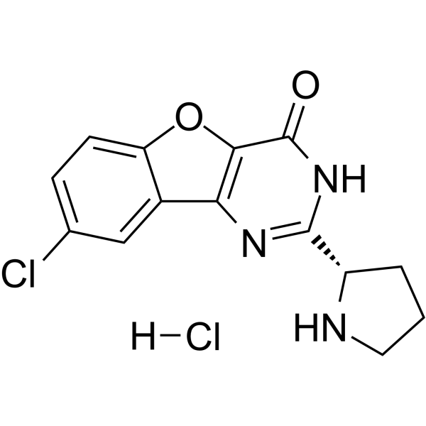
XL413 monohydrochloride
Based on 8 publication(s) in Google Scholar
XL413 monohydrochloride (BMS-863233 monohydrochloride) is an orally active, ATP-competitive, selective CDC7 kinase inhibitor. XL413 monohydrochloride reduces MCM2 phosphorylation levels, decreases DNA replication origin activation, and inhibits CD69 upregulation in stimulated lymphocytes. XL-413 possesses cell-dependent antiproliferative and pro-apoptotic activities. XL413 monohydrochloride attenuates ATR inhibitor-induced excessive origin activation, altered replication fork speed, and antiproliferative effects in sensitive cancer cells. XL413 monohydrochloride inhibits SARS-CoV-2 infection. XL413 monohydrochloride can be used for research related to cancer and SARS-CoV-2 infection.
For research use only. We do not sell to patients.
- Purity : 99.65%
- CAS No.: 2062200-97-7
- Formula: C14H13Cl2N3O2
- Molecular Weight:326.18
-
Storage:
4°C, sealed storage, away from moisture
* In solvent : -80°C, 1 year; -20°C, 6 months (sealed storage, away from moisture)

To place orders, for customer services and technical support, please contact: MedChemExpress USA
Tel: 609-228-6898 E-mail: [email protected] [email protected]


-
 Biological Activity
-
Chemical Information
-
Solvent & Solubility
- Protocols
- Purity & Documentation
- References
-
 Help & FAQs


-
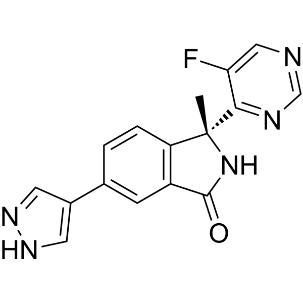
HY-100023
LY3177833CDC7 Inhibitor -
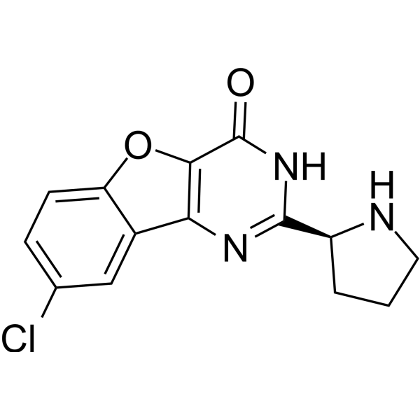
HY-15260
XL413CDC7 Inhibitor -
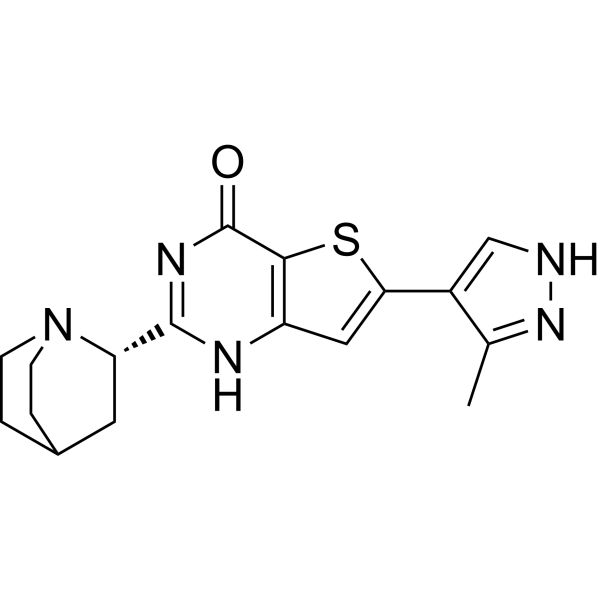
HY-100888
SimurosertibCDC7 Inhibitor -
HY-15144
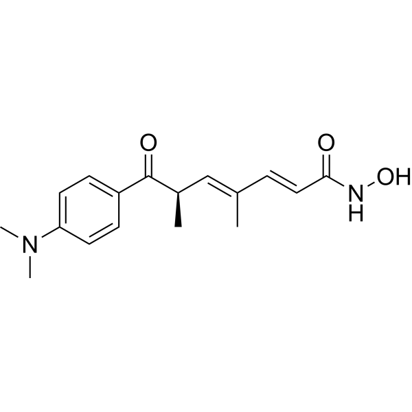
Trichostatin AHDAC Class I/II Inhibitor -
HY-15777
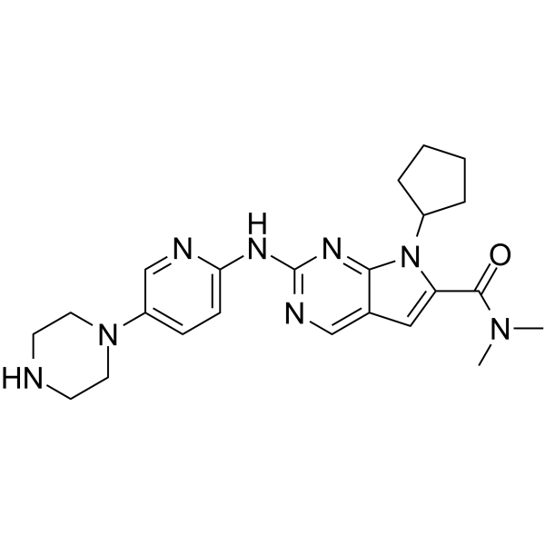
RibociclibCDK4/6 Inhibitor -
HY-11006
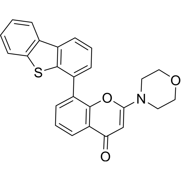
KU-57788DNA-PK Inhibitor -

HY-15763
ErastinFerroptosis Inducer -
HY-10201
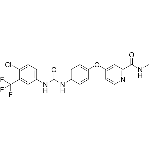
SorafenibMultikinase Inhibitor -
HY-10585
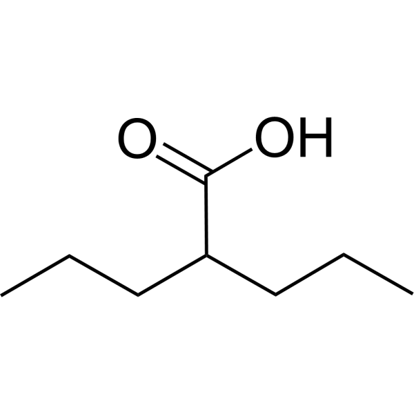
Valproic acidHDAC Inhibitor -
HY-50895
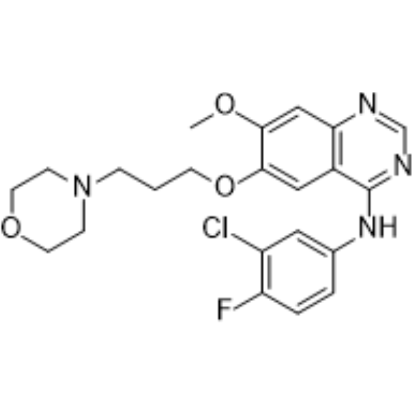
GefitinibEGFR Inhibitor -
HY-10087
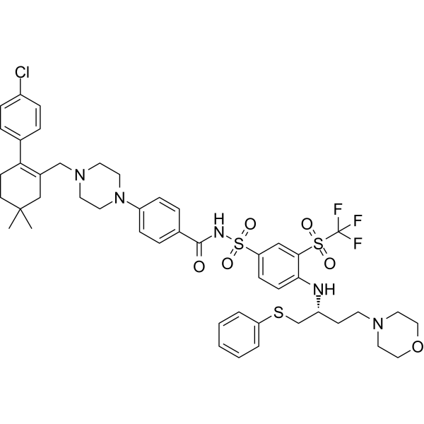
NavitoclaxBcl-2 Inhibitor -
HY-50878
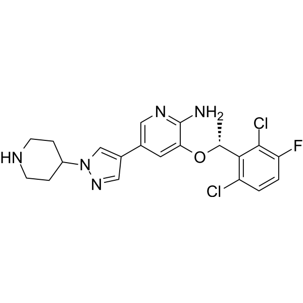
CrizotinibALK/c-Met/ROS1 Inhibitor
Publications Citing Use of MedChemExpress (MCE) XL413 monohydrochloride
More- Science. 2017 Dec 1;358(6367):eaan4368. [Abstract]
- MedComm (2020). 2025 May 15;6(6):e70150. [Abstract]
- J Transl Med. 2026 Jul 11.
- Sens Actuators B Chem. 15 May 2022, 131618.
- Oncogenesis. 2026 Apr 24;15(1):27. [Abstract]
- Am J Physiol Lung Cell Mol Physiol. 2018 Sep 1;315(3):L360-L370. [Abstract]
- bioRxiv. 2025 Dec 9.
- University of Washington. 2025.
 Customer Validation & Images

-
WB
All Caspase Isoforms
More

Biological Activity
Description
|
Cdc7 3.4 nM (IC50) |
Caspase 3 |
Caspase-7 |
In Vitro
XL-413 (1 µM; 1 h) monohydrochloride reduces baseline origin activation levels and reverses ATRi-induced alterations in origin density and replication fork speed in HCC1806 breast cancer cells; it decreases EdU incorporation and inhibits ATRi-induced replication origin hyperactivation in HCC1806 breast cancer cells[1].
XL-413 (0.25 µM; 72 h) monohydrochloride partially rescues ATRi-mediated antiproliferative effects in the sensitive breast cancer cell lines HCC1806 and HCC1569[1].
XL-413 (BMS-863233) monohydrochloride exhibits antiviral activity against infectious SARS-CoV-2 in Calu-1 cells and produces cytotoxic effects at higher concentrations[2].
XL-413 monohydrochloride shows no detectable modulation of DDX39B-related protein-protein interactions at concentrations of 5 μM and 50 μM in HEK293 cells[2].
XL-413 monohydrochloride exhibits potent inhibitory activity against purified recombinant DDK (Cdc7-Dbf4) kinase, with IC50 = 22.7 nM[4].
XL-413 (5 μM; 24-72 h) monohydrochloride abolishes DDK-dependent MCM2 phosphorylation in Colo-205 cells, whereas only a slight decrease in MCM2 phosphorylation is observed at 72 h in HCC1954 cells[4].
XL-413 monohydrochloride potently and selectively inhibits CDC7 kinase activity in Jurkat cells and OT-I CTLs; it inhibits CDC7 with an IC50 of 3.4 nM in biochemical assays[3].
XL-413 monohydrochloride inhibits the expression of the early T cell activation marker CD69 in anti-CD3-stimulated mouse lymphocytes[3].
XL-413 (5 μM; 72 h) monohydrochloride exhibits potent antiproliferative and pro-apoptotic activity against Colo-205 colorectal cancer cells (IC50 = 1.1 μM), but shows weak antiproliferative effects on HCC1954 breast cancer cells (IC50 = 22.9 μM)[4].
MedChemExpress (MCE) has not independently confirmed the accuracy of these methods. They are for reference only. Further protocols information, click here.
In Vivo
MedChemExpress (MCE) has not independently confirmed the accuracy of these methods. They are for reference only.


Chemical Information
-
CAS No. 2062200-97-7
-
Appearance Solid
-
Molecular Weight 326.18
-
Formula C14H13Cl2N3O2
-
Color White to off-white
-
SMILES
O=C1C(OC2=CC=C(Cl)C=C23)=C3N=C([C@H]4NCCC4)N1.[H]Cl
-
Synonyms
BMS-863233 monohydrochloride
-
Shipping
Room temperature in continental US; may vary elsewhere.
-
Storage
4°C, sealed storage, away from moisture
* In solvent : -80°C, 1 year; -20°C, 6 months (sealed storage, away from moisture)


Publications (8)
-
Journal Impact Factor
-
Most Recent
-
Science
2017 Dec 1;358(6367):eaan4368. PMID: 29191878 -
MedComm (2020)
2025 May 15;6(6):e70150. PMID: 40384988 -
-
-
Oncogenesis
2026 Apr 24;15(1):27. PMID: 42031714 -
Am J Physiol Lung Cell Mol Physiol
Cell division cycle 7 kinase is a negative regulator of cell-mediated collagen degradation. [Abstract]2018 Sep 1;315(3):L360-L370. PMID: 29792348
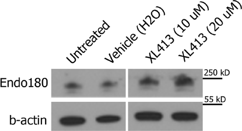
XL413 monohydrochloride purchased from MedChemExpress. Usage Cited in: Am J Physiol Lung Cell Mol Physiol. 2018 Sep 1;315(3):L360-L370. [Abstract]
Representative Western blot for Endo180 expression in U937 cells treated overnight with XL413.
-
-


Solvent & Solubility
In Vitro:
H2O : 10 mg/mL (30.66 mM; Need ultrasonic)
DMSO : 3.4 mg/mL (10.42 mM; Need ultrasonic and warming; Hygroscopic DMSO has a significant impact on the solubility of product, please use newly opened DMSO)
Please refer to the solubility information to select the appropriate solvent. Once prepared, please aliquot and store the solution to prevent product inactivation from repeated freeze-thaw cycles.
Storage method and period of stock solution: -80°C, 1 year; -20°C, 6 months (sealed storage, away from moisture). When stored at -80°C, please use it within 1 year. When stored at -20°C, please use it within 6 months.
* Note: If you choose water as the stock solution, please dilute it to the working solution, then filter and sterilize it with a 0.22 μm filter before use.
Please refer to the solubility information to select the appropriate solvent. Once prepared, please aliquot and store the solution to prevent product inactivation from repeated freeze-thaw cycles.
Storage method and period of stock solution: -80°C, 1 year; -20°C, 6 months (sealed storage, away from moisture). When stored at -80°C, please use it within 1 year. When stored at -20°C, please use it within 6 months.
* Note: If you choose water as the stock solution, please dilute it to the working solution, then filter and sterilize it with a 0.22 μm filter before use.
Concentration (start) × Volume (start) = Concentration (final) × Volume (final)

Protocols
-
Kinase activity and phosphorylation assays
Kinase activity assays measure the ability of kinases to transfer phosphate groups from ATP to specific substrates, while phosphorylation assays detect the presence and levels of phosphorylated proteins. Common methods include radiolabeled ATP incorporation (e. g. ,), ADP release detection via bioluminescence (e. g. ,[3]), enzyme-linked immunosorbent assays (ELISA) for phospho-specific epitopes (e. g. ,[6]), and microtiter-based formats for high-throughput screening (e. g. ,[8]). The ADP-Glo assay quantifies kinase activity by measuring ADP produced during phosphorylation using a luciferase-based system. Radiometric assays involve autoradiography or scintillation counting after incorporation of 32P-labeled ATP into substrate proteins. ELISA-based approaches rely on phospho-specific antibodies to detect activated kinases in cell lysates or purified samples.
-
Western Blot
Western blotting (WB) is a commonly used experimental method in molecular biology, biochemistry, and immunogenetics for identifying and quantifying target proteins. It combines gel electrophoresis with immunoassay, enabling researchers to analyze protein expression, post-translational modifications, and molecular weight.
-
Research Protocol for Infectious Diseases
Infectious-disease experiments test how pathogens interact with host barriers, innate immune receptors, inflammatory signaling, pathogen replication, and tissue injury; pattern-recognition receptors such as TLRs, RIG-I-like receptors, NOD-like receptors, and inflammasomes detect microbial molecules and activate NF-κB, interferon, and cytokine responses. The central hypothesis is that infection severity reflects the balance between pathogen burden and host response: protective inflammation restricts pathogen growth, whereas excessive or mislocalized inflammation contributes to tissue damage and disease phenotype. Unresolved questions include which host pathways are protective versus pathogenic, why some infection models fail to translate to human disease, and which combined readouts best predict clinically relevant infection outcomes.
-
Apoptosis
Apoptosis, also called programmed cell death, is generally characterized by distinct morphological characteristics.
-
PCNA Immunodetection Proliferation Assay
PCNA immunodetection measures proliferative activity by detecting proliferating cell nuclear antigen, a nuclear protein associated with DNA polymerase δ function and DNA replication. The assay readout is the proportion of PCNA-positive nuclei among total counted cells, but PCNA labeling is not identical to BrdU labeling because PCNA can mark late G1/early S-associated replication competence and may persist beyond active DNA synthesis depending on fixation and extraction conditions.
-
Apoptosis Solutions
Apoptosis is a regulated, generally non-lytic cell-death pathway that removes unwanted, damaged, infected, or abnormal cells through coordinated morphological changes, caspase activation, DNA fragmentation, and membrane remodeling. The intrinsic apoptosis pathway is controlled mainly by mitochondrial outer membrane permeabilization, BCL-2 family proteins, cytochrome c release, apoptosome formation, caspase-9 activation, and downstream executioner caspase-3/7 activation. The extrinsic apoptosis pathway is initiated by death receptors such as Fas, TNFR, and TRAIL receptors, which recruit adaptor proteins and activate caspase-8 before engaging executioner caspases or mitochondrial amplification through BID cleavage. Apoptosis is linked to many phenotypes, including cancer cell killing, tissue homeostasis, immune regulation, neurodegeneration, infection response, and treatment-induced cytotoxicity; unresolved questions include how apoptosis interacts with necroptosis, pyroptosis, ferroptos
-
Protocol for Kinase activity and phosphorylation assays
Kinase activity assays measure transfer of phosphate from ATP to a protein or peptide substrate, generating phosphorylated substrate, ADP, or incorporated radiolabeled phosphate as the readout; phosphorylation assays measure site-specific phosphorylation in cells or tissues as a proxy for kinase-pathway activation, inhibition, or substrate regulation. Phosphorylation can be detected by phospho-specific Western blot, immunoprecipitation kinase assay, phospho-immunofluorescence, phospho-flow cytometry, luminescent ADP detection, radiolabeled ATP incorporation, or reporter-based pathway assays, and these readouts can be applied to cancer cells, primary neurons, mouse tumors, organoids, inflammatory macrophages, ferroptosis studies, and mitophagy studies when the kinase target is biologically relevant.
-
Cell Viability Determination by MTT Colorimetric Assay
The following protocol uses the MTT colorimetric assay as a classic literature-established method for assessing cell viability/metabolic activity in cultured mammalian cells. MTT[3-(4,5-dimethylthiazol-2-yl)-2,5-diphenyltetrazolium bromide] is reduced by metabolically active cells to a colored formazan product; the amount of formazan is quantified spectrophotometrically and provides an indirect measure of metabolically active viable cells. Importantly, MTT reduction reflects cellular oxidoreductase/metabolic activity rather than an absolute direct count of living cells, so changes in cellular metabolism can alter the signal independently of cell number.


Purity & Documentation
-
Data Sheet (299 KB)
-
SDS (479 KB)
- English - EN (479 KB)
- Français - FR (479 KB)
- Deutsch - DE (479 KB)
- Norwegian - NO (479 KB)
- Español - ES (479 KB)
- Swedish - SV (479 KB)
- Italian - IT (479 KB)
- Korean - KR (479 KB)
- Portuguese - PT (479 KB)
-
Handling Instructions (2659 KB)
References
[2]. Liu X, et al. SARS-CoV-2-host proteome interactions for antiviral drug discovery. Molecular systems biology. 2021 Nov;17(11):e10396. [Content Brief]
[3]. Chen EW, et al. A Dual Inhibitor of Cdc7/Cdk9 Potently Suppresses T Cell Activation. Frontiers in immunology. 2019;10:1718. [Content Brief]
[4]. Sasi NK, et al. The potent Cdc7-Dbf4 (DDK) kinase inhibitor XL413 has limited activity in many cancer cell lines and discovery of potential new DDK inhibitor scaffolds. PLoS One. 2014 Nov 20;9(11):e113300. [Content Brief]


Complete Stock Solution Preparation Table
Please refer to the solubility information to select the appropriate solvent. Once prepared, please aliquot and store the solution to prevent product inactivation from repeated freeze-thaw cycles.
Storage method and period of stock solution: -80°C, 1 year; -20°C, 6 months (sealed storage, away from moisture). When stored at -80°C, please use it within 1 year. When stored at -20°C, please use it within 6 months.
| Optional Solvent | Concentration Solvent Mass | 1 mg | 5 mg | 10 mg | 25 mg |
|---|---|---|---|---|---|
| DMSO / H2O | 1 mM | 3.0658 mL | 15.3290 mL | 30.6579 mL | 76.6448 mL |
| 5 mM | 0.6132 mL | 3.0658 mL | 6.1316 mL | 15.3290 mL | |
| 10 mM | 0.3066 mL | 1.5329 mL | 3.0658 mL | 7.6645 mL | |
| H2O | 15 mM | 0.2044 mL | 1.0219 mL | 2.0439 mL | 5.1097 mL |
| 20 mM | 0.1533 mL | 0.7664 mL | 1.5329 mL | 3.8322 mL | |
| 25 mM | 0.1226 mL | 0.6132 mL | 1.2263 mL | 3.0658 mL | |
| 30 mM | 0.1022 mL | 0.5110 mL | 1.0219 mL | 2.5548 mL |
* Note: If you choose water as the stock solution, please dilute it to the working solution, then filter and sterilize it with a 0.22 μm filter before use.


XL413 monohydrochloride Related Classifications
HY-15260A Related Classifications
 Powered by Bioz