



ApoE Antibody (YA1516)
(Synonyms: APOE; Apolipoprotein E; Apo-E)
Based on 1 Customer Validation
ApoE Antibody (YA1516) is a Rabbit-derived and non-conjugated IgG monoclonal antibody, targeting to ApoE.
-
Host:
Rabbit
-
Isotype:
IgG
-
Application:
WB, IHC-P, ICC/IF, IP
-
Reactivity :
Human, Mouse
-
Formulation:
Supplied in PBS(pH 7.4), 150mM sodium chloride, 0.05% BSA, 0.02% sodium azide and 50% glycerol.
-
Conjugation:
Non-conjugated


To place orders, for customer services and technical support, please contact: MedChemExpress USA
Tel: 609-228-6898 E-mail: [email protected] [email protected]





Applications
| Application |
WB

WB: Western Blot
|
IHC-P
IHC-P: Immunohistochemistry-Paraffin
|
ICC/IF
ICC/IF: Immunocytochemistry/
Immunofluorescence |
IP
IP: Immunoprecipitation
|
|---|---|---|---|---|
| Dilution Ratio | 1:500-1:1000 | 1:50-1:100 | 1:50-1:200 | 1:50 |


Product Details
ApoE Antibody (YA1516) is a Rabbit-derived and non-conjugated IgG monoclonal antibody, targeting to ApoE.
-
Host Rabbit

-
Clonality Recombinant,Monoclonal

-
Species ReactivityHuman, Mouse
-
Observed Molecular WeightObserved band size: 36 kDa
Note: Due to possible protein modifications or aggregation, the molecular weight should be confirmed by actual measurement, and the predicted value is for reference only.

-
Calculated Molecular Weight Predicted band size: 36 kDa

Entrez Gene: 7804 Human ; 16975 Mouse ; 362558 Rat
SwissProt: Q14114 Human ; Q924X6 Mouse ;
OMIM: 608446 Human
A synthesized peptide derived from human Apolipoprotein E aa300-317.
Endogenous
Affinity Chromatography
Non-conjugated
Unmodified
IgG


Product Properties
-
Appearance
Solution
-
Formulation
Supplied in PBS(pH 7.4), 150mM sodium chloride, 0.05% BSA, 0.02% sodium azide and 50% glycerol.
-
Concentration
Batch-dependent, Please check the COA for the concentration of each lot. Check Lot Concentration
-
Storage & Stability
Stored at -20°C for 1 year. Avoid repeated freeze / thaw cycles.
-
Shipping
Shipping with blue ice.


Verification Images
-
 Immunohistochemical analysis of paraffin-embedded human glioma tissue using ApoE Antibody (HY-P81771, 1/100). The section was pretreated using heat mediated antigen retrieval with sodium citrate buffer (pH 6.0) for 8 minutes. The tissues were blocked with quick block buffer for 0.5 hours at room temperature, washed with PBS and PBST, and then incubated with the primary antibody overnight at 4℃. The detection was performed using an HRP conjugated compact polymer system. DAB was used as the chromogen. Tissues were counterstained with hematoxylin and mounted with DPX.
 -
 Immunohistochemical analysis of paraffin-embedded human liver cancer tissue using ApoE Antibody (HY-P81771, 1/100). The section was pretreated using heat mediated antigen retrieval with sodium citrate buffer (pH 6.0) for 8 minutes. The tissues were blocked with quick block buffer for 0.5 hours at room temperature, washed with PBS and PBST, and then incubated with the primary antibody overnight at 4℃. The detection was performed using an HRP conjugated compact polymer system. DAB was used as the chromogen. Tissues were counterstained with hematoxylin and mounted with DPX.
 -
 Immunohistochemical analysis of paraffin-embedded human testis cancer tissue using ApoE Antibody (HY-P81771, 1/100). The section was pretreated using heat mediated antigen retrieval with sodium citrate buffer (pH 6.0) for 8 minutes. The tissues were blocked with quick block buffer for 0.5 hours at room temperature, washed with PBS and PBST, and then incubated with the primary antibody overnight at 4℃. The detection was performed using an HRP conjugated compact polymer system. DAB was used as the chromogen. Tissues were counterstained with hematoxylin and mounted with DPX.
 -
 Immunohistochemical analysis of paraffin-embedded human skin cancer tissue using ApoE Antibody (HY-P81771, 1/100). The section was pretreated using heat mediated antigen retrieval with sodium citrate buffer (pH 6.0) for 8 minutes. The tissues were blocked with quick block buffer for 0.5 hours at room temperature, washed with PBS and PBST, and then incubated with the primary antibody overnight at 4℃. The detection was performed using an HRP conjugated compact polymer system. DAB was used as the chromogen. Tissues were counterstained with hematoxylin and mounted with DPX.
 -
 Immunohistochemical analysis of paraffin-embedded human lung cancer tissue using ApoE Antibody (HY-P81771, 1/100). The section was pretreated using heat mediated antigen retrieval with sodium citrate buffer (pH 6.0) for 8 minutes. The tissues were blocked with quick block buffer for 0.5 hours at room temperature, washed with PBS and PBST, and then incubated with the primary antibody overnight at 4℃. The detection was performed using an HRP conjugated compact polymer system. DAB was used as the chromogen. Tissues were counterstained with hematoxylin and mounted with DPX.
 -
 Immunohistochemical analysis of paraffin-embedded human liver tissue using ApoE Antibody (HY-P81771, 1/100). The section was pretreated using heat mediated antigen retrieval with sodium citrate buffer (pH 6.0) for 8 minutes. The tissues were blocked with quick block buffer for 0.5 hours at room temperature, washed with PBS and PBST, and then incubated with the primary antibody overnight at 4℃. The detection was performed using an HRP conjugated compact polymer system. DAB was used as the chromogen. Tissues were counterstained with hematoxylin and mounted with DPX.
 -
 Immunohistochemical analysis of paraffin-embedded human adrenal gland tissue using ApoE Antibody (HY-P81771, 1/100). The section was pretreated using heat mediated antigen retrieval with sodium citrate buffer (pH 6.0) for 8 minutes. The tissues were blocked with quick block buffer for 0.5 hours at room temperature, washed with PBS and PBST, and then incubated with the primary antibody overnight at 4℃. The detection was performed using an HRP conjugated compact polymer system. DAB was used as the chromogen. Tissues were counterstained with hematoxylin and mounted with DPX.
 -
 Immunohistochemical analysis of paraffin-embedded human cerebral cortex tissue using ApoE Antibody (HY-P81771, 1/100). The section was pretreated using heat mediated antigen retrieval with sodium citrate buffer (pH 6.0) for 8 minutes. The tissues were blocked with quick block buffer for 0.5 hours at room temperature, washed with PBS and PBST, and then incubated with the primary antibody overnight at 4℃. The detection was performed using an HRP conjugated compact polymer system. DAB was used as the chromogen. Tissues were counterstained with hematoxylin and mounted with DPX.
 -
 Immunohistochemical analysis of paraffin-embedded human cerebellum tissue using ApoE Antibody (HY-P81771, 1/100). The section was pretreated using heat mediated antigen retrieval with sodium citrate buffer (pH 6.0) for 8 minutes. The tissues were blocked with quick block buffer for 0.5 hours at room temperature, washed with PBS and PBST, and then incubated with the primary antibody overnight at 4℃. The detection was performed using an HRP conjugated compact polymer system. DAB was used as the chromogen. Tissues were counterstained with hematoxylin and mounted with DPX.
 -
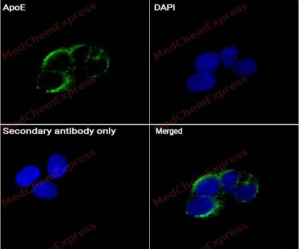 Immunocytochemistry analysis of 293T cells labeling ApoE Antibody (HY-P81771) at 1/50 dilution. Cells were fixed in 4% paraformaldehyde for 15 minutes at room temperature, permeabilized with 0.1% Triton X-100 for 10 minutes at room temperature, then blocked with BSA for Immunol Staining for 10 min at room temperature. Cells were then incubated with ApoE Antibody (HY-P81771) at 1/50 in BSA for Immunol Staining at 4 ℃ Overnight. AF488-conjugated AffiniPure Goat Anti-Rabbit IgG H&L(HY-P8002, Green) was used as the secondary antibody at 1/1,000 dilution. PBS instead of the primary antibody was used as the secondary antibody only control. The Nuclear counterstain was DAPI (Blue).
 -
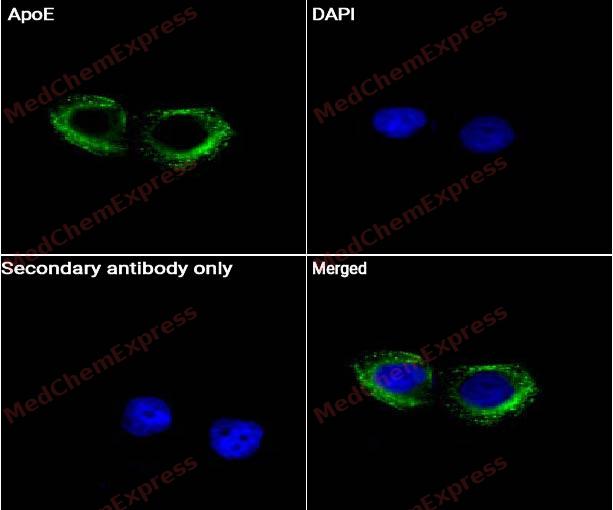 Immunocytochemistry analysis of 293T cells labeling ApoE Antibody (HY-P81771) at 1/200 dilution. Cells were fixed in 4% paraformaldehyde for 15 minutes at room temperature, permeabilized with 0.1% Triton X-100 for 10 minutes at room temperature, then blocked with BSA for Immunol Staining for 10 min at room temperature. Cells were then incubated with ApoE Antibody (HY-P81771) at 1/50 in BSA for Immunol Staining at 4 ℃ Overnight. AF488-conjugated AffiniPure Goat Anti-Rabbit IgG H&L(HY-P8002, Green) was used as the secondary antibody at 1/1,000 dilution. PBS instead of the primary antibody was used as the secondary antibody only control. The Nuclear counterstain was DAPI (Blue).


Background
-
Function
Apolipoprotein E (ApoE) is a key regulator of lipoprotein metabolism that mediates the hepatic clearance of diet-derived chylomicron remnants and liver-derived very-low-density lipoprotein (VLDL) remnants through interactions with members of the low-density lipoprotein receptor family[1][2]. ApoE deficiency disrupts this clearance pathway, resulting in marked hypercholesterolemia, accumulation of remnant lipoproteins, and enhanced susceptibility to atherosclerotic lesion formation[1][3][4]. Mechanistically, ApoE regulates cholesterol homeostasis and influences macrophage biology, linking lipid metabolism to vascular inflammation and atherogenesis[2][5]. In disease models, Apoe−/− mice develop spontaneous atherosclerosis even when maintained on a low-cholesterol diet and therefore represent one of the most widely used experimental systems for investigating cardiovascular disease mechanisms and therapeutic interventions[3][4][6]. Atherosclerotic lesions in these mice progress in a manner that reproduces many pathological features of human disease, making the model highly valuable for studies of plaque development and progression[6][2]. Compared with related human APOE isoforms, ApoE deficiency represents a complete loss-of-function state rather than an isoform-specific alteration, providing a robust platform for dissecting the physiological roles of ApoE in lipoprotein transport, monocyte/macrophage biology, and atherosclerosis[2][5]. For experimental applications, the Apoe−/− model is extensively used to evaluate genetic, nutritional, and pharmacological factors that modify atherosclerotic burden and vascular inflammation[6][2].
-
Subcellular Localization
Secreted; Secreted, extracellular space; Secreted, extracellular space, extracellular matrix; Extracellular vesicle; Endosome, multivesicular body
-
Expression
Tissue_specificity:Produced by various tissue and cell types, it is mainly found in lipid particles in plasma, interstitial fluid, and lymph (PubMed:25173806) . It is primarily synthesized by hepatocytes (PubMed:25173806) . It is also produced in large quantities in brain tissue, mainly by astrocytes and glial cells in the cerebral cortex, and also by neurons in the frontal cortex and hippocampus (PubMed:10027417, PubMed:3115992) . Cells in the peripheral nervous system also express it (PubMed:10027417, PubMed:25173806) . It is also expressed in the adrenal glands, testes, ovaries, skin, kidneys, spleen, adipose tissue, and macrophages in various tissues (PubMed:25173806) . -
Subunit
Homotetramer (PubMed:8340399). May interact with ABCA1; functionally associated with ABCA1 in the biogenesis of HDLs (PubMed:14754908). May interact with APP/A4 amyloid-beta peptide; the interaction is extremely stable in vitro but its physiological significance is unclear (PubMed:23620513, PubMed:8367470). May interact with MAPT (PubMed:7972031). May interact with MAP2 (PubMed:7891887). In the cerebrospinal fluid, interacts with secreted SORL1 (PubMed:30448281). Interacts with PMEL; this allows the loading of PMEL luminal fragment on ILVs to induce fibril nucleation
-
SwissProt ID
-
Synonyms
APOE; Apolipoprotein E; Apo-E
-
Research Field
Neuroscience


Documentation
-
Data Sheet (262 KB)
-
SDS (251 KB)
- English - EN (251 KB)
- Français - FR (251 KB)
- Deutsch - DE (251 KB)
- Norwegian - NO (251 KB)
- Español - ES (251 KB)
- Swedish - SV (251 KB)
- Italian - IT (251 KB)
- Korean - KR (251 KB)
- Portuguese - PT (251 KB)
-
User Guide for Antibodies (1077 KB)
References
[1]. Pendse AA, et al. Apolipoprotein E knock-out and knock-in mice: atherosclerosis, metabolic syndrome, and beyond. J Lipid Res. 2009 Apr;50 Suppl(Suppl):S178-82. [Content Brief]
[3]. Buzello M, et al. The apolipoprotein e knockout mouse: a model documenting accelerated atherogenesis in uremia. J Am Soc Nephrol. 2003 Feb;14(2):311-6. [Content Brief]
[4]. Lo Sasso G, et al. The Apoe(-/-) mouse model: a suitable model to study cardiovascular and respiratory diseases in the context of cigarette smoke exposure and harm reduction. J Transl Med. 2016 May 20;14(1):146. [Content Brief]
[6]. Meir KS, et al. Atherosclerosis in the apolipoprotein-E-deficient mouse: a decade of progress. Arterioscler Thromb Vasc Biol. 2004 Jun;24(6):1006-14. [Content Brief]
[7]. Getz GS, et al. ApoE knockout and knockin mice: the history of their contribution to the understanding of atherogenesis. J Lipid Res. 2016 May;57(5):758-66. [Content Brief]

 Powered by Bioz
