



CBL Antibody (YA3889)
(Synonyms: CBL; CBL2; NSLL; C-CBL; RNF55)Based on 1 Customer Validation
CBL Antibody (YA3889) is a Mouse-derived and non-conjugated IgG1 monoclonal antibody, targeting to CBL.
-
Host:
Mouse
-
Isotype:
IgG1
-
Application:
WB, IHC-P, ICC/IF, FC, ELISA
-
Reactivity :
Human, Mouse, Rat, Monkey, Rabbit
-
Formulation:
Supplied in PBS with 0.05% sodium azide.
-
Conjugation:
Non-conjugated


To place orders, for customer services and technical support, please contact: MedChemExpress USA
Tel: 609-228-6898 E-mail: [email protected] [email protected]





Applications
| Application |
WB

WB: Western Blot
|
IHC-P
IHC-P: Immunohistochemistry-Paraffin
|
ICC/IF
ICC/IF: Immunocytochemistry/
Immunofluorescence |
FC
FC: Flow Cytometry
|
ELISA
ELISA: Enzyme Linked Immunosorbent Assay
|
|---|---|---|---|---|---|
| Dilution Ratio | 1:500-1:2000 | 1:100-1:500 | 1:50-1:500 | 1:200-1:400 | 1:10000 |


Product Details
CBL Antibody (YA3889) is a Mouse-derived and non-conjugated IgG1 monoclonal antibody, targeting to CBL.
-
Host Mouse

-
Clonality Monoclonal

-
Species ReactivityHuman, Mouse, Rat, Monkey, Rabbit
-
Observed Molecular WeightObserved band size: 120 kDa
Note: Due to possible protein modifications or aggregation, the molecular weight should be confirmed by actual measurement, and the predicted value is for reference only.

-
Calculated Molecular Weight Predicted band size: 100 kDa

Purified recombinant fragment of human C-CBL aa 684-865.
affinity purified.
Non-conjugated
Unmodified
IgG1


Product Properties
-
Appearance
Solution
-
Formulation
Supplied in PBS with 0.05% sodium azide.
-
Concentration
Batch-dependent, Please check the COA for the concentration of each lot. Check Lot Concentration
-
Storage & Stability
Stored at -20°C for 1 year. Avoid repeated freeze / thaw cycles.
-
Shipping
Shipping with blue ice.


Verification Images
-
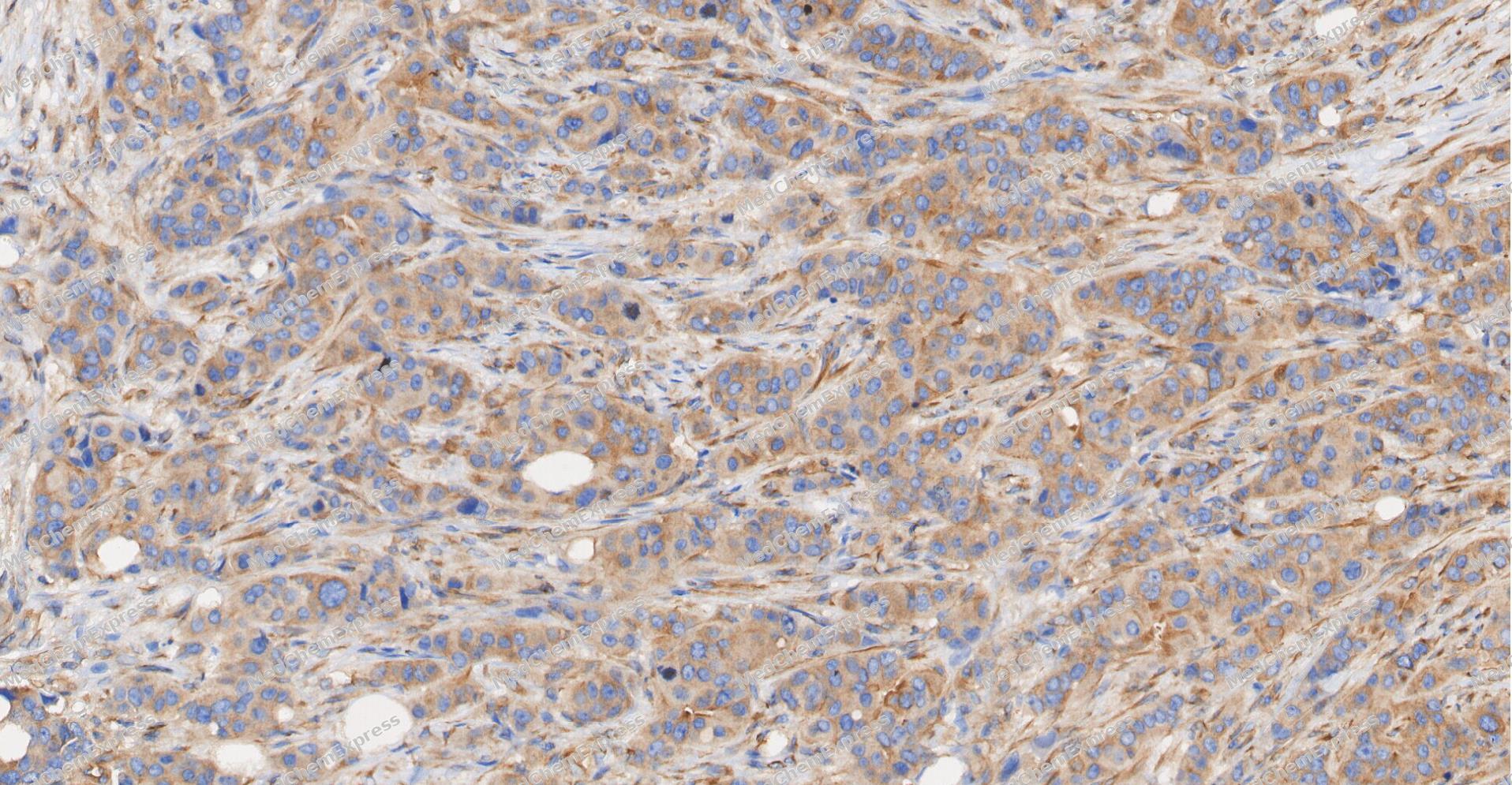 Immunohistochemical analysis of paraffin-embedded human Breast Cancer tissue using CBL antibody was performed. The section was pretreated using high-temperature and high-pressure mediated EDTA antigen retrieval buffer (pH 9.0), for 5 minutes. The tissues were incubated with primary antibody (HY-P84192, 1:600 dilution) at room temperature for 60 minutes. Detection was performed using an HRP conjugated compact polymer system. DAB was used as the chromogen. The tissues were counterstained with hematoxylin and mounted with neutral balsam mounting medium.
 -
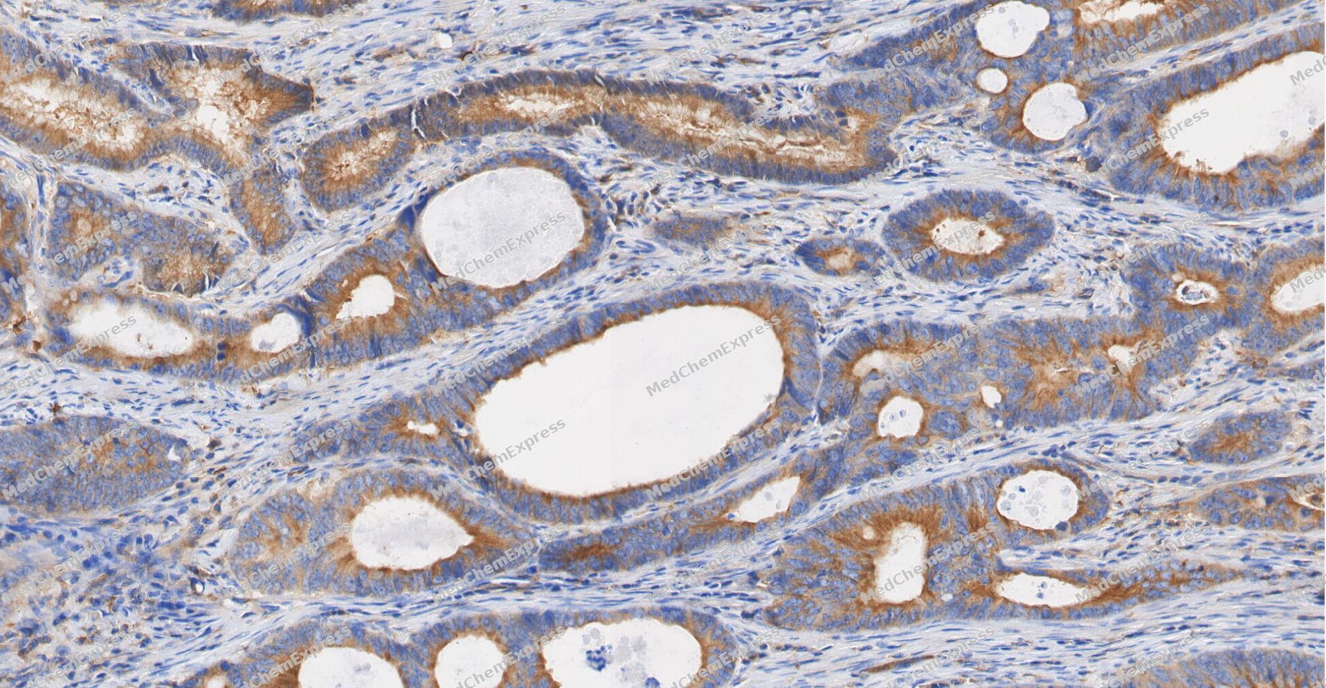 Immunohistochemical analysis of paraffin-embedded human Colon cancer tissue using CBL antibody was performed. The section was pretreated using high-temperature and high-pressure mediated EDTA antigen retrieval buffer (pH 9.0), for 5 minutes. The tissues were incubated with primary antibody (HY-P84192, 1:600 dilution) at room temperature for 60 minutes. Detection was performed using an HRP conjugated compact polymer system. DAB was used as the chromogen. The tissues were counterstained with hematoxylin and mounted with neutral balsam mounting medium.
 -
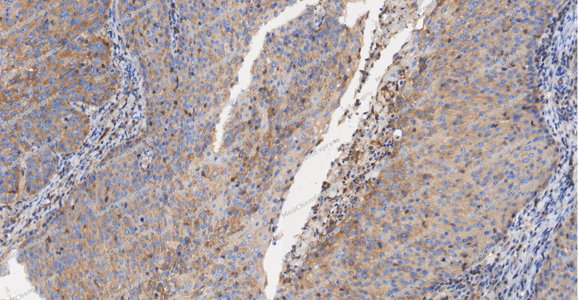 Immunohistochemical analysis of paraffin-embedded human Cervical cancer tissue using CBL antibody was performed. The section was pretreated using high-temperature and high-pressure mediated EDTA antigen retrieval buffer (pH 9.0), for 5 minutes. The tissues were incubated with primary antibody (HY-P84192, 1:600 dilution) at room temperature for 60 minutes. Detection was performed using an HRP conjugated compact polymer system. DAB was used as the chromogen. The tissues were counterstained with hematoxylin and mounted with neutral balsam mounting medium.
 -
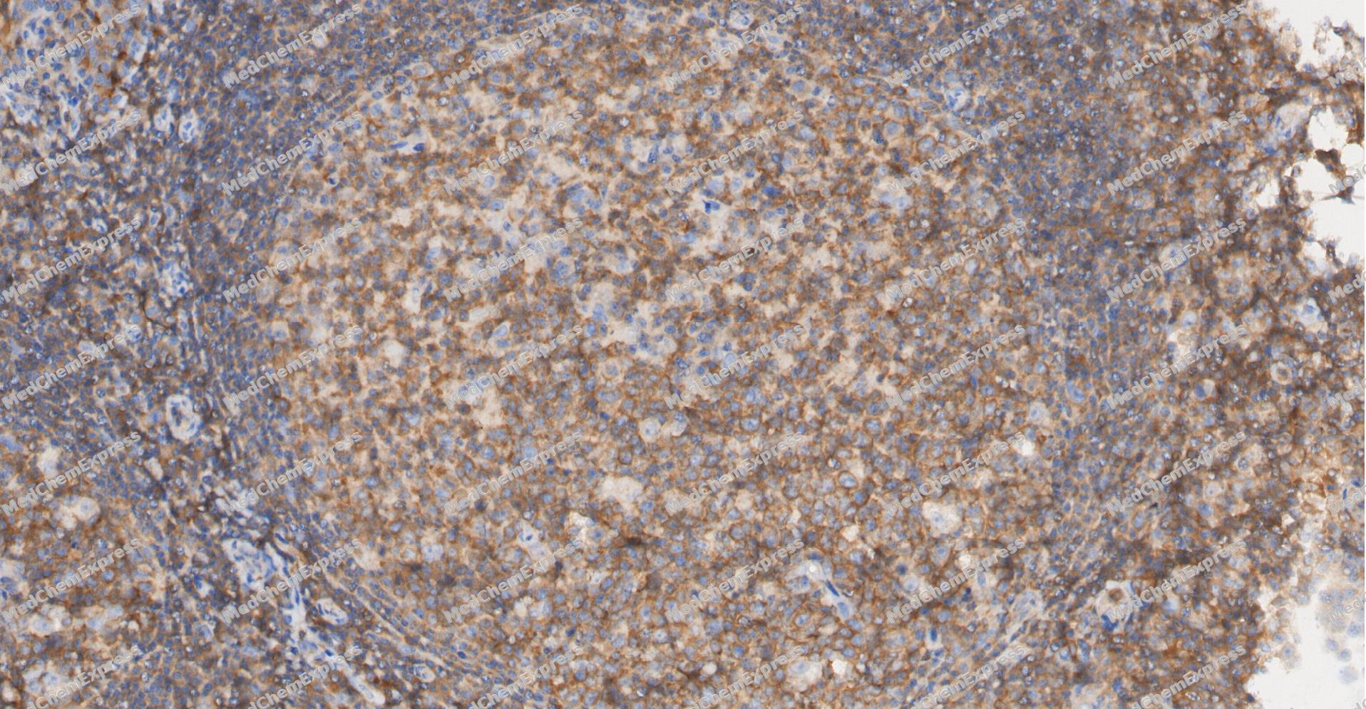 Immunohistochemical analysis of paraffin-embedded human Tonsil tissue using CBL antibody was performed. The section was pretreated using high-temperature and high-pressure mediated EDTA antigen retrieval buffer (pH 9.0), for 5 minutes. The tissues were incubated with primary antibody (HY-P84192, 1:600 dilution) at room temperature for 60 minutes. Detection was performed using an HRP conjugated compact polymer system. DAB was used as the chromogen. The tissues were counterstained with hematoxylin and mounted with neutral balsam mounting medium.
 -
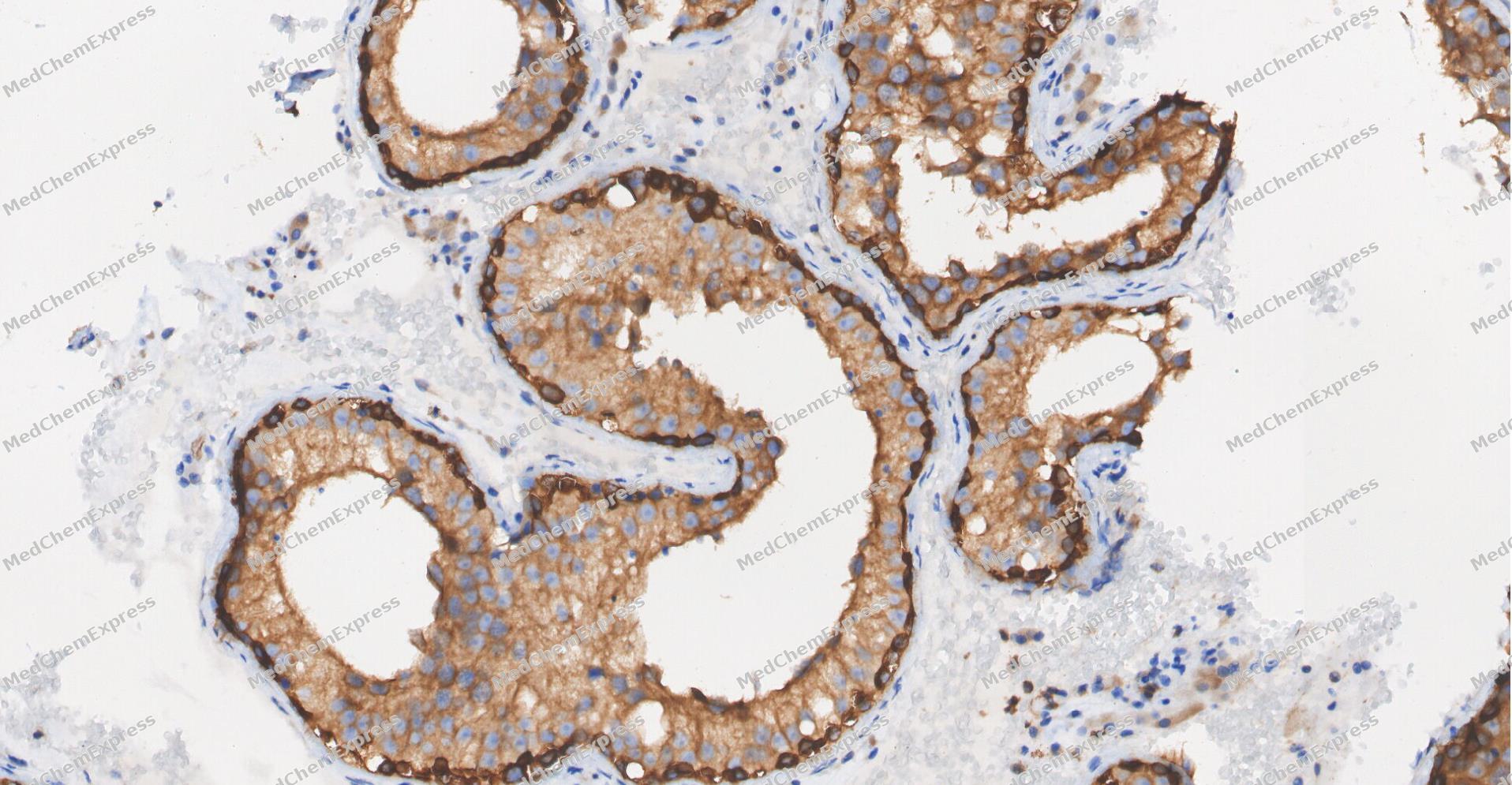 Immunohistochemical analysis of paraffin-embedded human Testis tissue using CBL antibody was performed. The section was pretreated using high-temperature and high-pressure mediated EDTA antigen retrieval buffer (pH 9.0), for 5 minutes. The tissues were incubated with primary antibody (HY-P84192, 1:600 dilution) at room temperature for 60 minutes. Detection was performed using an HRP conjugated compact polymer system. DAB was used as the chromogen. The tissues were counterstained with hematoxylin and mounted with neutral balsam mounting medium.
 -
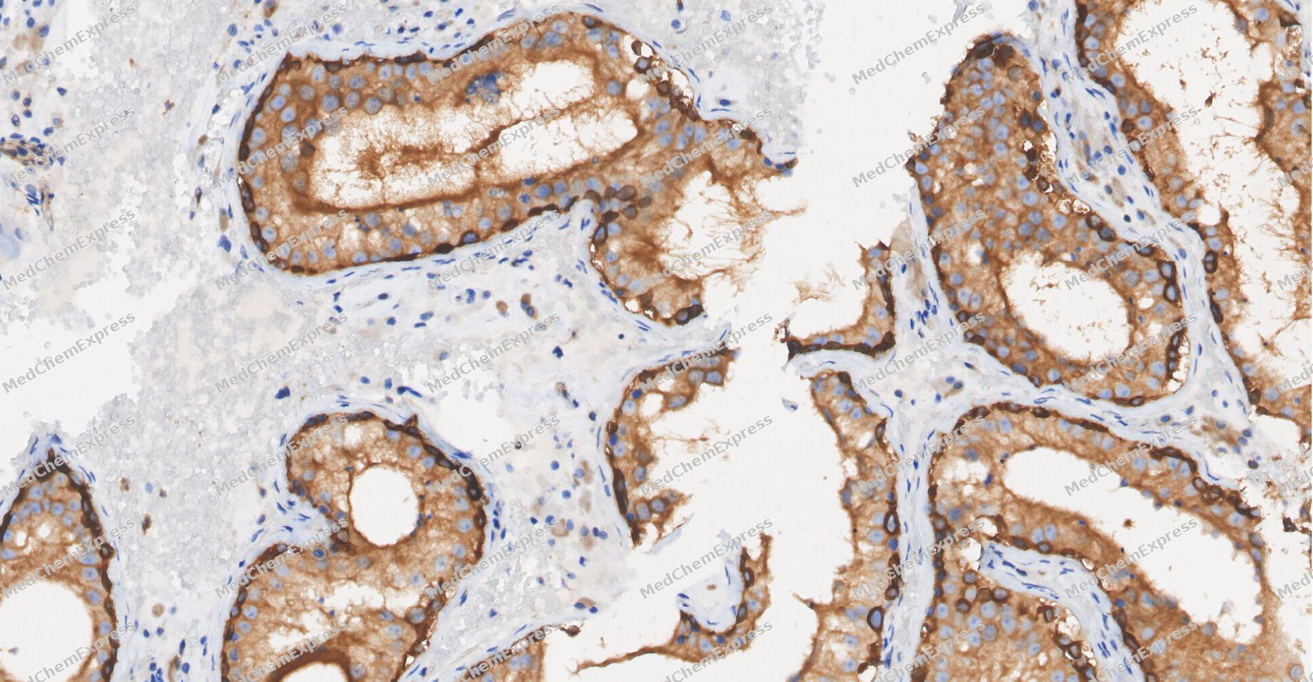 Immunohistochemical analysis of paraffin-embedded human Testis tissue using CBL antibody was performed. The section was pretreated using high-temperature and high-pressure mediated EDTA antigen retrieval buffer (pH 9.0), for 5 minutes. The tissues were incubated with primary antibody (HY-P84192, 1:600 dilution) at room temperature for 60 minutes. Detection was performed using an HRP conjugated compact polymer system. DAB was used as the chromogen. The tissues were counterstained with hematoxylin and mounted with neutral balsam mounting medium.
 -
 Immunohistochemical analysis of paraffin-embedded human glioma tissue using CBL Antibody (HY-P84192, 1/500). The section was pretreated using heat mediated antigen retrieval with sodium citrate buffer (pH 6.0) for 8 minutes. The tissues were blocked with quick block buffer for 0.5 hours at room temperature, washed with PBS and PBST, and then incubated with the primary antibody overnight at 4℃. The detection was performed using an HRP conjugated compact polymer system. DAB was used as the chromogen. Tissues were counterstained with hematoxylin and mounted with DPX.
 -
 Immunohistochemical analysis of paraffin-embedded human breast cancer tissue using CBL Antibody (HY-P84192, 1/500). The section was pretreated using heat mediated antigen retrieval with sodium citrate buffer (pH 6.0) for 8 minutes. The tissues were blocked with quick block buffer for 0.5 hours at room temperature, washed with PBS and PBST, and then incubated with the primary antibody overnight at 4℃. The detection was performed using an HRP conjugated compact polymer system. DAB was used as the chromogen. Tissues were counterstained with hematoxylin and mounted with DPX.
 -
 Immunohistochemical analysis of paraffin-embedded human urothelial cancer tissue using CBL Antibody (HY-P84192, 1/500). The section was pretreated using heat mediated antigen retrieval with sodium citrate buffer (pH 6.0) for 8 minutes. The tissues were blocked with quick block buffer for 0.5 hours at room temperature, washed with PBS and PBST, and then incubated with the primary antibody overnight at 4℃. The detection was performed using an HRP conjugated compact polymer system. DAB was used as the chromogen. Tissues were counterstained with hematoxylin and mounted with DPX.
 -
 Immunohistochemical analysis of paraffin-embedded human lung cancer tissue using CBL Antibody (HY-P84192, 1/500). The section was pretreated using heat mediated antigen retrieval with sodium citrate buffer (pH 6.0) for 8 minutes. The tissues were blocked with quick block buffer for 0.5 hours at room temperature, washed with PBS and PBST, and then incubated with the primary antibody overnight at 4℃. The detection was performed using an HRP conjugated compact polymer system. DAB was used as the chromogen. Tissues were counterstained with hematoxylin and mounted with DPX.
 -
 Immunohistochemical analysis of paraffin-embedded human stomach cancer tissue using CBL Antibody (HY-P84192, 1/500). The section was pretreated using heat mediated antigen retrieval with sodium citrate buffer (pH 6.0) for 8 minutes. The tissues were blocked with quick block buffer for 0.5 hours at room temperature, washed with PBS and PBST, and then incubated with the primary antibody overnight at 4℃. The detection was performed using an HRP conjugated compact polymer system. DAB was used as the chromogen. Tissues were counterstained with hematoxylin and mounted with DPX.
 -
 Immunohistochemical analysis of paraffin-embedded human liver cancer tissue using CBL Antibody (HY-P84192, 1/500). The section was pretreated using heat mediated antigen retrieval with sodium citrate buffer (pH 6.0) for 8 minutes. The tissues were blocked with quick block buffer for 0.5 hours at room temperature, washed with PBS and PBST, and then incubated with the primary antibody overnight at 4℃. The detection was performed using an HRP conjugated compact polymer system. DAB was used as the chromogen. Tissues were counterstained with hematoxylin and mounted with DPX.
 -
 Flow cytometric analysis of 1X10⁶ Raji cells labeling CBL Antibody (HY-P84192, red). Cells were fixed with 4% paraformaldehyde and permeabilised with 90% methanol. Then stained with the primary antibody at 1/400 dilution for an hour at 4℃. AF488-conjugated Goat Anti-Mouse IgG H&L (HY-P8005) was used as the secondary antibody at 1/1,000 dilution for 30 minutes at 4℃. Mouse IgG Isotype Control (HY-P80757, blue) was used as the isotype control, cells without incubation with primary antibody were used as the unlabeled control (black).
 -
 Immunocytochemistry analysis of Hela cells labeling CBL with CBL Antibody (HY-P84192) at 1/100 dilution. Cells were fixed in 4% paraformaldehyde for 15 minutes at room temperature, permeabilized with 0.1% Triton X-100 in PBS for 15 minutes at room temperature, then blocked with quick block buffer for 10 minutes at room temperature. Cells were then incubated with CBL Antibody (HY-P84192) at 1/100 dilution in quick block buffer overnight at 4 ℃.AF488-conjugated Goat Anti-Mouse IgG H&L (HY-P8005, Green) was used as the secondary antibody at 1/1,000 dilution. PBS instead of the primary antibody was used as the secondary antibody only control. The Nuclear counterstain was DAPI (Blue).
 -
 Immunocytochemistry analysis of Hela cells labeling CBL with CBL Antibody (HY-P84192) at 1/300 dilution. Cells were fixed in 4% paraformaldehyde for 15 minutes at room temperature, permeabilized with 0.1% Triton X-100 in PBS for 15 minutes at room temperature, then blocked with quick block buffer for 10 minutes at room temperature. Cells were then incubated with CBL Antibody (HY-P84192) at 1/300 dilution in quick block buffer overnight at 4 ℃.AF488-conjugated Goat Anti-Mouse IgG H&L (HY-P8005, Green) was used as the secondary antibody at 1/1,000 dilution. PBS instead of the primary antibody was used as the secondary antibody only control. The Nuclear counterstain was DAPI (Blue).


Background
-
Function
CBL is an E3 ubiquitin-protein ligase that acts as a negative regulator of many signaling pathways by mediating ubiquitination of cell surface receptors. Accepts ubiquitin from specific E2 ubiquitin-conjugating enzymes, and then transfers it to substrates promoting their degradation by the proteasome. Recognizes activated receptor tyrosine kinases, including KIT, FLT1, FGFR1, FGFR2, PDGFRA, PDGFRB, CSF1R, EPHA8 and KDR and mediates their ubiquitination to terminate signaling. Recognizes membrane-bound HCK, SRC and other kinases of the SRC family and mediates their ubiquitination and degradation. Ubiquitinates EGFR and SPRY2. Ubiquitinates NECTIN1 following association between NECTIN1 and herpes simplex virus 1/HHV-1 envelope glycoprotein D, leading to NECTIN1 removal from cell surface. Participates in signal transduction in hematopoietic cells. Plays an important role in the regulation of osteoblast differentiation and apoptosis. Essential for osteoclastic bone resorption. The 'Tyr-731' phosphorylated form induces the activation and recruitment of phosphatidylinositol 3-kinase to the cell membrane in a signaling pathway that is critical for osteoclast function. May be functionally coupled with the E2 ubiquitin-protein ligase UB2D3. In association with CBLB, required for proper feedback inhibition of ciliary platelet-derived growth factor receptor-alpha (PDGFRA) signaling pathway via ubiquitination and internalization of PDGFRA[1][2][3][4][5][6][7][8][9][10][11][12].
-
Subcellular Localization
Cytoplasm; Cell membrane; Cell projection, cilium; Golgi apparatus
-
Subunit
Forms homodimers; IFT20 promotes the formation of stable homodimers (PubMed:29237719). Interacts (phosphorylated at Tyr-731) with PIK3R1. Associates with NCK via its SH3 domain. The phosphorylated C-terminus interacts with CD2AP via its second SH3 domain. Binds to UBE2L3. Interacts with adapters SLA, SLA2 and with the phosphorylated C-terminus of SH2B2. Interacts with EGFR, SYK and ZAP70 via the highly conserved Cbl-N region. Also interacts with SORBS1 and INPPL1/SHIP2. Interacts with phosphorylated LAT2 (By similarity). Interacts with CBLB (PubMed:29237719). Interacts with ALK, AXL, BLK, FGR and FGFR2. Interacts with CSF1R, EPHB1, FLT1, KDR, PDGFRA and PDGFRB; regulates receptor degradation through ubiquitination. Interacts with HCK and LYN. Interacts with ATX2 (By similarity). Interacts with TEK/TIE2 (tyrosine phosphorylated). Interacts with SH3KBP1 and this interaction is inhibited in the presence of SHKBP1 or ARAP1 (By similarity). Interacts with SIGLEC10 (By similarity). Interacts with IFT20 (PubMed:29237719). Interacts with SPRY2; the interaction inhibits CBL-mediated ubiquitination of EGFR (PubMed:17974561). Interacts (phosphorylated at Tyr-774) with tensin TNS4 (via SH2 domain); the interaction is enhanced in the presence of EGF and reduces interaction of CBL with EGFR (PubMed:23774213). Interacts with EGFR; the interaction is reduced in the presence of TNS4 (PubMed:23774213). Interacts with CD5 (PubMed:23376399). Interacts with CD93 (PubMed:26848865)
-
SwissProt ID
-
Synonyms
CBL; CBL2; NSLL; C-CBL; RNF55


Documentation
-
Data Sheet (235 KB)
-
SDS (251 KB)
- English - EN (251 KB)
- Français - FR (251 KB)
- Deutsch - DE (251 KB)
- Norwegian - NO (251 KB)
- Español - ES (251 KB)
- Swedish - SV (251 KB)
- Italian - IT (251 KB)
- Korean - KR (251 KB)
- Portuguese - PT (251 KB)
-
User Guide for Antibodies (1077 KB)

 Powered by Bioz
