



Syndecan 1 Antibody (YA1490)
(Synonyms: SDC1; SDC; Syndecan-1; SYND1; CD138)Based on 1 publication(s) in Google Scholar
Syndecan 1 Antibody (YA1490) is a Rabbit-derived and non-conjugated IgG monoclonal antibody, targeting to Syndecan 1.
-
Host:
Rabbit
-
Isotype:
IgG
-
Application:
WB, IHC-P, ICC/IF, FC
-
Reactivity :
Human, Mouse, Rat
-
Formulation:
1.Supplied in PBS with 0.05% sodium azide.
2.Supplied in TBS (pH7.4), 0.05% BSA, 40% Glycerol. Preservative: 0.05% Sodium Azide.
Please refer to the lot-specific COA for specific buffer information.
-
Conjugation:
Non-conjugated


To place orders, for customer services and technical support, please contact: MedChemExpress USA
Tel: 609-228-6898 E-mail: [email protected] [email protected]



Publications Citing Use of MedChemExpress (MCE) Syndecan 1 Antibody (YA1490)
More

Applications
| Application |
WB

WB: Western Blot
|
ICC/IF
ICC/IF: Immunocytochemistry/
Immunofluorescence |
IF-Tissue
IF-Tissue: Immunofluorescence-Tissue
|
IHC-P
IHC-P: Immunohistochemistry-Paraffin
|
FC
FC: Flow Cytometry
|
|---|---|---|---|---|---|
| Dilution Ratio | 1:2000 | 1:100 | 1:50 | 1:50-1:200 | 1:1000 |


Product Details
Syndecan 1 Antibody (YA1490) is a Rabbit-derived and non-conjugated IgG monoclonal antibody, targeting to Syndecan 1.
-
Host Rabbit

-
Clonality Monoclonal,Recombinant

-
Species ReactivityHuman, Mouse, Rat
-
Observed Molecular WeightObserved band size: 37/100 kDa
Note: Due to possible protein modifications or aggregation, the molecular weight should be confirmed by actual measurement, and the predicted value is for reference only.

-
Calculated Molecular Weight Predicted band size: 33 kDa

Entrez Gene: 6382 Human ; 20969 Mouse ; 25216 Rat
SwissProt: P18827 Human ; P18828 Mouse ; P26260 Rat
OMIM: 186355 Human
Recombinant protein within Human Syndecan 1 aa 13-274 / 310.
Endogenous
affinity purified
Non-conjugated
Unmodified
IgG


Product Properties
-
Appearance
Solution
-
Formulation
1.Supplied in PBS with 0.05% sodium azide.
2.Supplied in TBS (pH7.4), 0.05% BSA, 40% Glycerol. Preservative: 0.05% Sodium Azide.
Please refer to the lot-specific COA for specific buffer information. -
Concentration
Batch-dependent, Please check the COA for the concentration of each lot. Check Lot Concentration
-
Storage & Stability
Stored at -20°C for 1 year. Avoid repeated freeze / thaw cycles.
-
Shipping
Shipping with blue ice.


Publications (1)
-
Journal Impact Factor
-
Most Recent
-
Probiotics Antimicrob Proteins
The Therapeutic Potential of Lactobacillus crispatus for Chronic Endometritis: A Comprehensive Clinical Trial and Experimental Investigation. [Abstract]2024 Aug 22. PMID: 39172215


Verification Images
-
 Immunohistochemical analysis of paraffin-embedded mouse small intestine tissue using Syndecan 1 Antibody (HY-P81745, 1/200). The section was pretreated using heat mediated antigen retrieval with sodium citrate buffer (pH 6.0) for 8 minutes. The tissues were blocked with quick block buffer for 0.5 hours at room temperature, washed with PBS and PBST, and then incubated with the primary antibody overnight at 4℃. The detection was performed using an HRP conjugated compact polymer system. DAB was used as the chromogen. Tissues were counterstained with hematoxylin and mounted with DPX.
 -
 Immunohistochemical analysis of paraffin-embedded mouse rectum tissue using Syndecan 1 Antibody (HY-P81745, 1/200). The section was pretreated using heat mediated antigen retrieval with sodium citrate buffer (pH 6.0) for 8 minutes. The tissues were blocked with quick block buffer for 0.5 hours at room temperature, washed with PBS and PBST, and then incubated with the primary antibody overnight at 4℃. The detection was performed using an HRP conjugated compact polymer system. DAB was used as the chromogen. Tissues were counterstained with hematoxylin and mounted with DPX.
 -
 Immunohistochemical analysis of paraffin-embedded mouse kidney tissue using Syndecan 1 Antibody (HY-P81745, 1/200). The section was pretreated using heat mediated antigen retrieval with sodium citrate buffer (pH 6.0) for 8 minutes. The tissues were blocked with quick block buffer for 0.5 hours at room temperature, washed with PBS and PBST, and then incubated with the primary antibody overnight at 4℃. The detection was performed using an HRP conjugated compact polymer system. DAB was used as the chromogen. Tissues were counterstained with hematoxylin and mounted with DPX.
 -
 Immunohistochemical analysis of paraffin-embedded mouse vagina tissue using Syndecan 1 Antibody (HY-P81745, 1/200). The section was pretreated using heat mediated antigen retrieval with sodium citrate buffer (pH 6.0) for 8 minutes. The tissues were blocked with quick block buffer for 0.5 hours at room temperature, washed with PBS and PBST, and then incubated with the primary antibody overnight at 4℃. The detection was performed using an HRP conjugated compact polymer system. DAB was used as the chromogen. Tissues were counterstained with hematoxylin and mounted with DPX.
 -
 Immunohistochemical analysis of paraffin-embedded mouse colon tissue using Syndecan 1 Antibody (HY-P81745, 1/200). The section was pretreated using heat mediated antigen retrieval with sodium citrate buffer (pH 6.0) for 8 minutes. The tissues were blocked with quick block buffer for 0.5 hours at room temperature, washed with PBS and PBST, and then incubated with the primary antibody overnight at 4℃. The detection was performed using an HRP conjugated compact polymer system. DAB was used as the chromogen. Tissues were counterstained with hematoxylin and mounted with DPX.
 -
 Immunohistochemical analysis of paraffin-embedded mouse stomach tissue using Syndecan 1 Antibody (HY-P81745, 1/200). The section was pretreated using heat mediated antigen retrieval with sodium citrate buffer (pH 6.0) for 8 minutes. The tissues were blocked with quick block buffer for 0.5 hours at room temperature, washed with PBS and PBST, and then incubated with the primary antibody overnight at 4℃. The detection was performed using an HRP conjugated compact polymer system. DAB was used as the chromogen. Tissues were counterstained with hematoxylin and mounted with DPX.
 -
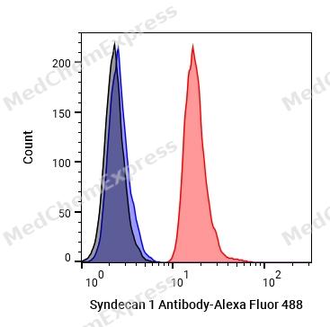 Flow cytometric analysis of 1X10^6 Hela cells labeling Syndecan 1 Antibody (HY-P81745, red). Cells were stained with the primary antibody at 1/100 dilution for an hour at 4℃. Alexa Fluor® 488-conjugated AffiniPure Goat Anti-Rabbit IgG H&L (HY-P8002) was used as the secondary antibody at 1/1,000 dilution for 30 minutes at 4℃. Rabbit IgG Isotype Control (HY-P80879, blue) was used as the isotype control, cells without incubation with primary antibody were used as the unlabeled control (black).
 -
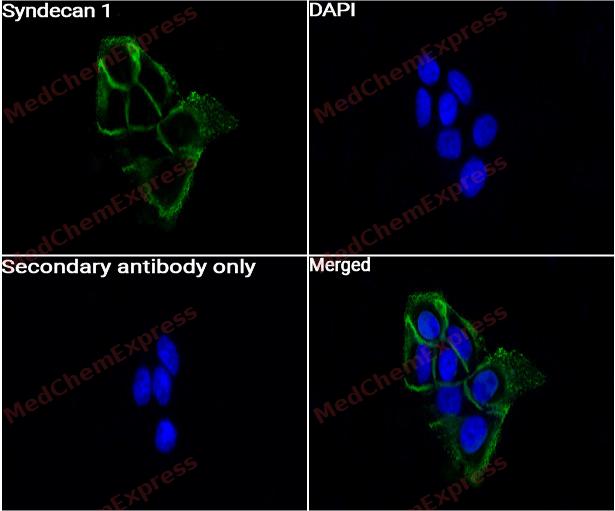 Immunocytochemistry analysis of A431 cells labeling Syndecan 1 with Syndecan 1 Antibody (HY-P81745) at 1/50 dilution. Cells were fixed in 4% paraformaldehyde for 15 minutes at room temperature, permeabilized with 0.1% Triton X-100 for 10 minutes at room temperature, then blocked with BSA for Immunol Staining for 10 min at room temperature. Cells were then incubated with Syndecan 1 Antibody (HY-P81745) at 1/50 dilution in BSA for Immunol Staining at 4 ℃ overnight. AF488-conjugated AffiniPure Goat Anti-Rabbit IgG H&L(HY-P8002, Green) was used as the secondary antibody at 1/1,000 dilution. PBS instead of the primary antibody was used as the secondary antibody only control. The Nuclear counterstain was DAPI (Blue).
 -
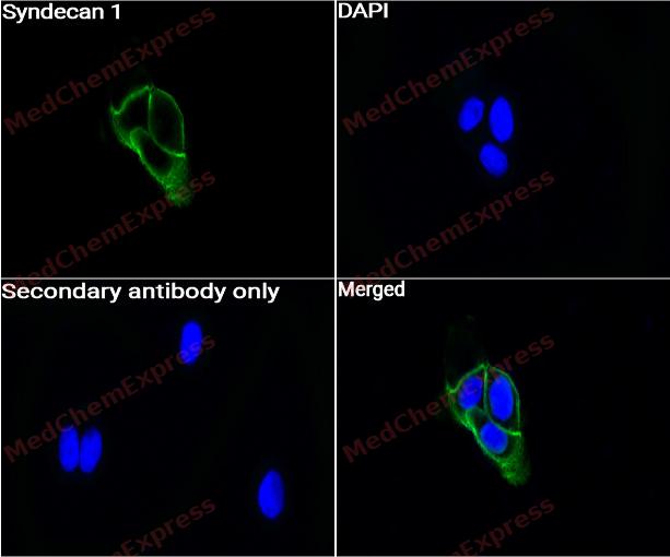 Immunocytochemistry analysis of A431 cells labeling Syndecan 1 with Syndecan 1 Antibody (HY-P81745) at 1/100 dilution. Cells were fixed in 4% paraformaldehyde for 15 minutes at room temperature, permeabilized with 0.1% Triton X-100 for 10 minutes at room temperature, then blocked with BSA for Immunol Staining for 10 min at room temperature. Cells were then incubated with Syndecan 1 Antibody (HY-P81745) at 1/100 dilution in BSA for Immunol Staining at 4 ℃ overnight. AF488-conjugated AffiniPure Goat Anti-Rabbit IgG H&L(HY-P8002, Green) was used as the secondary antibody at 1/1,000 dilution. PBS instead of the primary antibody was used as the secondary antibody only control. The Nuclear counterstain was DAPI (Blue).


Background
-
Function
Syndecan 1 cell surface proteoglycan that contains both heparan sulfate and chondroitin sulfate and that links the cytoskeleton to the interstitial matrix. Regulates exosome biogenesis in concert with SDCBP and PDCD6IP. Able to induce its own expression in dental mesenchymal cells and also in the neighboring dental epithelial cells via an MSX1-mediated pathway[1].
-
Subcellular Localization
Membrane; Single-pass type I membrane protein; Secreted; Secreted, extracellular exosome
-
Expression
Tissue_specificity:Protein levels were detected in the placenta (PubMed: 32337544) . Protein levels were detected in fibroblasts (PubMed: 36213313) .
Positive sample: HeLa cell lysate, A431 cell lysate, Daudi cell lysate, Raji cell lysate, Ramos cell lysate, A431, Hela, HepG2, human tonsil tissue, human kidney tissue, -
Subunit
Interacts with CDCP1. Interacts (via C-terminus) with TIAM1 (via PDZ domain). Interacts with MDK (By similarity)
-
SwissProt ID
-
Synonyms
SDC1; SDC; Syndecan-1; SYND1; CD138
-
Research Field
Cell Biology
Documentation
-
Data Sheet (234 KB)
-
SDS (251 KB)
- English - EN (251 KB)
- Français - FR (251 KB)
- Deutsch - DE (251 KB)
- Norwegian - NO (251 KB)
- Español - ES (251 KB)
- Swedish - SV (251 KB)
- Italian - IT (251 KB)
- Korean - KR (251 KB)
- Portuguese - PT (251 KB)
-
User Guide for Antibodies (1077 KB)

 Powered by Bioz
