



CXCR3 Antibody (YA4052)
(Synonyms: CXCR3; GPR9; MigR; CD182; Mig-R; CKR-L2; CMKAR3; IP10-R)
Based on 1 Customer Validation
CXCR3 Antibody (YA4052) is a Mouse-derived and non-conjugated IgG1 monoclonal antibody, targeting to CXCR3.
-
Host:
Mouse
-
Isotype:
IgG
-
Application:
WB, IHC-P, FC, ELISA
-
Reactivity :
Human
-
Formulation:
Supplied in PBS with 0.05% sodium azide
-
Conjugation:
Non-conjugated


To place orders, for customer services and technical support, please contact: MedChemExpress USA
Tel: 609-228-6898 E-mail: [email protected] [email protected]





Applications
| Application |
WB

WB: Western Blot
|
IHC-P
IHC-P: Immunohistochemistry-Paraffin
|
FC
FC: Flow Cytometry
|
ELISA
ELISA: Enzyme Linked Immunosorbent Assay
|
|---|---|---|---|---|
| Dilution Ratio | 1:500-1:2000 | 1:200-1:1000 | 1:200-1:400 | 1:10000 |


Product Details
CXCR3 Antibody (YA4052) is a Mouse-derived and non-conjugated IgG1 monoclonal antibody, targeting to CXCR3.
-
Host Mouse

-
Clonality Monoclonal

-
Species ReactivityHuman
-
Observed Molecular WeightObserved band size: 50 kDa
Note: Due to possible protein modifications or aggregation, the molecular weight should be confirmed by actual measurement, and the predicted value is for reference only.

-
Calculated Molecular Weight Predicted band size: 40.7 kDa

Purified recombinant fragment of human CD183 (AA: extra mix) expressed in E. Coli.
affinity purified.
Non-conjugated
Unmodified
IgG


Product Properties
-
Appearance
Solution
-
Formulation
Supplied in PBS with 0.05% sodium azide
-
Concentration
Batch-dependent, Please check the COA for the concentration of each lot. Check Lot Concentration
-
Storage & Stability
Stored at -20°C for 1 year. Avoid repeated freeze / thaw cycles.
-
Shipping
Shipping with blue ice.


Verification Images
-
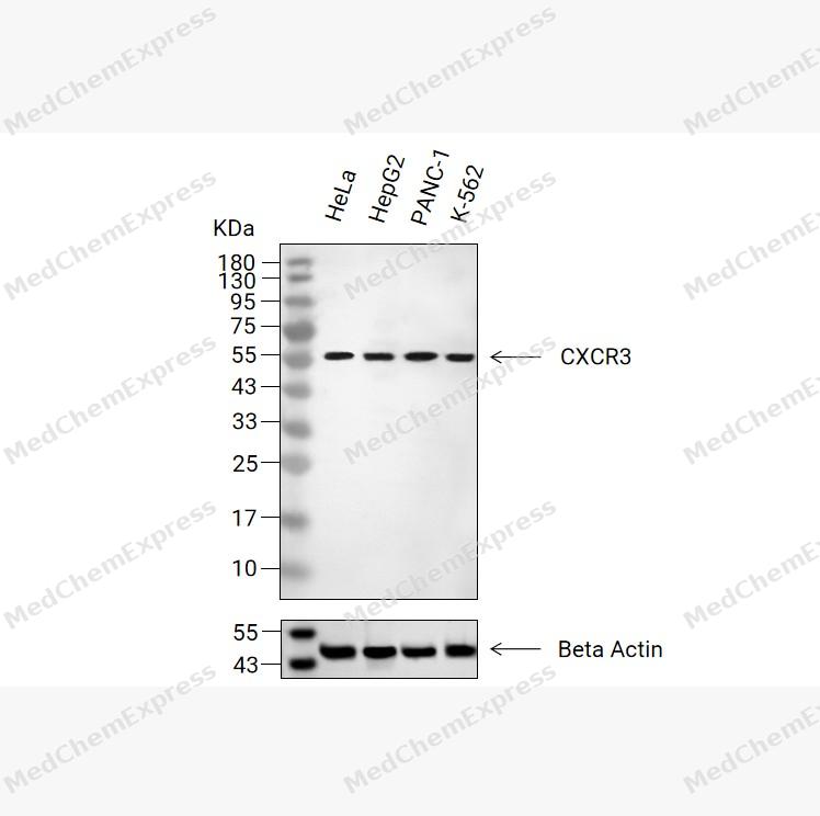 Western blot analysis of extracts from HeLa(lane 2(20μg), HepG2(lane 3(20μg), PANC-1(lane 4(20μg), K-562(lane 5(20μg) using CXCR3 Antibody (HY-P84355). Proteins were transferred to a PVDF membrane and blocked with 5% non-fat milk in TBST for 2 hours at room temperature. The primary antibody (1/1000) and Loading control antibody (Beta Actin, HY-P80993, 1/10,000) was used in 5% non-fat milk in TBST at 4°C overnight. Goat Anti-Mouse IgG-HRP Secondary Antibody (HY-P8004, 1/10,000) was used for 1 hour at room temperature.
 -
 Immunohistochemical analysis of paraffin-embedded human tonsil tissue using CXCR3 Antibody (HY-P84355, 1/500). The section was pretreated using heat mediated antigen retrieval with sodium citrate buffer (pH 6.0) for 8 minutes. The tissues were blocked with quick block buffer for 0.5 hours at room temperature, washed with PBS and PBST, and then incubated with the primary antibody overnight at 4℃. The detection was performed using an HRP conjugated compact polymer system. DAB was used as the chromogen. Tissues were counterstained with hematoxylin and mounted with DPX.
 -
 Immunohistochemical analysis of paraffin-embedded human lymph node tissue using CXCR3 Antibody (HY-P84355, 1/500). The section was pretreated using heat mediated antigen retrieval with sodium citrate buffer (pH 6.0) for 8 minutes. The tissues were blocked with quick block buffer for 0.5 hours at room temperature, washed with PBS and PBST, and then incubated with the primary antibody overnight at 4℃. The detection was performed using an HRP conjugated compact polymer system. DAB was used as the chromogen. Tissues were counterstained with hematoxylin and mounted with DPX.
 -
 Immunohistochemical analysis of paraffin-embedded human liver cancer tissue using CXCR3 Antibody (HY-P84355, 1/500). The section was pretreated using heat mediated antigen retrieval with sodium citrate buffer (pH 6.0) for 8 minutes. The tissues were blocked with quick block buffer for 0.5 hours at room temperature, washed with PBS and PBST, and then incubated with the primary antibody overnight at 4℃. The detection was performed using an HRP conjugated compact polymer system. DAB was used as the chromogen. Tissues were counterstained with hematoxylin and mounted with DPX.
 -
 Immunohistochemical analysis of paraffin-embedded human colon tissue using CXCR3 Antibody (HY-P84355, 1/500). The section was pretreated using heat mediated antigen retrieval with sodium citrate buffer (pH 6.0) for 8 minutes. The tissues were blocked with quick block buffer for 0.5 hours at room temperature, washed with PBS and PBST, and then incubated with the primary antibody overnight at 4℃. The detection was performed using an HRP conjugated compact polymer system. DAB was used as the chromogen. Tissues were counterstained with hematoxylin and mounted with DPX.
 -
 Immunohistochemical analysis of paraffin-embedded human spleen tissue using CXCR3 Antibody (HY-P84355, 1/500). The section was pretreated using heat mediated antigen retrieval with sodium citrate buffer (pH 6.0) for 8 minutes. The tissues were blocked with quick block buffer for 0.5 hours at room temperature, washed with PBS and PBST, and then incubated with the primary antibody overnight at 4℃. The detection was performed using an HRP conjugated compact polymer system. DAB was used as the chromogen. Tissues were counterstained with hematoxylin and mounted with DPX.
 -
 Immunohistochemical analysis of paraffin-embedded human lung tissue using CXCR3 Antibody (HY-P84355, 1/500). The section was pretreated using heat mediated antigen retrieval with sodium citrate buffer (pH 6.0) for 8 minutes. The tissues were blocked with quick block buffer for 0.5 hours at room temperature, washed with PBS and PBST, and then incubated with the primary antibody overnight at 4℃. The detection was performed using an HRP conjugated compact polymer system. DAB was used as the chromogen. Tissues were counterstained with hematoxylin and mounted with DPX.


Background
-
Function
CD183 (CXCR3) is a CXC chemokine receptor primarily expressed on activated T cells, Th1 cells, NK cells, and NKT cells. By responding to CXCL9, CXCL10, and CXCL11 signals, CD183 mediates the migration of immune cells to sites of inflammation and tumors; it serves as a key marker for studying Th1 immunity, anti-tumor immunity, immunity against infection, and autoimmune diseases.
-
Subcellular Localization
Cell membrane; Multi-pass membrane protein; Cell membrane; Multi-pass membrane protein
-
Expression
Tissue_specificity:Isomer 1 and isomer 2 are primarily expressed in the heart, kidneys, liver, and skeletal muscle. Isomer 1 is also expressed in the placenta. Isomer 2 is expressed in endothelial cells. It is also expressed in T cells (protein level) . -
Isoforms & Post-Translational Modification
CD183 (CXCR3) has 3 isomers: P49682-1: 40660 Da (predicted); P49682-2: 45523 Da (predicted); P49682-3: 28715 Da (predicted).
Sulfation on Tyr-27 and Tyr-29 is essential for CXCL10 binding and subsequent signal transduction induction;N-glycosylated -
Subunit
Homomer. Forms heteromers with ACKR4
-
SwissProt ID
-
Synonyms
CXCR3; GPR9; MigR; CD182; Mig-R; CKR-L2; CMKAR3; IP10-R


Documentation
-
Data Sheet (263 KB)
-
SDS (252 KB)
- English - EN (252 KB)
- Français - FR (252 KB)
- Deutsch - DE (252 KB)
- Norwegian - NO (252 KB)
- Español - ES (252 KB)
- Swedish - SV (252 KB)
- Italian - IT (252 KB)
- Korean - KR (252 KB)
- Portuguese - PT (252 KB)
-
User Guide for Antibodies (1077 KB)
References
[1]. Huo R, et al. CXCR3 inhibitors for therapeutic interventions: current status and perspectives. Front Pharmacol. 2025 Jul 25;16:1556196. [Content Brief]
[2]. Boyé K, et al. Ligand activation induces different conformational changes in CXCR3 receptor isoforms as evidenced by plasmon waveguide resonance (PWR). Sci Rep. 2017 Sep 6;7(1):10703. [Content Brief]
[3]. Caccuri F, et al. The Synthetic Dipeptide Pidotimod Shows a Chemokine-Like Activity through CXC Chemokine Receptor 3 (CXCR3). Int J Mol Sci. 2019 Oct 24;20(21):5287. [Content Brief]
[4]. Eiger DS, et al. Location bias contributes to functionally selective responses of biased CXCR3 agonists. Nat Commun. 2022 Oct 4;13(1):5846. [Content Brief]
[5]. Wang X, et al. Effects of CXCL4/CXCR3 on the lipopolysaccharide-induced injury in human umbilical vein endothelial cells. J Cell Physiol. 2019 Dec;234(12):22378-22385. [Content Brief]
[6]. Torraca V, et al. The CXCR3-CXCL11 signaling axis mediates macrophage recruitment and dissemination of mycobacterial infection. Dis Model Mech. 2015 Mar;8(3):253-69. [Content Brief]
[7]. Wu Q, et al. Altered CXCR3 isoform expression regulates prostate cancer cell migration and invasion. Mol Cancer. 2012 Jan 11;11:3. [Content Brief]
[8]. Sheng W, et al. Astrocyte-derived CXCL10 exacerbates endothelial cells pyroptosis and blood-brain barrier disruption via CXCR3/cGAS/AIM2 pathway after intracerebral hemorrhage. Cell Death Discov. 2025 Aug 8;11(1):373. [Content Brief]
[9]. Lee J, et al. CXCR3-CXCL11 Signaling Restricts Angiogenesis and Promotes Pericyte Recruitment. Arterioscler Thromb Vasc Biol. 2024 Dec;44(12):2577-2595. [Content Brief]
[10]. Huang YV, et al. Novel Therapeutic Approach Targeting CXCR3 to Treat Immunotherapy Myocarditis. Circ Res. 2025 Feb 28;136(5):473-490. [Content Brief]
[11]. Bodnar RJ, et al. Differential regulation of pericyte function by the CXC receptor 3. Wound Repair Regen. 2015 Nov-Dec;23(6):785-96. [Content Brief]

 Powered by Bioz
