



CEACAM1 Antibody (YA3788)
(Synonyms: CEACAM1; BGP; BGP1; BGPI; CD66a)
Based on 1 Customer Validation
CEACAM1 Antibody (YA3788) is a Mouse-derived and non-conjugated IgG2b monoclonal antibody, targeting to CEACAM1.
-
Host:
Mouse
-
Isotype:
IgG
-
Application:
WB, IHC-P, FC, ELISA
-
Reactivity :
Human
-
Formulation:
Supplied in PBS with 0.05% sodium azide
-
Conjugation:
Non-conjugated


To place orders, for customer services and technical support, please contact: MedChemExpress USA
Tel: 609-228-6898 E-mail: [email protected] [email protected]





Applications
| Application |
WB

WB: Western Blot
|
IHC-P
IHC-P: Immunohistochemistry-Paraffin
|
FC
FC: Flow Cytometry
|
ELISA
ELISA: Enzyme Linked Immunosorbent Assay
|
|---|---|---|---|---|
| Dilution Ratio | 1:500-1:2000 | 1:200-1:1000 | 1:200-1:400 | 1:10000 |


Product Details
CEACAM1 Antibody (YA3788) is a Mouse-derived and non-conjugated IgG2b monoclonal antibody, targeting to CEACAM1.
-
Host Mouse

-
Clonality Monoclonal

-
Species ReactivityHuman
-
Observed Molecular WeightObserved band size: 120-150 kDa
Note: Due to possible protein modifications or aggregation, the molecular weight should be confirmed by actual measurement, and the predicted value is for reference only.

-
Calculated Molecular Weight Predicted band size: 58 kDa

Purified recombinant fragment of human CD66A (AA: extra 65-201) expressed in E. Coli.
affinity purified.
Non-conjugated
Unmodified
IgG


Product Properties
-
Appearance
Solution
-
Formulation
Supplied in PBS with 0.05% sodium azide
-
Concentration
Batch-dependent, Please check the COA for the concentration of each lot. Check Lot Concentration
-
Storage & Stability
Stored at -20°C for 1 year. Avoid repeated freeze / thaw cycles.
-
Shipping
Shipping with blue ice.


Verification Images
-
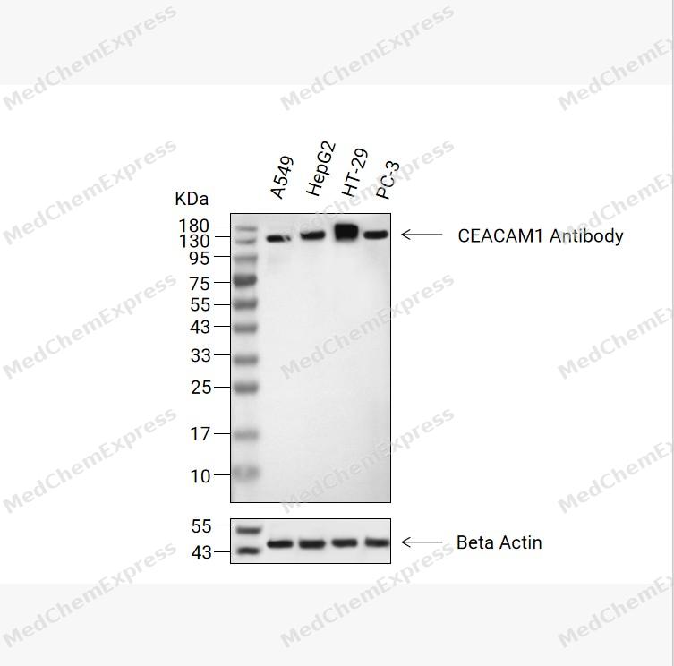 Western blot analysis of extracts from A549(lane 2(20μg), HepG2(lane 3(20μg), HT-29(lane 4(20μg), PC-3(lane 5(20μg) using CEACAM1 Antibody (HY-P84091). Proteins were transferred to a PVDF membrane and blocked with 5% non-fat milk in TBST for 2 hours at room temperature. The primary antibody (1/1000) and Loading control antibody (Beta Actin, HY-P80993, 1/10,000) was used in 5% non-fat milk in TBST at 4°C overnight. Goat Anti-Mouse IgG-HRP Secondary Antibody (HY-P8004, 1/10,000) was used for 1 hour at room temperature.
 -
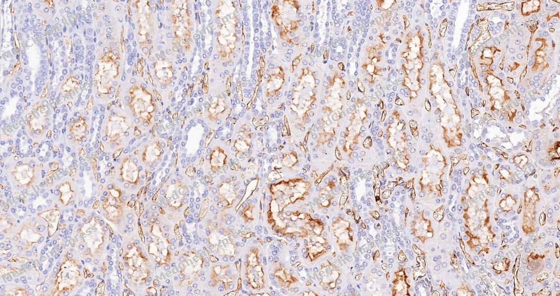 Immunohistochemical analysis of paraffin-embedded Human kidney tissue using CEACAM1 Antibody (HY-P84091, 1/500) . The section was pretreated using heat mediated antigen retrieval with sodium citrate buffer (PH 6.0)for 8 minutes. The tissues were blocked with quick block buffer for 0.5 hours at room temperature, washed with PBS and PBST, and then incubated with the primary antibody overnight at 4℃. The detection was performed using an HRP conjugated compact polymer system. DAB was used as the chromogen. Tissues were counterstained with hematoxylin and mounted with DPX.
 -
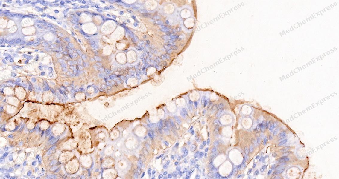 Immunohistochemical analysis of paraffin-embedded Human colon tissue using CEACAM1 Antibody (HY-P84091, 1/500) . The section was pretreated using heat mediated antigen retrieval with sodium citrate buffer (PH 6.0)for 8 minutes. The tissues were blocked with quick block buffer for 0.5 hours at room temperature, washed with PBS and PBST, and then incubated with the primary antibody overnight at 4℃. The detection was performed using an HRP conjugated compact polymer system. DAB was used as the chromogen. Tissues were counterstained with hematoxylin and mounted with DPX.
 -
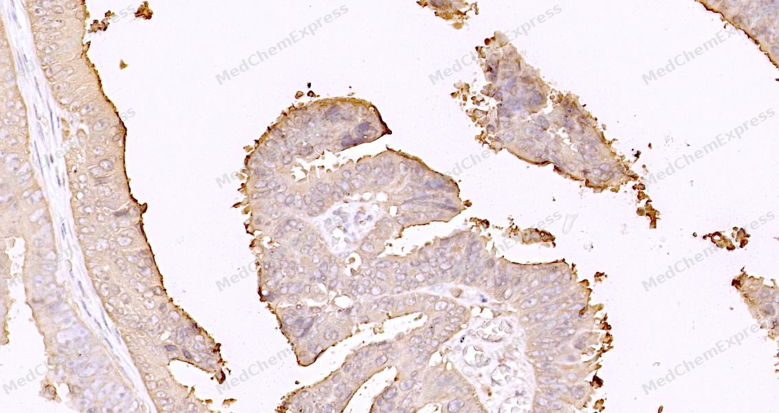 Immunohistochemical analysis of paraffin-embedded Human Colon Cancer tissue using CEACAM1 Antibody (HY-P84091, 1/500) . The section was pretreated using heat mediated antigen retrieval with sodium citrate buffer (PH 6.0)for 8 minutes. The tissues were blocked with quick block buffer for 0.5 hours at room temperature, washed with PBS and PBST, and then incubated with the primary antibody overnight at 4℃. The detection was performed using an HRP conjugated compact polymer system. DAB was used as the chromogen. Tissues were counterstained with hematoxylin and mounted with DPX.
 -
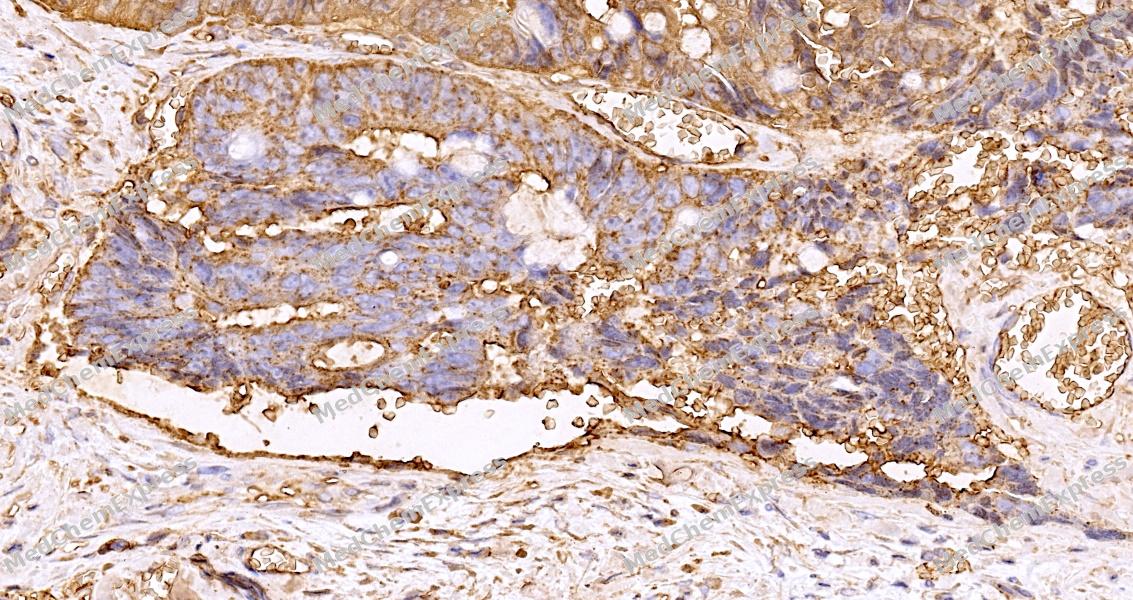 Immunohistochemical analysis of paraffin-embedded Human Rectal Cancer tissue using CEACAM1 Antibody (HY-P84091, 1/500) . The section was pretreated using heat mediated antigen retrieval with sodium citrate buffer (PH 6.0)for 8 minutes. The tissues were blocked with quick block buffer for 0.5 hours at room temperature, washed with PBS and PBST, and then incubated with the primary antibody overnight at 4℃. The detection was performed using an HRP conjugated compact polymer system. DAB was used as the chromogen. Tissues were counterstained with hematoxylin and mounted with DPX.
 -
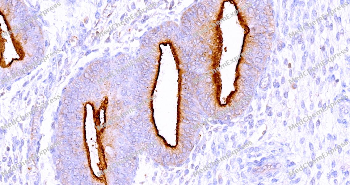 Immunohistochemical analysis of paraffin-embedded human endometrium tissue using CEACAM1 Antibody (HY-P84091, 1/500) . The section was pretreated using heat mediated antigen retrieval with sodium citrate buffer (PH 6.0)for 8 minutes. The tissues were blocked with quick block buffer for 0.5 hours at room temperature, washed with PBS and PBST, and then incubated with the primary antibody overnight at 4℃. The detection was performed using an HRP conjugated compact polymer system. DAB was used as the chromogen. Tissues were counterstained with hematoxylin and mounted with DPX.
 -
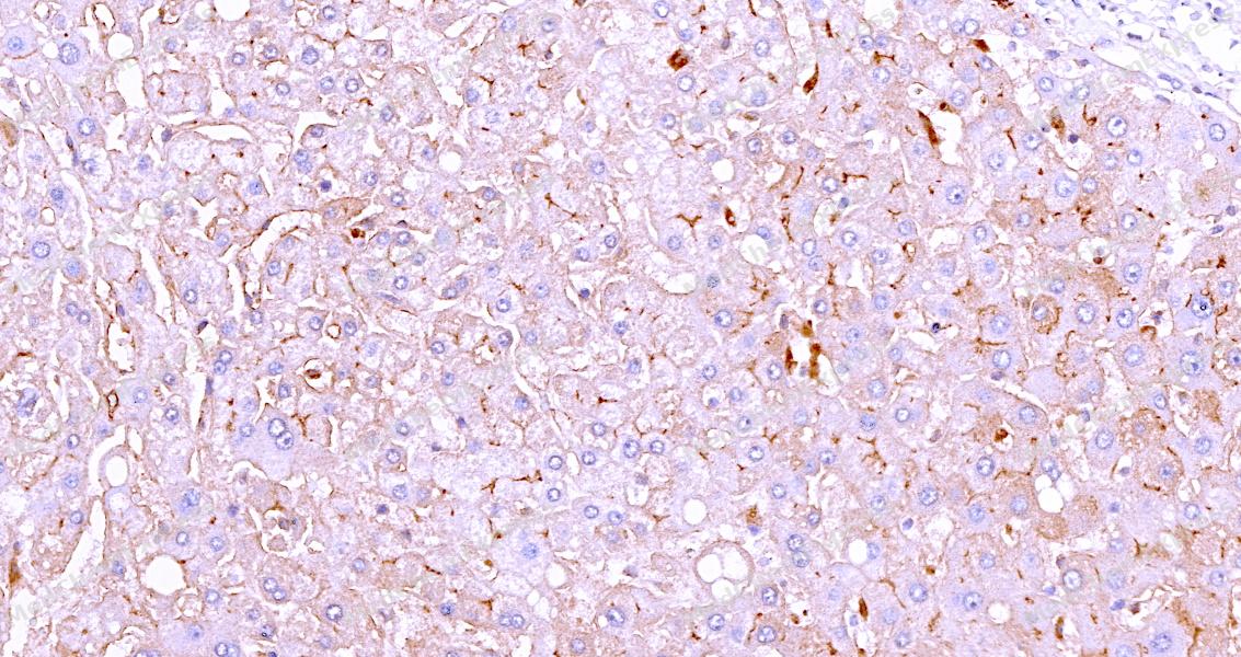 Immunohistochemical analysis of paraffin-embedded human liver tissue using CEACAM1 Antibody (HY-P84091, 1/500) . The section was pretreated using heat mediated antigen retrieval with sodium citrate buffer (PH 6.0)for 8 minutes. The tissues were blocked with quick block buffer for 0.5 hours at room temperature, washed with PBS and PBST, and then incubated with the primary antibody overnight at 4℃. The detection was performed using an HRP conjugated compact polymer system. DAB was used as the chromogen. Tissues were counterstained with hematoxylin and mounted with DPX.


Background
-
Function
Carcinoembryonic antigen-related cell adhesion molecule 1 (CEACAM1), also known as CD66a, BGP, or C-CAM, is an extensively studied, multifunctional transmembrane protein which is the primordial member of the carcinoembryonic antigen cell adhesion molecul
-
Subcellular Localization
Cell membrane; Single-pass type I membrane protein; Lateral cell membrane; Apical cell membrane; Basal cell membrane; Cell junction; Cell junction, adherens junction; Secreted; Secreted; Secreted; Cell membrane; Single-pass type I membrane protein; Cell membrane; Single-pass type I membrane protein; Cell membrane; Single-pass type I membrane protein; Cell membrane; Single-pass type I membrane protein; Cytoplasmic vesicle, secretory vesicle membrane; Lateral cell membrane; Apical cell membrane; Basal cell membrane; Cell junction; Cell junction, adherens junction; Cell projection, microvillus membrane; Single-pass type I membrane protein; Apical cell membrane; Single-pass type I membrane protein
-
Expression
Tissue_specificity:It is expressed in colonic columnar epithelial cells (protein level) (PubMed: 10436421) . The predominant form expressed on T cells is the one containing a long cytoplasmic domain (PubMed: 18424730) . It is also expressed in granulocytes and lymphocytes. Leukocytes express only isoforms 6 and 1 (PubMed: 11994468) .
Induction:Induced in primary T cells by activation with IL-2 -
Isoforms & Post-Translational Modification
P13688 has 11 isomers: P13688-1: 57560 Da (predicted); P13688-2: 45950 Da (predicted); P13688-3: 35302 Da (predicted); P13688-4: 38578 Da (predicted); P13688-5: 50349 Da (predicted); P13688-6: 46910 Da (predicted); P13688-7: 27431 Da (predicted); P13688-8: 50521 Da (predicted); P13688-9: 43062 Da (predicted); P13688-10: 51147 Da (predicted); P13688-11: 39871 Da (predicted).
Phosphorylated on serine and tyrosine (By similarity). Isoform 1 is phosphorylated on tyrosine by Src family kinases like SRC and LCK and by receptor like CSF3R, EGFR and INSR upon stimulation (PubMed:15467833, PubMed:18424730, PubMed:7478590). Phosphorylated at Ser-508; mediates activity. Phosphorylated at Tyr-493; regulates activity (By similarity). Phosphorylated at Tyr-493 by EGFR and INSR upon stimulation; this phosphorylation is Ser-508-phosphorylation-dependent; mediates cellular internalization; increases interaction with downstream proteins like SHC1 and FASN (By similarity). Phosphorylated at Tyr-493 and Tyr-520 by LCK; mediates PTPN6 association and is regulated by homophilic ligation of CEACAM1 in the absence of T cell activation (PubMed:18424730). Phosphorylated at Tyr-520; mediates interaction with PTPN11 (By similarity);Phosphorylated on serine and threonine -
Subunit
Monomer. Oligomer. Heterodimer. Homodimer (PubMed:26483485). Cis-dimer/oligomer (via Ig-like C2-type and/or via cytoplasmic domains); induced by trans-homophilic cell adhesion through an allosteric mechanism transmitted by the Ig-like V-type domain, and is regulated by intracellular calcium and calmodulin. Interacts (via cytoplasmic domain) with calmodulin in a calcium dependent manner; reduces homophilic cell adhesion through dissociation of dimer (By similarity). Isoform 1 interacts (via cytoplasmic domain) with PTPN11 (preferentially) and PTPN6; cis-homodimer form is preferred; this interaction is decreased by formation of Isoform 1 /Isoform 8 cis-heterodimers and is dependent on the monomer/dimer equilibrium; this interaction is phosphorylation-dependent (PubMed:23696226). Isoform 1 interacts with LYN (By similarity). Isoform 1 interacts (via cytoplasmic domain) with SRC (via SH2 domain); this interaction is regulated by trans-homophilic cell adhesion (PubMed:7478590). Isoform 1 interacts (via cytoplasmic domain) with LCK; mediates phosphorylation at Tyr-493 and Tyr-520 resulting in PTPN6 association. Isoform 1 interacts with PTPN6; this interaction is phosphorylation-dependent and causes a profound decrease in TCR stimulation-induced CD247 and ZAP70 phosphorylation. Isoform 1 interacts with TCR/CD3 complex through TCR beta chain and CD3E; colocalizes at the cell surface and upon stimulation of the TCR/CD3 complex recruits PTPN6 in the TCR/CD3 complex, resulting in dephosphorylation of CD247 and ZAP70 (PubMed:18424730). Isoform 1 interacts (via cytoplasmic domain) with SHC1 (via SH2 domain); SHC1 mediates interaction with INSR or EGFR in a Ser-508 phosphorylation-dependent manner (By similarity). Isoform 1 interacts with EGFR; the interaction is indirect (PubMed:15467833). Isoform 1 interacts with CSF3R; down-regulates the CSF3R-STAT3 pathway through recruitment of PTPN6 that dephosphorylates CSF3R (By similarity). Isoform 1 (phosphorylated form) interacts with TLR4 and SYK; recruits PTPN6 that dephosphorylates SYK, reducing the production of reactive oxygen species (ROS) and lysosome disruption, leading to a reduction of the inflammasome activity (By similarity). Isoform 1 interacts with FLNA; inhibits cell migration and cell scattering by interfering with the interaction of FLNA with RALA (PubMed:16291724). Isoform 1 interacts (via cytoplasmic domain) with PXN; the interaction is phosphotyrosyl-dependent (PubMed:11035932). Isoform 1 interacts with KLRK1; recruits PTPN6 that dephosphorylates VAV1 (PubMed:23696226). Isoform 1 interacts with CEACAM8 (PubMed:11994468). Isoform 1 interacts with FASN; this interaction is insulin and phosphorylation-dependent; reduces fatty-acid synthase activity (By similarity). Interacts (via Ig-like V-type) with HAVCR2 (via Ig-like V-type); facilitates the maturation and cell surface expression of HAVCR2 thereby regulating T cell tolerance induction (PubMed:25363763). Isoform 8 interacts (via the cytoplasmic domain) with ANXA2; this interaction is regulated by phosphorylation and appears in the AIIt complex (PubMed:14522961). Interacts (via Lewis X moieties) with CD209 (via C-type lectin domain); this interaction is regulated by the glycosylation pattern of CEACAM1 on cell types and regulates contact between dendritic cells and neutrophils (PubMed:16246332)
-
SwissProt ID
-
Synonyms
CEACAM1; BGP; BGP1; BGPI; CD66a


Documentation
-
Data Sheet (234 KB)
-
SDS (251 KB)
- English - EN (251 KB)
- Français - FR (251 KB)
- Deutsch - DE (251 KB)
- Norwegian - NO (251 KB)
- Español - ES (251 KB)
- Swedish - SV (251 KB)
- Italian - IT (251 KB)
- Korean - KR (251 KB)
- Portuguese - PT (251 KB)
-
User Guide for Antibodies (1077 KB)

 Powered by Bioz
