



CDK1 Antibody (YA516)
(Synonyms: CDK1; CDC2; CDC28A; CDKN1; P34CDC2; Cyclin-dependent kinase 1; CDK1; Cell division control protein 2 homolog; Cell division protein kinase 1; p34 protein kinase)Based on 3 publication(s) in Google Scholar
CDK1 Antibody (YA516) is a Rabbit-derived and non-conjugated IgG monoclonal antibody, targeting to CDK1.
-
Host:
Rabbit
-
Isotype:
IgG
-
Application:
WB, IP
-
Reactivity :
Human
-
Formulation:
Supplied in 50 mM Tris-Glycine (pH 7.4), 0.15 M NaCl, 40% Glycerol and 0.05% BSA. Preservative: 0.01% Sodium azide
-
Conjugation:
Non-conjugated


To place orders, for customer services and technical support, please contact: MedChemExpress USA
Tel: 609-228-6898 E-mail: [email protected] [email protected]



Publications Citing Use of MedChemExpress (MCE) CDK1 Antibody (YA516)
More

Applications
| Application |
WB

WB: Western Blot
|
IP
IP: Immunoprecipitation
|
|---|---|---|
| Dilution Ratio | 1:500-1:1000 | 1:20 |


Product Details
CDK1 Antibody (YA516) is a Rabbit-derived and non-conjugated IgG monoclonal antibody, targeting to CDK1.
-
Host Rabbit

-
Clonality Recombinant,Monoclonal

-
Species ReactivityHuman
-
Observed Molecular WeightObserved band size: 34 kDa
Note: Due to possible protein modifications or aggregation, the molecular weight should be confirmed by actual measurement, and the predicted value is for reference only.

-
Calculated Molecular Weight Predicted band size: 34 kDa

Synthetic peptide corresponding to Human CDK1.The exact sequence is proprietary to MCE.
Endogenous
affinity purified
Non-conjugated
Unmodified
IgG


Product Properties
-
Appearance
Solution
-
Formulation
Supplied in 50 mM Tris-Glycine (pH 7.4), 0.15 M NaCl, 40% Glycerol and 0.05% BSA. Preservative: 0.01% Sodium azide
-
Concentration
Batch-dependent, Please check the COA for the concentration of each lot. Check Lot Concentration
-
Storage & Stability
Stored at -20°C for 1 year. Avoid repeated freeze / thaw cycles.
-
Shipping
Shipping with blue ice.


Publications (3)
-
Journal Impact Factor
-
Most Recent
-
J Inflamm Res
Sodium Propionate Alleviates Atopic Dermatitis by Inhibiting Ferroptosis via Activation of LTBP2/FABP4 Signaling Pathway. [Abstract]2024 Nov 29:17:10047-10064. PMID: 39634285 -
Histol Histopathol
Targeting of CDK1 and PRMT5 as a potential therapeutic combination for non-small cell lung cancer. [Abstract]2026 Jul;41(7):1275-1288. PMID: 41487038


Verification Images
-
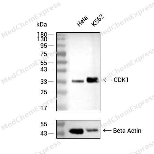 Western blot analysis of extracts from Hela (lane 2(20μg) and K562(lane 3(20μg) using CDK1 (HY-P80611) Rabbit mAb. Proteins were transferred to a PVDF membrane and blocked with 5% non-fat milk in TBST for 2 hour at room temperature. The primary antibody (1/1000) and Loading control antibody (Beta Actin, HY-P80438, 1/10000) was used in 5% non-fat milk in TBST at 4°C overnight. Goat Anti-Mouse/Rabbit IgG-HRP Secondary Antibody (1/10000) was used for 1 hour at room temperature.


Background
-
Function
CDK1 (cyclin-dependent kinase 1) is a highly conserved serine/threonine kinase that functions as a central regulator of eukaryotic cell-cycle progression through cyclin-dependent substrate phosphorylation, particularly during G2/M transition and mitosis[1][2]. Mechanistically, CDK1 forms active complexes with cyclins and coordinates centrosome dynamics, chromosome segregation, spindle organization, and mitotic progression through phosphorylation-dependent signaling networks[2][3]. CDK1 also regulates translational control during mitosis by directly phosphorylating key components of the protein synthesis machinery, including 4E-BP1, thereby linking cell-cycle progression with cellular growth programs[4][5]. In disease models, aberrant CDK1 activity contributes to tumor-cell proliferation, and elevated dependence on CDK1-mediated mitotic signaling has supported its investigation as a therapeutic target across multiple malignancies[6][7]. Compared with related cyclin-dependent kinase isoforms, CDK1 is distinguished by its indispensable role in driving mitotic entry and execution, making it a critical node for cell-cycle control and experimental interrogation of mitotic mechanisms[1][2]. For experimental applications, the ATP-competitive inhibitor RO-3306 is widely used to achieve reversible CDK1 suppression, inducing G2/M arrest and enabling mechanistic studies of mitotic regulation, DNA-damage responses, apoptosis, and cancer-cell vulnerability[6][8]. Furthermore, pharmacological inhibition of CDK1 blocks phosphorylation-dependent oncogenic functions of downstream substrates, including SET isoform 1, which regulates mitotic fidelity, cell migration, invasion, and tumor growth in cellular and xenograft models[3][6].
-
Subcellular Localization
Nucleus; Cytoplasm; Mitochondrion; Cytoplasm, cytoskeleton, microtubule organizing center, centrosome; Cytoplasm, cytoskeleton, spindle
-
Expression
Tissue_specificity:Found in breast cancer tissue
Induction:Follows a cyclic expression; during interphase, accumulates gradually following G1, S to reach a critical threshold at the end of G2, which promotes self-activation and triggers onset of mitosis. Induced transiently by TGFB1 at an early phase of TGFB1-mediated apoptosis, but later repressed. Triggered by CKS1B during mitotic entry in breast cancer cells. Down-regulated under genotoxic stresses triggered by PKR/EIF2AK2-mediated phosphorylation -
Isoforms & Post-Translational Modification
P06493 has 2 isomers: P06493-1: 34095 Da (predicted); P06493-2: 27503 Da (predicted).
Phosphorylation at Thr-161 by CAK/CDK7 activates kinase activity (PubMed:20360007). Phosphorylation at Thr-14 and Tyr-15 by PKMYT1 prevents nuclear translocation (PubMed:7569953). Phosphorylation at Tyr-15 by WEE1 and WEE2 inhibits the protein kinase activity and acts as a negative regulator of entry into mitosis (G2 to M transition) (PubMed:20360007). Phosphorylation by PKMYT1 and WEE1 takes place during mitosis to keep CDK1-cyclin-B complexes inactive until the end of G2 (PubMed:20360007, PubMed:7569953). By the end of G2, PKMYT1 and WEE1 are inactivated, but CDC25A and CDC25B are activated (PubMed:20360007). Dephosphorylation by active CDC25A and CDC25B at Thr-14 and Tyr-15, leads to CDK1 activation at the G2-M transition (PubMed:20360007). Phosphorylation at Tyr-15 by WEE2 during oogenesis is required to maintain meiotic arrest in oocytes during the germinal vesicle (GV) stage, a long period of quiescence at dictyate prophase I, leading to prevent meiotic reentry (PubMed:29606300). Phosphorylation by WEE2 is also required for metaphase II exit during egg activation to ensure exit from meiosis in oocytes and promote pronuclear formation (PubMed:29606300). Phosphorylated at Tyr-4 by PKR/EIF2AK2 upon genotoxic stress (PubMed:20395957). This phosphorylation triggers CDK1 polyubiquitination and subsequent proteolysis, thus leading to G2 arrest (PubMed:20395957). In response to UV irradiation, phosphorylation at Tyr-15 by PRKCD activates the G2/M DNA damage checkpoint (PubMed:19917613);Polyubiquitinated upon genotoxic stress -
Subunit
Forms a stable but non-covalent complex with a regulatory subunit and with a cyclin (PubMed:20360007). The cyclin subunit imparts substrate specificity to the complex (PubMed:20360007). Interacts with cyclins-B (CCNB1, CCNB2 and CCNB3) to form a serine/threonine kinase holoenzyme complex also known as maturation promoting factor (MPF) (PubMed:20360007, PubMed:2188730, PubMed:2344612). Promotes G2-M transition when in complex with a cyclin-B (PubMed:20360007). Can also form CDK1-cylin-D and CDK1-cyclin-E complexes that phosphorylate RB1 in vitro (PubMed:17459720). Associates with cyclins-A and B1 during S-phase in regenerating hepatocytes (PubMed:20360007). Interacts with DLGAP5 (PubMed:15145941). Binds to the CDK inhibitors CDKN1A/p21 and CDKN1B/p27. Interacts with catalytically active CCNB1 and RALBP1 during mitosis to form an endocytotic complex during interphase (PubMed:12775724). Interacts with FANCC (PubMed:9242535). Interacts with CEP63; this interaction recruits CDK1 to centrosomes (PubMed:21406398). Interacts with CENPA (PubMed:25556658). Interacts with NR1D1 (PubMed:27238018). Interacts with proteasome subunit PSMA8; to participate in meiosis progression during spermatogenesis (By similarity)
-
SwissProt ID
-
Synonyms
CDK1; CDC2; CDC28A; CDKN1; P34CDC2; Cyclin-dependent kinase 1; CDK1; Cell division control protein 2 homolog; Cell division protein kinase 1; p34 protein kinase
-
Research Field
Cell Biology
Documentation
-
Data Sheet (262 KB)
-
SDS (252 KB)
- English - EN (252 KB)
- Français - FR (252 KB)
- Deutsch - DE (252 KB)
- Norwegian - NO (252 KB)
- Español - ES (252 KB)
- Swedish - SV (252 KB)
- Italian - IT (252 KB)
- Korean - KR (252 KB)
- Portuguese - PT (252 KB)
-
User Guide for Antibodies (1077 KB)
References
[1]. Wikipedia.
[2]. Łukasik P, et al. Cyclin-Dependent Kinases (CDK) and Their Role in Diseases Development-Review. Int J Mol Sci. 2021 Mar 13;22(6):2935. [Content Brief]
[3]. Yin L, et al. Cyclin-dependent kinase 1-mediated phosphorylation of SET at serine 7 is essential for its oncogenic activity. Cell Death Dis. 2019 May 16;10(6):385. [Content Brief]
[4]. Jin Z, et al. Compartmentation of Metabolism of the C12-, C9-, and C5-n-dicarboxylates in Rat Liver, Investigated by Mass Isotopomer Analysis: ANAPLEROSIS FROM DODECANEDIOATE. J Biol Chem. 2015 Jul 24;290(30):18671-7. [Content Brief]
[5]. Velásquez C, et al. Mitotic protein kinase CDK1 phosphorylation of mRNA translation regulator 4E-BP1 Ser83 may contribute to cell transformation. Proc Natl Acad Sci U S A. 2016 Jul 26;113(30):8466-71. [Content Brief]
[6]. Kojima K, et al. Cyclin-dependent kinase 1 inhibitor RO-3306 enhances p53-mediated Bax activation and mitochondrial apoptosis in AML. Cancer Sci. 2009 Jun;100(6):1128-36. [Content Brief]
[7]. Shi Z, et al. From Structure Modification to Drug Launch: A Systematic Review of the Ongoing Development of Cyclin-Dependent Kinase Inhibitors for Multiple Cancer Therapy. J Med Chem. 2022 May 12;65(9):6390-6418. [Content Brief]

 Powered by Bioz
