



CDK4 Antibody (YA3764)
(Synonyms: CMM3; PSK-J3)
Based on 1 Customer Validation
CDK4 Antibody (YA3764) is a Mouse-derived and non-conjugated IgG2a monoclonal antibody, targeting to CDK4.
-
Host:
Mouse
-
Isotype:
IgG
-
Application:
WB, IHC-P, FC, ELISA
-
Reactivity :
Human, Mouse, Monkey
-
Formulation:
Supplied in PBS with 0.05% sodium azide
-
Conjugation:
Non-conjugated


To place orders, for customer services and technical support, please contact: MedChemExpress USA
Tel: 609-228-6898 E-mail: [email protected] [email protected]





Applications
| Application |
WB

WB: Western Blot
|
IHC-P
IHC-P: Immunohistochemistry-Paraffin
|
FC
FC: Flow Cytometry
|
ELISA
ELISA: Enzyme Linked Immunosorbent Assay
|
|---|---|---|---|---|
| Dilution Ratio | 1:500-1:2000 | 1:200-1:1000 | 1:200-1:400 | 1:10000 |


Product Details
CDK4 Antibody (YA3764) is a Mouse-derived and non-conjugated IgG2a monoclonal antibody, targeting to CDK4.
-
Host Mouse

-
Clonality Monoclonal

-
Species ReactivityHuman, Mouse, Monkey
-
Observed Molecular WeightObserved band size: 34 kDa
Note: Due to possible protein modifications or aggregation, the molecular weight should be confirmed by actual measurement, and the predicted value is for reference only.

-
Calculated Molecular Weight Predicted band size: 34 kDa

Purified recombinant fragment of human CDK4 (AA: 77-303) expressed in E. Coli.
affinity purified.
Non-conjugated
Unmodified
IgG


Product Properties
-
Appearance
Solution
-
Formulation
Supplied in PBS with 0.05% sodium azide
-
Concentration
Batch-dependent, Please check the COA for the concentration of each lot. Check Lot Concentration
-
Storage & Stability
Stored at -20°C for 1 year. Avoid repeated freeze / thaw cycles.
-
Shipping
Shipping with blue ice.


Verification Images
-
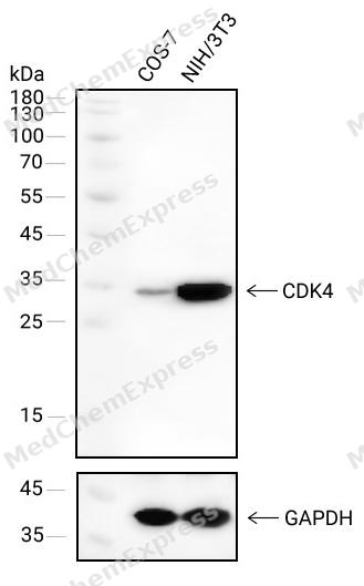 Western blot analysis of extracts from COS-7(lane 1(40μg) )and NIH/3T3(lane 2(40μg) ) using CDK4 antibody. Proteins were transferred to a NC membrane and blocked with 5% Skim milk in TBST for 2 hour at room temperature. The primary antibody ( 1/1000) and Loading control antibody (GAPDH, 1/1000) was used in 5% Skim milk in TBST at 4°C overnight. Goat Anti-Mouse IgG-HRP Secondary Antibody (1/10,000) was used for 1 hour at room temperature.
 -
 Western blot analysis was performed on protein extracts (25 μg) from A549 (lane 2),A431 (lane 3), Mouse heart (lane 4) and Raw264.7 (lane 5) using CDK4 antibody. Proteins were transferred onto a 0.45 μm PVDF membrane using the Trans-Blot® Turbo™ system for 13 min. The membrane was then blocked with 5% nonfat milk in TBST (HY-K1025) for 1 h at room temperature. The primary antibody (1:1000) and loading control antibody GAPDH Antibody (HRP) (HY-P80954A) (1:5000) were diluted in 5% nonfat milk in TBST and incubated with the membrane overnight at 4°C. After washing, the membrane of primary antibody was incubated with HRP-conjugated goat anti-rabbit/mouse IgG secondary antibody (HY-P8001/HY-P8004) (1:5000) diluted in 5% nonfat milk in TBST for 1 h at room temperature. Protein bands were visualized using an Ultra High Sensitivity ECL detection kit (HY-K1005).
 -
 Immunohistochemical analysis of paraffin-embedded human pancreatic cancer using CDK4 antibody. The section was pre-treated using heat mediated antigen retrieval with EDTA (pH 9.0) for 14 minutes. The tissues were blocked in 5% BSA for 30 minutes at room temperature, washed with TBST, and then probed with the primary antibody (HY-P84067, 1/500) for 1 hour at room temperature. The detection was performed using Polymer HRP-conjugated Goat Anti-Mouse/Rabbit lgG(H&L) secondary antibody (HY-P83652). DAB was used as the chromogen. Tissues were counterstained with hematoxylin and mounted with DPX.
 -
 Immunohistochemical analysis of paraffin-embedded human placenta using CDK4 antibody. The section was pre-treated using heat mediated antigen retrieval with EDTA (pH 9.0) for 14 minutes. The tissues were blocked in 5% BSA for 30 minutes at room temperature, washed with TBST, and then probed with the primary antibody (HY-P84067, 1/500) for 1 hour at room temperature. The detection was performed using Polymer HRP-conjugated Goat Anti-Mouse/Rabbit lgG(H&L) secondary antibody (HY-P83652). DAB was used as the chromogen. Tissues were counterstained with hematoxylin and mounted with DPX.
 -
 Immunohistochemical analysis of paraffin-embedded human non-small cell lung cancer using CDK4 antibody. The section was pre-treated using heat mediated antigen retrieval with EDTA (pH 9.0) for 14 minutes. The tissues were blocked in 5% BSA for 30 minutes at room temperature, washed with TBST, and then probed with the primary antibody (HY-P84067, 1/500) for 1 hour at room temperature. The detection was performed using Polymer HRP-conjugated Goat Anti-Mouse/Rabbit lgG(H&L) secondary antibody (HY-P83652). DAB was used as the chromogen. Tissues were counterstained with hematoxylin and mounted with DPX.
 -
 Immunohistochemical analysis of paraffin-embedded human melanoma using CDK4 antibody. The section was pre-treated using heat mediated antigen retrieval with EDTA (pH 9.0) for 14 minutes. The tissues were blocked in 5% BSA for 30 minutes at room temperature, washed with TBST, and then probed with the primary antibody (HY-P84067, 1/500) for 1 hour at room temperature. The detection was performed using Polymer HRP-conjugated Goat Anti-Mouse/Rabbit lgG(H&L) secondary antibody (HY-P83652). DAB was used as the chromogen. Tissues were counterstained with hematoxylin and mounted with DPX.
 -
 Immunohistochemical analysis of paraffin-embedded human lymph node using CDK4 antibody. The section was pre-treated using heat mediated antigen retrieval with EDTA (pH 9.0) for 14 minutes. The tissues were blocked in 5% BSA for 30 minutes at room temperature, washed with TBST, and then probed with the primary antibody (HY-P84067, 1/500) for 1 hour at room temperature. The detection was performed using Polymer HRP-conjugated Goat Anti-Mouse/Rabbit lgG(H&L) secondary antibody (HY-P83652). DAB was used as the chromogen. Tissues were counterstained with hematoxylin and mounted with DPX.
 -
 Immunohistochemical analysis of paraffin-embedded human breast cancer using CDK4 antibody. The section was pre-treated using heat mediated antigen retrieval with EDTA (pH 9.0) for 14 minutes. The tissues were blocked in 5% BSA for 30 minutes at room temperature, washed with TBST, and then probed with the primary antibody (HY-P84067, 1/500) for 1 hour at room temperature. The detection was performed using Polymer HRP-conjugated Goat Anti-Mouse/Rabbit lgG(H&L) secondary antibody (HY-P83652). DAB was used as the chromogen. Tissues were counterstained with hematoxylin and mounted with DPX.
 -
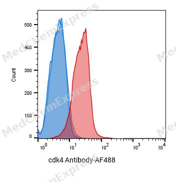 Flow cytometric analysis of 1X10^6 Hela cells labeling CDK4 Antibody(red). Cells were fixed with 4% paraformaldehyde and permeabilised with 0.2% Triton X-100. Then stained with the primary antibody at 1/400 dilution overnight at 4℃. AF 488 Goat Anti-mouse IgG H&L was used as the secondary antibody at 1/1,000 dilution for 45 minutes at room temperature. Mouse IgG Isotype Control (blue) was used as the isotype control, cells without incubation with primary antibody were used as the unlabeled control (black).


Background
-
Function
CDK4 is a cyclin-dependent serine/threonine kinase that partners with D-type cyclins to drive G1-phase progression before DNA synthesis[1]. Mechanistically, cyclin D-CDK4/6 phosphorylates RB, weakens RB-dependent transcriptional repression, and enables late-G1 signaling toward E2F-linked S-phase entry[2]. In cancer models, disruption of the CDK-RB1-E2F pathway supports malignant proliferation, and HR^+/HER2^- breast cancer provides a clinically established setting for CDK4/6 inhibition[3]. Compared with related isoforms, CDK4 and CDK6 share cell-cycle functions, but CDK4 is described as a prominent oncogenic driver in breast cancer, whereas CDK6 has a crucial role in hematopoietic stem-cell differentiation[3][4]. For experimental and translational applications, palbociclib, ribociclib, and abemaciclib inhibit CDK4/6, reduce RB phosphorylation, induce G1 arrest, and support endocrine-therapy combinations in HR^+/HER2^- advanced breast cancer studies[3][5][6][7].
-
Subcellular Localization
Cytoplasm; Nucleus; Nucleus membrane
-
Isoforms & Post-Translational Modification
P11802 has 2 isomers: P11802-1: 33730 Da (predicted); P11802-2: 20725 Da (predicted).
Phosphorylation at Thr-172 is required for enzymatic activity. Phosphorylated, in vitro, at this site by CCNH-CDK7, but, in vivo, appears to be phosphorylated by a proline-directed kinase. In the cyclin D-CDK4-CDKN1B complex, this phosphorylation and consequent CDK4 enzyme activity, is dependent on the tyrosine phosphorylation state of CDKN1B. Thus, in proliferating cells, CDK4 within the complex is phosphorylated on Thr-172 in the T-loop. In resting cells, phosphorylation on Thr-172 is prevented by the non-tyrosine-phosphorylated form of CDKN1B -
Subunit
Component of the D-CDK4 complex, composed of CDK4 and some D-type G1 cyclin (CCND1, CCND2 or CCND3). Interacts directly in the complex with CCND1, CCND2 or CCND3. Interacts with SEI1 and ZNF655. Forms a ternary complex, cyclin D-CDK4-CDKN1B, involved in modulating CDK4 enzymatic activity. Interacts directly with CDKN1B (phosphorylated on 'Tyr-88' and 'Tyr-89'); the interaction allows assembly of the cyclin D-CDK4 complex, Thr-172 phosphorylation, nuclear translocation and enhances the cyclin D-CDK4 complex activity. CDK4 activity is either inhibited or enhanced depending on stoichiometry of complex. The non-tyrosine-phosphorylated form of CDKN1B prevents T-loop phosphorylation of CDK4 producing inactive CDK4. Interacts (unphosphorylated form) with CDK2. Also forms ternary complexes with CDKN1A or CDKN2A. Interacts directly with CDKN1A (via its N-terminal); the interaction promotes the assembly of the cyclin D-CDK4 complex, its nuclear translocation and promotes the cyclin D-dependent enzyme activity of CDK4. Interacts with CCND1; the interaction is prevented with the binding of CCND1 to INSM1 during cell cycle progression. Probably forms a complex composed of chaperones HSP90 and HSP70, co-chaperones CDC37, PPP5C, TSC1 and client protein TSC2, CDK4, AKT, RAF1 and NR3C1; this complex does not contain co-chaperones STIP1/HOP and PTGES3/p23 (PubMed:29127155). Interacts with CEBPA (when phosphorylated) (PubMed:15107404). Interacts with FNIP1 and FNIP2 (PubMed:27353360)
-
SwissProt ID
-
Synonyms
CMM3; PSK-J3


Documentation
References
[1]. Baker SJ, et al. CDK4: a master regulator of the cell cycle and its role in cancer. Genes Cancer. 2022 Aug 25;13:21-45. [Content Brief]
[2]. Harbour JW, et al. Cdk phosphorylation triggers sequential intramolecular interactions that progressively block Rb functions as cells move through G1. Cell. 1999 Sep 17;98(6):859-69. [Content Brief]
[3]. Braal CL, et al. Inhibiting CDK4/6 in Breast Cancer with Palbociclib, Ribociclib, and Abemaciclib: Similarities and Differences. Drugs. 2021 Feb;81(3):317-331. [Content Brief]
[4]. Fassl A, et al. CDK4 and CDK6 kinases: From basic science to cancer therapy. Science. 2022 Jan 14;375(6577):eabc1495. [Content Brief]
[5]. Finn RS, et al. Palbociclib and Letrozole in Advanced Breast Cancer. N Engl J Med. 2016 Nov 17;375(20):1925-1936. [Content Brief]
[6]. Hortobagyi GN, et al. Ribociclib as First-Line Therapy for HR-Positive, Advanced Breast Cancer. N Engl J Med. 2016 Nov 3;375(18):1738-1748. [Content Brief]
[7]. Goetz MP, et al. MONARCH 3: Abemaciclib As Initial Therapy for Advanced Breast Cancer. J Clin Oncol. 2017 Nov 10;35(32):3638-3646. [Content Brief]

 Powered by Bioz
