



CEBP beta Antibody (YA1835)
(Synonyms: CEBPB; LAP; TCF5; PP9092; CCAAT/enhancer-binding protein beta; C/EBP beta; Liver activator protein; Nuclear factor NF-IL6; Transcription factor 5; TCF-5)
Based on 1 Customer Validation
CEBP beta Antibody (YA1835) is a Rabbit-derived and non-conjugated IgG monoclonal antibody, targeting to CEBP beta.
-
Host:
Rabbit
-
Isotype:
IgG
-
Application:
WB, ICC/IF, IP, FC
-
Reactivity :
Human, Mouse, Rat
-
Formulation:
Supplied in Rabbit IgG in 10mM phosphate buffered saline , pH 7.4, 150mM sodium chloride, 0.05% BSA, 0.02% sodium azide and 50% glycerol.
-
Conjugation:
Non-conjugated


To place orders, for customer services and technical support, please contact: MedChemExpress USA
Tel: 609-228-6898 E-mail: [email protected] [email protected]





Applications
| Application |
WB

WB: Western Blot
|
ICC/IF
ICC/IF: Immunocytochemistry/
Immunofluorescence |
IP
IP: Immunoprecipitation
|
FC
FC: Flow Cytometry
|
|---|---|---|---|---|
| Dilution Ratio | 1:500-1:1000 | 1:50-1:200 | 1:50 | 1:50-1:100 |


Product Details
CEBP beta Antibody (YA1835) is a Rabbit-derived and non-conjugated IgG monoclonal antibody, targeting to CEBP beta.
-
Host Rabbit

-
Clonality Recombinant,Monoclonal

-
Species ReactivityHuman, Mouse, Rat
-
Observed Molecular WeightObserved band size: 36 kDa
Note: Due to possible protein modifications or aggregation, the molecular weight should be confirmed by actual measurement, and the predicted value is for reference only.

-
Calculated Molecular Weight Predicted band size: 36 kDa

Entrez Gene: 1051 Human ; 12608 Mouse ; 24253 Rat
SwissProt: P17676 Human ; P28033 Mouse ; P21272 Rat
OMIM: 189965 Human
A synthesized peptide derived from human CEBP Beta aa300-345/345.
Endogenous
Affinity Chromatography
Non-conjugated
Unmodified
IgG


Product Properties
-
Appearance
Solution
-
Formulation
Supplied in Rabbit IgG in 10mM phosphate buffered saline , pH 7.4, 150mM sodium chloride, 0.05% BSA, 0.02% sodium azide and 50% glycerol.
-
Concentration
Batch-dependent, Please check the COA for the concentration of each lot. Check Lot Concentration
-
Storage & Stability
Stored at -20°C for 1 year. Avoid repeated freeze / thaw cycles.
-
Shipping
Shipping with blue ice.


Verification Images
-
 Western blot analysis of extracts from NIH/3T3(lane 2(20ug) and NIH/3T3(lane 3(40ug) using CEBPB Antibody (HY-P82090) Rabbit mAb. Proteins were transferred to a PVDF membrane and blocked with 5% non-fat milk in TBST for 2 hour at room temperature. The primary antibody (1/1000) and Loading control antibody (Beta Actin, HY-P80438, 1/10000) was used in 5% non-fat milk in TBST at 4°C overnight. Goat Anti-Mouse/Rabbit IgG-HRP Secondary Antibody (1/10000) was used for 1 hour at room temperature.
 -
 Flow cytometric analysis of 1X10⁶ HeLa cells labeling CEBP beta Antibody (HY-P82090, red). Cells were fixed with 4% paraformaldehyde and permeabilised with 90% methanol. Then stained with the primary antibody at 1/100 dilution for an hour at 4℃. AF488-conjugated Goat Anti-Rabbit IgG H&L (HY-P8002) was used as the secondary antibody at 1/1,000 dilution for 30 minutes at 4℃. Rabbit IgG Isotype Control (HY-P80879, blue) was used as the isotype control, cells without incubation with primary antibody were used as the unlabeled control (black).
 -
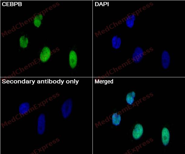 Immunocytochemistry analysis of NIH-3T3 cells labeling CEBPB with CEBPB antibody (HY-P82090) at 1/50 dilution. Cells were fixed in 4% paraformaldehyde for 15 minutes at room temperature, permeabilized with 0.1% Triton X-100 for 10 minutes at room temperature, then blocked with QuickBlock™ Blocking Buffer for Immunol Staining for 10 min at room temperature. Cells were then incubated with CEBPB antibody (HY-P82090) at 1/50 dilution in QuickBlock™ Blocking Buffer for Immunol Staining at 4 ℃. Alexa Fluor® 488-conjugated AffiniPure Goat Anti-Rabbit IgG H&L(HY-P8002, Green) was used as the secondary antibody at 1/1,000 dilution. PBS instead of the primary antibody was used as the secondary antibody only control. The Nuclear counterstain was DAPI (Blue).
 -
 Immunocytochemistry analysis of HeLa cells labeling CEBPB with CEBPB antibody (HY-P82090) at 1/50 dilution. Cells were fixed in 4% paraformaldehyde for 15 minutes at room temperature, permeabilized with 0.1% Triton X-100 for 10 minutes at room temperature, then blocked with QuickBlock™ Blocking Buffer for Immunol Staining for 10 min at room temperature. Cells were then incubated with CEBPB antibody (HY-P82090) at 1/50 dilution in QuickBlock™ Blocking Buffer for Immunol Staining at 4 ℃. Alexa Fluor® 488-conjugated AffiniPure Goat Anti-Rabbit IgG H&L(HY-P8002, Green) was used as the secondary antibody at 1/1,000 dilution. PBS instead of the primary antibody was used as the secondary antibody only control. The Nuclear counterstain was DAPI (Blue).
 -
 Immunocytochemistry analysis of HeLa cells labeling CEBPB with CEBPB antibody (HY-P82090) at 1/50 dilution. Cells were fixed in 4% paraformaldehyde for 15 minutes at room temperature, permeabilized with 0.1% Triton X-100 for 10 minutes at room temperature, then blocked with BSA for Immunol Staining for 10 min at room temperature. Cells were then incubated with CEBPB antibody (HY-P82090) at 1/50 dilution in BSA for Immunol Staining at 4 ℃,Stay overnight. AF488-conjugated AffiniPure Goat Anti-Rabbit IgG H&L(HY-P8002, Green) was used as the secondary antibody at 1/1,000 dilution. PBS instead of the primary antibody was used as the secondary antibody only control. The Nuclear counterstain was DAPI (Blue).
 -
 Immunocytochemistry analysis of NIH-3T3 cells labeling CEBPB with CEBPB antibody (HY-P82090) at 1/50 dilution. Cells were fixed in 4% paraformaldehyde for 15 minutes at room temperature, permeabilized with 0.1% Triton X-100 for 10 minutes at room temperature, then blocked with BSA for Immunol Staining for 10 min at room temperature. Cells were then incubated with CEBPB antibody (HY-P82090) at 1/50 dilution in BSA for Immunol Staining at 4℃,Stay overnight. AF488-conjugated AffiniPure Goat Anti-Rabbit IgG H&L(HY-P8002, Green) was used as the secondary antibody at 1/1,000 dilution. PBS instead of the primary antibody was used as the secondary antibody only control. The Nuclear counterstain was DAPI (Blue).


Background
-
Function
CEBP beta is important transcription factor regulating the expression of genes involved in immune and inflammatory responses. Also plays a significant role in adipogenesis, as well as in the gluconeogenic pathway, liver regeneration, and hematopoiesis. The consensus recognition site is 5'-T[TG]NNGNAA[TG]-3'. Its functional capacity is governed by protein interactions and post-translational protein modifications. During early embryogenesis, plays essential and redundant roles with CEBPA. Has a promitotic effect on many cell types such as hepatocytes and adipocytes but has an antiproliferative effect on T-cells by repressing MYC expression, facilitating differentiation along the T-helper 2 lineage. Binds to regulatory regions of several acute-phase and cytokines genes and plays a role in the regulation of acute-phase reaction and inflammation. Also plays a role in intracellular bacteria killing. During adipogenesis, is rapidly expressed and, after activation by phosphorylation, induces CEBPA and PPARG, which turn on the series of adipocyte genes that give rise to the adipocyte phenotype. The delayed transactivation of the CEBPA and PPARG genes by CEBPB appears necessary to allow mitotic clonal expansion and thereby progression of terminal differentiation. Essential for female reproduction because of a critical role in ovarian follicle development. Restricts osteoclastogenesis: together with NFE2L1; represses expression of DSPP during odontoblast differentiation; Essential for gene expression induction in activated macrophages. Plays a major role in immune responses such as CD4(+) T-cell response, granuloma formation and endotoxin shock. Not essential for intracellular bacteria killing; Acts as a dominant negative through heterodimerization with isoform 2. Promotes osteoblast differentiation and osteoclastogenesis[1][2][3][4][5][6].
-
Subcellular Localization
Nucleus; Cytoplasm
-
Expression
Tissue_specificity:Expressed at low levels in the lung, kidney and spleen
Induction:By ER stress -
Isoforms & Post-Translational Modification
P17676 has 3 isomers: P17676-1: 36106 Da (predicted); P17676-2: 33592 Da (predicted); P17676-3: 15938 Da (predicted).
Methylated. Methylation at Arg-3 by CARM1 and at Lys-43 by EHMT2 inhibit transactivation activity. Methylation is probably inhibited by phosphorylation at Thr-235;Sumoylated by polymeric chains of SUMO2 or SUMO3 (PubMed:12810706). Sumoylation at Lys-174 is required for inhibition of T-cells proliferation. In adipocytes, sumoylation at Lys-174 by PIAS1 leads to ubiquitination and subsequent proteasomal degradation. Desumoylated by SENP2, which abolishes ubiquitination and stabilizes protein levels (By similarity);Ubiquitinated, leading to proteasomal degradation;Phosphorylated at Thr-235 by MAPK and CDK2, serves to prime phosphorylation at Thr-226 and Ser-231 by GSK3B and acquire DNA-binding as well as transactivation activities, required to induce adipogenesis. MAPK and CDK2 act sequentially to maintain Thr-235 in the primed phosphorylated state during mitotical cloning expansion and thereby progression of terminal differentiation. Phosphorylation at Thr-266 enhances transactivation activity. Phosphorylation at Ser-325 in response to calcium increases transactivation activity. Phosphorylated at Thr-235 by RPS6KA1 (PubMed:11684016);O-glycosylated, glycosylation at Ser-227 and Ser-228 prevents phosphorylation on Thr-235, Ser-231 and Thr-226 and DNA binding activity which delays the adipocyte differentiation program;Acetylated. Acetylation at Lys-43 is an important and dynamic regulatory event that contributes to its ability to transactivate target genes, including those associated with adipogenesis and adipocyte function. Deacetylation by HDAC1 represses its transactivation activity. Acetylated by KAT2A and KAT2B within a cluster of lysine residues between amino acids 129-133, this acetylation is strongly induced by glucocorticoid treatment and enhances transactivation activity -
Subunit
Binds DNA as a homodimer and as a heterodimer (PubMed:11018027, PubMed:11257229, PubMed:11792321).
-
SwissProt ID
-
Synonyms
CEBPB; LAP; TCF5; PP9092; CCAAT/enhancer-binding protein beta; C/EBP beta; Liver activator protein; Nuclear factor NF-IL6; Transcription factor 5; TCF-5
-
Research Field
Epigenetics and Nuclear Signaling


Documentation
-
Data Sheet (235 KB)
-
SDS (252 KB)
- English - EN (252 KB)
- Français - FR (252 KB)
- Deutsch - DE (252 KB)
- Norwegian - NO (252 KB)
- Español - ES (252 KB)
- Swedish - SV (252 KB)
- Italian - IT (252 KB)
- Korean - KR (252 KB)
- Portuguese - PT (252 KB)
-
User Guide for Antibodies (1077 KB)

 Powered by Bioz
