



Desmoglein 3 Antibody (YA3960)
(Synonyms: PVA; CDHF6)Based on 1 Customer Validation
Desmoglein 3 Antibody (YA3960) is a Mouse-derived and non-conjugated IgG1 monoclonal antibody, targeting to Desmoglein 3.
-
Host:
Mouse
-
Isotype:
IgG1
-
Application:
WB, IHC-P, FC, ELISA
-
Reactivity :
Human
-
Formulation:
Supplied in PBS with 0.05% sodium azide.
-
Conjugation:
Non-conjugated


To place orders, for customer services and technical support, please contact: MedChemExpress USA
Tel: 609-228-6898 E-mail: [email protected] [email protected]





Applications
| Application |
WB

WB: Western Blot
|
IHC-P
IHC-P: Immunohistochemistry-Paraffin
|
FC
FC: Flow Cytometry
|
ELISA
ELISA: Enzyme Linked Immunosorbent Assay
|
|---|---|---|---|---|
| Dilution Ratio | 1:500-1:2000 | 1:200-1:1000 | 1:200-1:400 | 1:10000 |


Product Details
Desmoglein 3 Antibody (YA3960) is a Mouse-derived and non-conjugated IgG1 monoclonal antibody, targeting to Desmoglein 3.
-
Host Mouse

-
Clonality Monoclonal

-
Species ReactivityHuman
-
Observed Molecular WeightObserved band size: 145 kDa
Note: Due to possible protein modifications or aggregation, the molecular weight should be confirmed by actual measurement, and the predicted value is for reference only.

-
Calculated Molecular Weight Predicted band size: 108 kDa

Purified recombinant fragment of human DSG3 (AA: 55-159) expressed in E. Coli.
affinity purified.
Non-conjugated
Unmodified
IgG1


Product Properties
-
Appearance
Solution
-
Formulation
Supplied in PBS with 0.05% sodium azide.
-
Concentration
Batch-dependent, Please check the COA for the concentration of each lot. Check Lot Concentration
-
Storage & Stability
Stored at -20°C for 1 year. Avoid repeated freeze / thaw cycles.
-
Shipping
Shipping with blue ice.


Verification Images
-
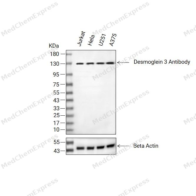 Western blot analysis of extracts from Jurkat (lane 2(20μg), Hela (lane 3(20μg), U251 (lane 4(20μg), A375 (lane 5(20μg) using Desmoglein 3 Antibody. Proteins were transferred to a PVDF membrane and blocked with 5% BSA in TBST for 2 hour at room temperature. The primary antibody (1/1000) and Loading control antibody (Beta Actin, HY-P80438, 1/3000) was used in 5% BSA in TBST at 4°C overnight. Goat Anti-Mouse/Rabbit IgG-HRP Secondary Antibody (HY-P8004/HY-P8001, 1/10,000) was used for 1 hour at room temperature.
 -
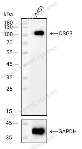 Western blot analysis of extracts from A431(lane 1(40μg) ) using DSG3 antibody(HY-P84263). Proteins were transferred to a NC membrane and blocked with 5% Skim milk in TBST for 2 hour at room temperature. The primary antibody ( 1/1000) and Loading control antibody (GAPDH,1/1000) was used in 5% Skim milk in TBST at 4°C overnight. Goat Anti-Mouse IgG-HRP Secondary Antibody (1/10
 -
 Immunohistochemical analysis of paraffin-embedded human oral mucosa tissue using Desmoglein 3 Antibody (HY-P84263, 1/800). The section was pretreated using heat mediated antigen retrieval with sodium citrate buffer (pH 6.0) for 8 minutes. The tissues were blocked with quick block buffer for 0.5 hours at room temperature, washed with PBS and PBST, and then incubated with the primary antibody overnight at 4℃. The detection was performed using an HRP conjugated compact polymer system. DAB was used as the chromogen. Tissues were counterstained with hematoxylin and mounted with DPX.
 -
 Immunohistochemical analysis of paraffin-embedded human tonsil tissue using Desmoglein 3 Antibody (HY-P84263, 1/800). The section was pretreated using heat mediated antigen retrieval with sodium citrate buffer (pH 6.0) for 8 minutes. The tissues were blocked with quick block buffer for 0.5 hours at room temperature, washed with PBS and PBST, and then incubated with the primary antibody overnight at 4℃. The detection was performed using an HRP conjugated compact polymer system. DAB was used as the chromogen. Tissues were counterstained with hematoxylin and mounted with DPX.
 -
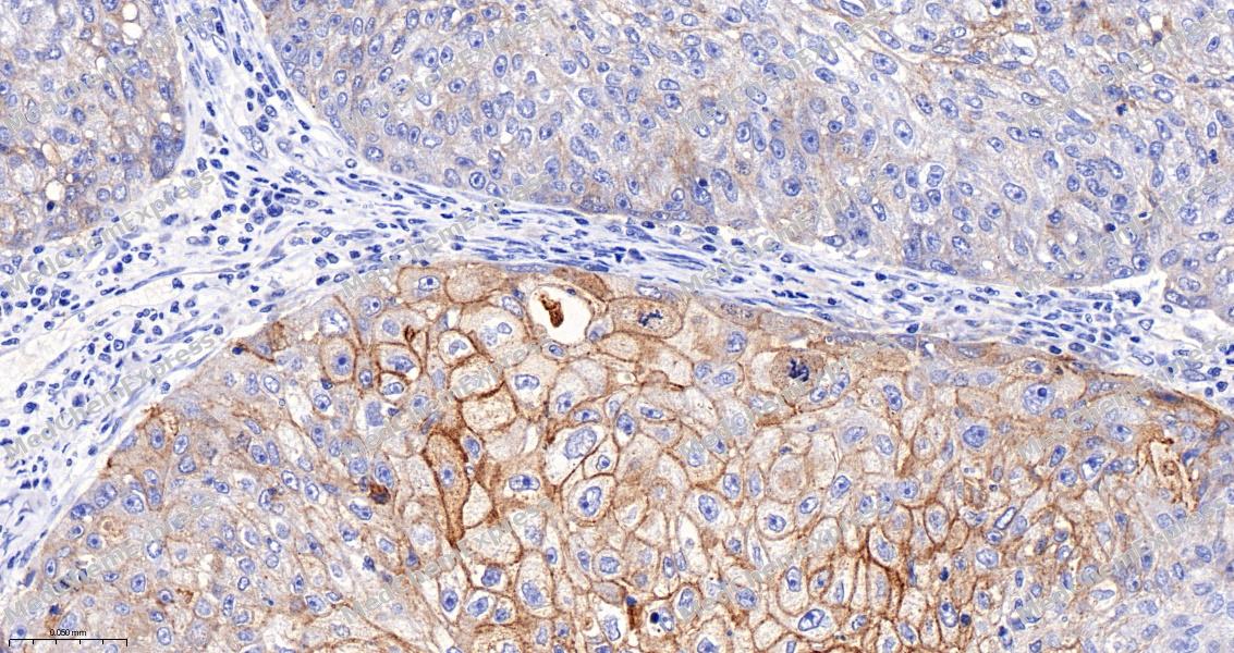 Immunohistochemical analysis of paraffin-embedded human oral cancer tissue using Desmoglein 3 Antibody (HY-P84263, 1/800). The section was pretreated using heat mediated antigen retrieval with sodium citrate buffer (pH 6.0) for 8 minutes. The tissues were blocked with quick block buffer for 0.5 hours at room temperature, washed with PBS and PBST, and then incubated with the primary antibody overnight at 4℃. The detection was performed using an HRP conjugated compact polymer system. DAB was used as the chromogen. Tissues were counterstained with hematoxylin and mounted with DPX.
 -
 Immunohistochemical analysis of paraffin-embedded human cervical cancer tissue using Desmoglein 3 Antibody (HY-P84263, 1/800). The section was pretreated using heat mediated antigen retrieval with sodium citrate buffer (pH 6.0) for 8 minutes. The tissues were blocked with quick block buffer for 0.5 hours at room temperature, washed with PBS and PBST, and then incubated with the primary antibody overnight at 4℃. The detection was performed using an HRP conjugated compact polymer system. DAB was used as the chromogen. Tissues were counterstained with hematoxylin and mounted with DPX.
 -
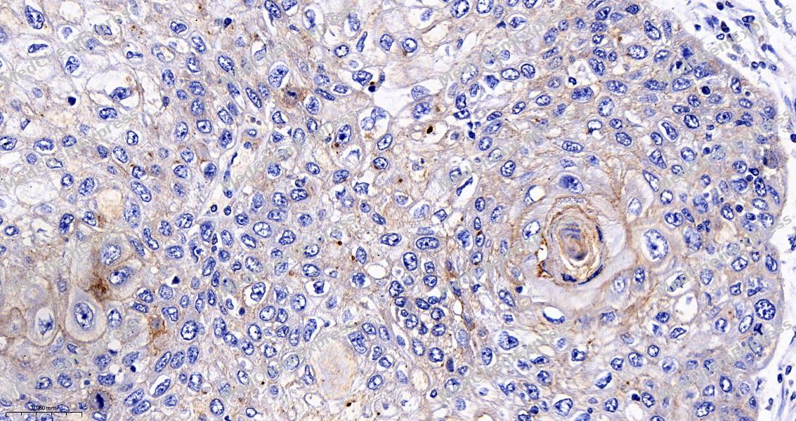 Immunohistochemical analysis of paraffin-embedded human skin cancer tissue using Desmoglein 3 Antibody (HY-P84263, 1/800). The section was pretreated using heat mediated antigen retrieval with sodium citrate buffer (pH 6.0) for 8 minutes. The tissues were blocked with quick block buffer for 0.5 hours at room temperature, washed with PBS and PBST, and then incubated with the primary antibody overnight at 4℃. The detection was performed using an HRP conjugated compact polymer system. DAB was used as the chromogen. Tissues were counterstained with hematoxylin and mounted with DPX.
 -
 Immunohistochemical analysis of paraffin-embedded human urothelial cancer tissue using Desmoglein 3 Antibody (HY-P84263, 1/800). The section was pretreated using heat mediated antigen retrieval with sodium citrate buffer (pH 6.0) for 8 minutes. The tissues were blocked with quick block buffer for 0.5 hours at room temperature, washed with PBS and PBST, and then incubated with the primary antibody overnight at 4℃. The detection was performed using an HRP conjugated compact polymer system. DAB was used as the chromogen. Tissues were counterstained with hematoxylin and mounted with DPX.
 -
 Immunohistochemical analysis of paraffin-embedded human thyroid cancer tissue using Desmoglein 3 Antibody (HY-P84263, 1/800). The section was pretreated using heat mediated antigen retrieval with sodium citrate buffer (pH 6.0) for 8 minutes. The tissues were blocked with quick block buffer for 0.5 hours at room temperature, washed with PBS and PBST, and then incubated with the primary antibody overnight at 4℃. The detection was performed using an HRP conjugated compact polymer system. DAB was used as the chromogen. Tissues were counterstained with hematoxylin and mounted with DPX.
 -
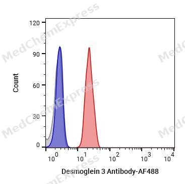 Flow cytometric analysis of 1X10^6 A431 cells labeling DSG3 Antibody(HY-P84263
 -
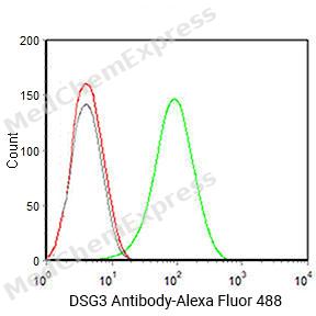 Flow cytometric analysis of 1X10^6 A431 cells labeling DSG3 Antibody(30627P,green). Cells were fixed with 4% paraformaldehyde and permeabilised with 0.2% Triton X-100. Then stained with the primary antibody at 1/400 dilution overnight at 4℃.Alexa Fluor® 488 Goat Anti-mouse IgG H&L (invitrogen A11001) was used as the secondary antibody at 1/1,000 dilution for 45 minutes at room temperature. Mouse IgG Isotype Control (Invitrogen 14-4714-B2, gray) was used as the isotype control, cells without incubation with primary antibody were used as the unlabeled control (red).


Background
-
Function
Desmoglein 3 is an A component of desmosome cell-cell junctions which are required for positive regulation of cellular adhesion. Required for adherens and desmosome junction assembly in response to mechanical force in keratinocytes. Required for desmosome-mediated cell-cell adhesion of cells surrounding the telogen hair club and the basal layer of the outer root sheath epithelium, consequently is essential for the anchoring of telogen hairs in the hair follicle. Required for the maintenance of the epithelial barrier via promoting desmosome-mediated intercellular attachment of suprabasal epithelium to basal cells. May play a role in the protein stability of the desmosome plaque components DSP, JUP, PKP1, PKP2 and PKP3. Required for YAP1 localization at the plasma membrane in keratinocytes in response to mechanical strain, via the formation of an interaction complex composed of DSG3, PKP1 and YWHAG. May also be involved in the positive regulation of YAP1 target gene transcription and as a result cell proliferation. Positively regulates cellular contractility and cell junction formation via organization of cortical F-actin bundles and anchoring of actin to tight junctions, in conjunction with RAC1. The cytoplasmic pool of DSG3 is required for the localization of CDH1 and CTNNB1 at developing adherens junctions, potentially via modulation of SRC activity. Inhibits keratinocyte migration via suppression of p38MAPK signaling, may therefore play a role in moderating wound healing[1][2][3][4][5].
-
Subcellular Localization
Cell membrane; Single-pass type I membrane protein; Cell junction, desmosome; Cytoplasm; Cell junction, tight junction; Cell junction
-
Expression
Tissue_specificity:Expressed throughout the basal and spinous layer of the epidermis with weak expression in the granular layer (at protein level) (PubMed:19717567, PubMed:7665906) . Also expressed in the buccal mucosa, esophagus and cervix (at protein level) (PubMed:7665906)
Induction:Induced in the hours following cyclic mechanical strain in keratinocytes -
Subunit
Homodimer (PubMed:19717567). Part of a complex that contains DSG3, PKP1, YAP1 and YWHAG; the complex is required for localization of DSG3 and YAP1 to the cell membrane in keratinocytes (PubMed:31835537). Interacts with PKP2 (PubMed:11790773). Interacts with CTNND1; the interaction facilitates DSG3 localization and retention at cell-cell junctions (PubMed:18343367). Interacts with CDH1; the interaction is required for CDH1 localization to developing adherens junctions (PubMed:22294297). Interacts with RAC1; the interaction is required for DSG3 translocation to cell-cell junctions, organization of cortical F-actin bundles and actin anchoring at cell-cell junctions (PubMed:22796473). Interacts with DSC3; the interaction may limit the interaction of DSC3 with p38MAPK family members and therefore repress p38MAPK signaling activation (By similarity)
-
SwissProt ID
-
Synonyms
PVA; CDHF6


Documentation
-
Data Sheet (258 KB)
-
SDS (251 KB)
- English - EN (251 KB)
- Français - FR (251 KB)
- Deutsch - DE (251 KB)
- Norwegian - NO (251 KB)
- Español - ES (251 KB)
- Swedish - SV (251 KB)
- Italian - IT (251 KB)
- Korean - KR (251 KB)
- Portuguese - PT (251 KB)
-
User Guide for Antibodies (1077 KB)

 Powered by Bioz
