



EAAT3 Antibody (YA2365)
(Synonyms: SLC1A1; EAAC1; EAAT3; Excitatory amino acid transporter 3; Excitatory amino-acid carrier 1; Neuronal and epithelial glutamate transporter; Sodium-dependent glutamate/aspartate transporter 3; Solute carrier family 1 member 1)
Based on 1 Customer Validation
EAAT3 Antibody (YA2365) is a Rabbit-derived and non-conjugated IgG monoclonal antibody, targeting to EAAT3.
-
Host:
Rabbit
-
Isotype:
IgG
-
Application:
WB, IHC-P, ICC/IF
-
Reactivity :
Human, Mouse, Rat
-
Formulation:
Supplied in 50mM Tris-Glycine(pH 7.4), 0.15M NaCl, 40%Glycerol, 0.01% sodium azide and 0.05% BSA.
-
Conjugation:
Non-conjugated


To place orders, for customer services and technical support, please contact: MedChemExpress USA
Tel: 609-228-6898 E-mail: [email protected] [email protected]





Applications
| Application |
WB

WB: Western Blot
|
IHC-P
IHC-P: Immunohistochemistry-Paraffin
|
ICC/IF
ICC/IF: Immunocytochemistry/
Immunofluorescence |
|---|---|---|---|
| Dilution Ratio | 1:500-1:1000 | 1:50-1:100 | 1:50-1:200 |


Product Details
EAAT3 Antibody (YA2365) is a Rabbit-derived and non-conjugated IgG monoclonal antibody, targeting to EAAT3.
-
Host Rabbit

-
Clonality Recombinant,Monoclonal

-
Species ReactivityHuman, Mouse, Rat
-
Observed Molecular WeightObserved band size: 70 kDa
Note: Due to possible protein modifications or aggregation, the molecular weight should be confirmed by actual measurement, and the predicted value is for reference only.

-
Calculated Molecular Weight Predicted band size: 57 kDa

Entrez Gene: 6505 Human ; 20510 Mouse ; 25550 Rat
SwissProt: P43005 Human ; P51906 Mouse ; P51907 Rat
OMIM: 222730 Human
A synthesized peptide derived from human EAAT3 aa150-200/524.
Endogenous
Affinity Chromatography
Non-conjugated
Unmodified
IgG


Product Properties
-
Appearance
Solution
-
Formulation
Supplied in 50mM Tris-Glycine(pH 7.4), 0.15M NaCl, 40%Glycerol, 0.01% sodium azide and 0.05% BSA.
-
Concentration
Batch-dependent, Please check the COA for the concentration of each lot. Check Lot Concentration
-
Storage & Stability
Stored at -20°C for 1 year. Avoid repeated freeze / thaw cycles.
-
Shipping
Shipping with blue ice.


Verification Images
-
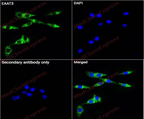 Immunocytochemistry analysis of SH-SY5Y cells labeling EAAT3 Antibody (HY-P82620) at 1/50 dilution. Cells were fixed in 4% paraformaldehyde for 15 minutes at room temperature, permeabilized with 0.1% Triton X-100 for 10 minutes at room temperature, then blocked with QuickBlock™ Blocking Buffer for Immunol Staining for 10 min at room temperature. Cells were then incubated with EAAT3 Antibody (HY-P82620) at 1/50 dilution in QuickBlock™ Blocking Buffer for Immunol Staining at 4 ℃. Alexa Fluor® 488-conjugated AffiniPure Goat Anti-Rabbit IgG H&L(HY-P8002, Green) was used as the secondary antibody at 1/1,000 dilution. PBS instead of the primary antibody was used as the secondary antibody only control. The Nuclear counterstain was DAPI (Blue).
 -
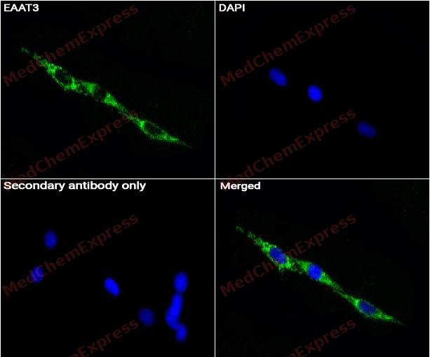 Immunocytochemistry analysis of SH-SY5Y cells labeling EAAT3 Antibody (HY-P82620) at 1/200 dilution. Cells were fixed in 4% paraformaldehyde for 15 minutes at room temperature, permeabilized with 0.1% Triton X-100 for 10 minutes at room temperature, then blocked with QuickBlock™ Blocking Buffer for Immunol Staining for 10 min at room temperature. Cells were then incubated with EAAT3 Antibody (HY-P82620) at 1/200 dilution in QuickBlock™ Blocking Buffer for Immunol Staining at 4 ℃. Alexa Fluor® 488-conjugated AffiniPure Goat Anti-Rabbit IgG H&L(HY-P8002, Green) was used as the secondary antibody at 1/1,000 dilution. PBS instead of the primary antibody was used as the secondary antibody only control. The Nuclear counterstain was DAPI (Blue).
 -
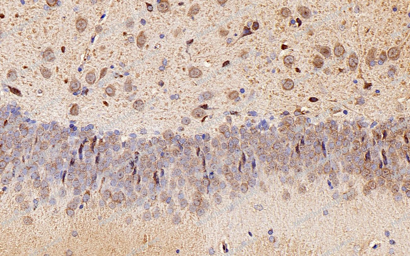 Immunohistochemical analysis of paraffin-embedded Rat brain tissue using EAAT3 Antibody (YA2365). The section was pre-treated using heat mediated antigen retrieval with Tris-EDTA buffer (pH 9.0) for 8 minutes. The tissues were blocked in QuickBlock for 20 minutes at room temperature, washed with ddH2O and PBS, and then probed with the primary antibody (HY-P82620, 1/100) in 4℃ overnight. The detection was performed using an HRP conjugated compact polymer system. DAB was used as the chromogen. Tissues were counterstained with hematoxylin and mounted with DPX.
 -
Immunohistochemical analysis of paraffin-embedded Rat brain tissue using EAAT3 Antibody (YA2365). The section was pre-treated using heat mediated antigen retrieval with Tris-EDTA buffer (pH 9.0) for 8 minutes. The tissues were blocked in QuickBlock for 20 minutes at room temperature, washed with ddH2O and PBS, and then probed with the primary antibody (HY-P82620, 1/100) in 4℃ overnight. The detection was performed using an HRP conjugated compact polymer system. DAB was used as the chromogen. Tissues were counterstained with hematoxylin and mounted with DPX.


Background
-
Function
The neuronal excitatory amino acid transporter 3 (EAAT3, also known as EAAC1) mediates Na+-dependent uptake of glutamate and the uncharged amino acid L-cysteine, which is essential for neuronal glutathione synthesis and prevention of oxidative stress[1][2][3]. Mechanistically, EAAT3 exhibits a high affinity for cysteine via thiolate formation in its binding site, allowing predominantly unidirectional transport without significant substrate release[1]. EAAT3 contributes to maintaining low extracellular glutamate concentrations at the synapse, thereby regulating excitatory neurotransmission and limiting excitotoxicity[3][4]. In disease models, EAAT3 expression is altered in the striatum of schizophrenia and bipolar disorder, implicating neuronal glutamate transport dysfunction in psychiatric pathology[5][6]. Compared with related isoforms, EAAT3 uniquely transports cysteine efficiently, whereas glial transporters EAAT1 and EAAT2 exhibit negligible cysteine transport[1][2]. EAAT3 also displays partial sensitivity to membrane lipid composition, associating with cholesterol-rich lipid rafts, which influences transporter localization and function[7]. Pharmacologically, EAAT3 can be inhibited by broad-spectrum EAAT inhibitors such as threo-beta-hydroxyaspartate, though selective activation remains challenging due to the scarcity of effective agonists[8][9][10]. Therefore, EAAT3 serves as a critical regulator of neuronal redox balance and glutamatergic signaling, providing a distinct target for studies of oxidative stress, synaptic modulation, and neuropsychiatric disease mechanisms[1][2][3][5].
-
Subcellular Localization
Cell membrane; Multi-pass membrane protein; Apical cell membrane; Multi-pass membrane protein; Synapse, synaptosome; Early endosome membrane; Late endosome membrane; Recycling endosome membrane
-
Expression
Tissue_specificity:Expressed in all tissues tested including liver, muscle, testis, ovary, retinoblastoma cell line, neurons and brain (in which there was dense expression in substantia nigra, red nucleus, hippocampus and in cerebral cortical layers) -
Subunit
Homotrimer (PubMed:33658209). Interacts with ARL6IP5. Interacts with RTN2 (via N-terminus); the interaction promotes cell surface expression of SLC1A1.
-
SwissProt ID
-
Synonyms
SLC1A1; EAAC1; EAAT3; Excitatory amino acid transporter 3; Excitatory amino-acid carrier 1; Neuronal and epithelial glutamate transporter; Sodium-dependent glutamate/aspartate transporter 3; Solute carrier family 1 member 1
-
Research Field
Neuroscience


Documentation
-
Data Sheet (263 KB)
-
SDS (252 KB)
- English - EN (252 KB)
- Français - FR (252 KB)
- Deutsch - DE (252 KB)
- Norwegian - NO (252 KB)
- Español - ES (252 KB)
- Swedish - SV (252 KB)
- Italian - IT (252 KB)
- Korean - KR (252 KB)
- Portuguese - PT (252 KB)
-
User Guide for Antibodies (1077 KB)
References
[1]. Shigeri Y, et al. Effects of threo-beta-hydroxyaspartate derivatives on excitatory amino acid transporters (EAAT4 and EAAT5). J Neurochem. 2001 Oct;79(2):297-302. [Content Brief]
[2]. van Veggel L, et al. Still in Search for an EAAT Activator: GT949 Does Not Activate EAAT2, nor EAAT3 in Impedance and Radioligand Uptake Assays. ACS Chem Neurosci. 2024 Apr 3;15(7):1424-1431. [Content Brief]
[3]. O'Kane RL, et al. Na(+)-dependent glutamate transporters (EAAT1, EAAT2, and EAAT3) of the blood-brain barrier. A mechanism for glutamate removal. J Biol Chem. 1999 Nov 5;274(45):31891-5. [Content Brief]
[4]. Kovermann P, et al. Cellular Physiology and Pathophysiology of EAAT Anion Channels. Front Cell Neurosci. 2022 Jan 6;15:815279. [Content Brief]
[5]. Rabezanahary H, et al. Live virus neutralizing antibodies against pre and post Omicron strains in food and retail workers in Québec, Canada. Heliyon. 2024 May 21;10(10):e31026. [Content Brief]
[6]. Watts SD, et al. Cysteine transport through excitatory amino acid transporter 3 (EAAT3). PLoS One. 2014 Oct 2;9(10):e109245. [Content Brief]
[7]. Chen Y, et al. The glutamate transporters EAAT2 and EAAT3 mediate cysteine uptake in cortical neuron cultures. J Neurochem. 2003 Mar;84(6):1332-9. [Content Brief]
[8]. Jensen AA, et al. Pharmacological characterization of human excitatory amino acid transporters EAAT1, EAAT2 and EAAT3 in a fluorescence-based membrane potential assay. Biochem Pharmacol. 2004 Jun 1;67(11):2115-27. [Content Brief]
[9]. McCullumsmith RE, et al. Striatal excitatory amino acid transporter transcript expression in schizophrenia, bipolar disorder, and major depressive disorder. Neuropsychopharmacology. 2002 Mar;26(3):368-75. [Content Brief]
[10]. Butchbach ME, et al. Association of excitatory amino acid transporters, especially EAAT2, with cholesterol-rich lipid raft microdomains: importance for excitatory amino acid transporter localization and function. J Biol Chem. 2004 Aug 13;279(33):34388-96. [Content Brief]

 Powered by Bioz
