



Estrogen Receptor alpha Antibody (YA3629)
(Synonyms: ER; ESR; Era; ESRA; ESTRR; NR3A1)Based on 1 publication(s) in Google Scholar
Estrogen Receptor alpha Antibody (YA3629) is a Mouse-derived and non-conjugated IgG1 monoclonal antibody, targeting to Estrogen Receptor alpha.
-
Host:
Mouse
-
Isotype:
IgG1
-
Application:
WB, IHC-P, ICC/IF, FC, ELISA
-
Reactivity :
Human, Mouse, Rat, Rabbit
-
Formulation:
Supplied in PBS with 0.05% sodium azide
-
Conjugation:
Non-conjugated


To place orders, for customer services and technical support, please contact: MedChemExpress USA
Tel: 609-228-6898 E-mail: [email protected] [email protected]



Publications Citing Use of MedChemExpress (MCE) Estrogen Receptor alpha Antibody (YA3629)
More

Applications
| Application |
WB

WB: Western Blot
|
IHC-P
IHC-P: Immunohistochemistry-Paraffin
|
ICC/IF
ICC/IF: Immunocytochemistry/
Immunofluorescence |
FC
FC: Flow Cytometry
|
ELISA
ELISA: Enzyme Linked Immunosorbent Assay
|
|---|---|---|---|---|---|
| Dilution Ratio | 1:500-1:2000 | 1:100-1:500 | 1:50-1:500 | 1:200-1:400 | 1:10000 |


Product Details
Estrogen Receptor alpha Antibody (YA3629) is a Mouse-derived and non-conjugated IgG1 monoclonal antibody, targeting to Estrogen Receptor alpha.
-
Host Mouse

-
Clonality Monoclonal

-
Species ReactivityHuman, Mouse, Rat, Rabbit
-
Observed Molecular WeightObserved band size: 66 kDa
Note: Due to possible protein modifications or aggregation, the molecular weight should be confirmed by actual measurement, and the predicted value is for reference only.

-
Calculated Molecular Weight Predicted band size: 66 kDa

Purified recombinant fragment of human ESR1 (AA: 2-185) expressed in E. Coli.
affinity purified.
Non-conjugated
Unmodified
IgG1


Product Properties
-
Appearance
Liquid
-
Formulation
Supplied in PBS with 0.05% sodium azide
-
Concentration
Batch-dependent, Please check the COA for the concentration of each lot. Check Lot Concentration
-
Storage & Stability
Stored at -20°C for 1 year. Avoid repeated freeze / thaw cycles.
-
Shipping
Shipping with blue ice.


Publications (1)
-
Journal Impact Factor
-
Most Recent


Verification Images
-
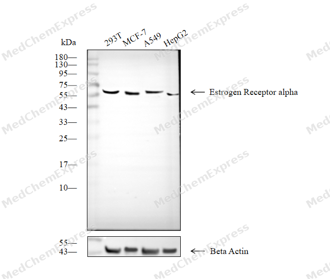 Western blot analysis of extracts from 293T (lane 2(20μg), MCF-7 (lane 3(20μg), A549 (lane 4(20μg) and HepG2 (lane 5(20μg) using Estrogen Receptor alpha Antibody (HY-P83932). Proteins were transferred to a PVDF membrane and blocked with 5% non-fat milk in TBST for 2 hour at room temperature. The primary antibody (1/1000) and Loading control antibody (Beta Actin, HY-P80993, 1/10000) was used in 5% non-fat milk in TBST at 4°C overnight. Goat Anti-Mouse IgG-HRP Secondary Antibody (1/10000) was used for 1 hour at room temperature.
 -
 Immunohistochemical analysis of paraffin-embedded human fallopian tube tissue using Estrogen Receptor alpha Antibody (HY-P83932, 1/200). The section was pretreated using heat mediated antigen retrieval with sodium citrate buffer (pH 6.0) for 8 minutes. The tissues were blocked with quick block buffer for 0.5 hours at room temperature, washed with PBS and PBST, and then incubated with the primary antibody overnight at 4℃. The detection was performed using an HRP conjugated compact polymer system. DAB was used as the chromogen. Tissues were counterstained with hematoxylin and mounted with DPX.
 -
 Immunohistochemical analysis of paraffin-embedded human breast cancer tissue using Estrogen Receptor alpha Antibody (HY-P83932, 1/200). The section was pretreated using heat mediated antigen retrieval with sodium citrate buffer (pH 6.0) for 8 minutes. The tissues were blocked with quick block buffer for 0.5 hours at room temperature, washed with PBS and PBST, and then incubated with the primary antibody overnight at 4℃. The detection was performed using an HRP conjugated compact polymer system. DAB was used as the chromogen. Tissues were counterstained with hematoxylin and mounted with DPX.
 -
 Immunohistochemical analysis of paraffin-embedded human endometrium cancer tissue using Estrogen Receptor alpha Antibody (HY-P83932, 1/200). The section was pretreated using heat mediated antigen retrieval with sodium citrate buffer (pH 6.0) for 8 minutes. The tissues were blocked with quick block buffer for 0.5 hours at room temperature, washed with PBS and PBST, and then incubated with the primary antibody overnight at 4℃. The detection was performed using an HRP conjugated compact polymer system. DAB was used as the chromogen. Tissues were counterstained with hematoxylin and mounted with DPX.
 -
 Immunohistochemical analysis of paraffin-embedded human lung cancer tissue using Estrogen Receptor alpha Antibody (HY-P83932, 1/200). The section was pretreated using heat mediated antigen retrieval with sodium citrate buffer (pH 6.0) for 8 minutes. The tissues were blocked with quick block buffer for 0.5 hours at room temperature, washed with PBS and PBST, and then incubated with the primary antibody overnight at 4℃. The detection was performed using an HRP conjugated compact polymer system. DAB was used as the chromogen. Tissues were counterstained with hematoxylin and mounted with DPX.
 -
 Immunohistochemical analysis of paraffin-embedded rat cervix tissue using Estrogen Receptor alpha Antibody (HY-P83932, 1/200). The section was pretreated using heat mediated antigen retrieval with sodium citrate buffer (pH 6.0) for 8 minutes. The tissues were blocked with quick block buffer for 0.5 hours at room temperature, washed with PBS and PBST, and then incubated with the primary antibody overnight at 4℃. The detection was performed using an HRP conjugated compact polymer system. DAB was used as the chromogen. Tissues were counterstained with hematoxylin and mounted with DPX.
 -
 Immunohistochemical analysis of paraffin-embedded rat endometrium tissue using Estrogen Receptor alpha Antibody (HY-P83932, 1/200). The section was pretreated using heat mediated antigen retrieval with sodium citrate buffer (pH 6.0) for 8 minutes. The tissues were blocked with quick block buffer for 0.5 hours at room temperature, washed with PBS and PBST, and then incubated with the primary antibody overnight at 4℃. The detection was performed using an HRP conjugated compact polymer system. DAB was used as the chromogen. Tissues were counterstained with hematoxylin and mounted with DPX.
 -
 Immunohistochemical analysis of paraffin-embedded rat fallopian tube tissue using Estrogen Receptor alpha Antibody (HY-P83932, 1/200). The section was pretreated using heat mediated antigen retrieval with sodium citrate buffer (pH 6.0) for 8 minutes. The tissues were blocked with quick block buffer for 0.5 hours at room temperature, washed with PBS and PBST, and then incubated with the primary antibody overnight at 4℃. The detection was performed using an HRP conjugated compact polymer system. DAB was used as the chromogen. Tissues were counterstained with hematoxylin and mounted with DPX.
 -
 Immunocytochemistry analysis of MCF-7 cells labeling Estrogen Receptor alpha with Estrogen Receptor alpha Antibody (HY-P83932) at 1/100 dilution. Cells were fixed in 4% paraformaldehyde for 15 minutes at room temperature, permeabilized with 0.1% Triton X-100 in PBS for 15 minutes at room temperature, then blocked with quick block buffer for 10 minutes at room temperature. Cells were then incubated with Estrogen Receptor alpha Antibody (HY-P83932) at 1/100 dilution in quick block buffer overnight at 4 ℃.AF488-conjugated Goat Anti-Mouse IgG H&L (HY-P8005, Green) was used as the secondary antibody at 1/1,000 dilution. PBS instead of the primary antibody was used as the secondary antibody only control. The Nuclear counterstain was DAPI (Blue).
 -
 Immunocytochemistry analysis of MCF-7 cells labeling Estrogen Receptor alpha with Estrogen Receptor alpha Antibody (HY-P83932) at 1/200 dilution. Cells were fixed in 4% paraformaldehyde for 15 minutes at room temperature, permeabilized with 0.1% Triton X-100 in PBS for 15 minutes at room temperature, then blocked with quick block buffer for 10 minutes at room temperature. Cells were then incubated with Estrogen Receptor alpha Antibody (HY-P83932) at 1/200 dilution in quick block buffer overnight at 4 ℃.AF488-conjugated Goat Anti-Mouse IgG H&L (HY-P8005, Green) was used as the secondary antibody at 1/1,000 dilution. PBS instead of the primary antibody was used as the secondary antibody only control. The Nuclear counterstain was DAPI (Blue).


Background
-
Function
Estrogen Receptor alpha is a Nuclear hormone receptor. The steroid hormones and their receptors are involved in the regulation of eukaryotic gene expression and affect cellular proliferation and differentiation in target tissues. Ligand-dependent nuclear transactivation involves either direct homodimer binding to a palindromic estrogen response element (ERE) sequence or association with other DNA-binding transcription factors, such as AP-1/c-Jun, c-Fos, ATF-2, Sp1 and Sp3, to mediate ERE-independent signaling. Ligand binding induces a conformational change allowing subsequent or combinatorial association with multiprotein coactivator complexes through LXXLL motifs of their respective components. Mutual transrepression occurs between the estrogen receptor (ER) and NF-kappa-B in a cell-type specific manner. Decreases NF-kappa-B DNA-binding activity and inhibits NF-kappa-B-mediated transcription from the IL6 promoter and displace RELA/p65 and associated coregulators from the promoter. Recruited to the NF-kappa-B response element of the CCL2 and IL8 promoters and can displace CREBBP. Present with NF-kappa-B components RELA/p65 and NFKB1/p50 on ERE sequences. Can also act synergistically with NF-kappa-B to activate transcription involving respective recruitment adjacent response elements; the function involves CREBBP. Can activate the transcriptional activity of TFF1. Also mediates membrane-initiated estrogen signaling involving various kinase cascades. Essential for MTA1-mediated transcriptional regulation of BRCA1 and BCAS3. Maintains neuronal survival in response to ischemic reperfusion injury when in the presence of circulating estradiol (17-beta-estradiol/E2); Involved in activation of NOS3 and endothelial nitric oxide production. Isoforms lacking one or several functional domains are thought to modulate transcriptional activity by competitive ligand or DNA binding and/or heterodimerization with the full-length receptor. Binds to ERE and inhibits isoform 1[1][2][3].
-
Subcellular Localization
Nucleus; Cytoplasm; Cell membrane; Peripheral membrane protein; Cytoplasmic side; Nucleus; Cytoplasm; Cell membrane; Peripheral membrane protein; Cytoplasmic side; Cell membrane; Single-pass type I membrane protein; Nucleus; Golgi apparatus; Cell membrane
-
Expression
Tissue_specificity:Widely expressed (PubMed: 10970861) . Not expressed in the pituitary gland (PubMed: 10970861) ; Widely expressed, but not expressed in the pituitary gland. -
Isoforms & Post-Translational Modification
P03372 has 4 isomers: P03372-1: 66216 Da (predicted); P03372-2: 53687 Da (predicted); P03372-3: 47629 Da (predicted); P03372-4: 35298 Da (predicted).
Phosphorylated by cyclin A/CDK2 and CK1. Phosphorylation probably enhances transcriptional activity. Self-association induces phosphorylation. Dephosphorylation at Ser-118 by PPP5C inhibits its transactivation activity. Phosphorylated by LMTK3 in vitro;Glycosylated; contains N-acetylglucosamine, probably O-linked;Ubiquitinated; regulated by LATS1 via DCAF1 it leads to ESR1 proteasomal degradation (PubMed:21602804, PubMed:28068668). Deubiquitinated by OTUB1 (PubMed:19383985). Ubiquitinated by STUB1/CHIP; in the CA1 hippocampal region following loss of endogenous circulating estradiol (17-beta-estradiol/E2) (By similarity). Ubiquitinated by UBR5, leading to its degradation: UBR5 specifically recognizes and binds ligand-bound ESR1 when it is not associated with coactivators (NCOAs) (PubMed:37478846). In presence of NCOAs, the UBR5-degron is not accessible, preventing its ubiquitination and degradation (PubMed:37478846);Dimethylated by PRMT1 at Arg-260. The methylation may favor cytoplasmic localization (PubMed:18657504, PubMed:24498420). Demethylated by JMJD6 at Arg-260 (PubMed:24498420);Palmitoylated (isoform 3). Not biotinylated (isoform 3);Palmitoylated by ZDHHC7 and ZDHHC21. Palmitoylation is required for plasma membrane targeting and for rapid intracellular signaling via ERK and AKT kinases and cAMP generation, but not for signaling mediated by the nuclear hormone receptor -
Subunit
Binds DNA as a homodimer. Can form a heterodimer with ESR2. Interacts with FOXC2, MAP1S, SLC30A9, UBE1C and NCOA3 coactivator (By similarity). Interacts with PELP1, the interaction is enhanced by 17-beta-estradiol, the interaction increases ESR1 transcriptional activity (PubMed:11481323, PubMed:14963108). Interacts with EP300; the interaction is estrogen-dependent and enhanced by CITED1. Interacts with CITED1; the interaction is estrogen-dependent. Interacts with NCOA5 and NCOA6 coactivators. Interacts with NCOA7; the interaction is a ligand-inducible. Interacts with PHB2, and UBE1C. Interacts with AKAP13. Interacts with CUEDC2. Interacts with KDM5A. Interacts with SMARD1. Interacts with HEXIM1. Interacts with PBXIP1. Interaction with MUC1 is stimulated by 7 beta-estradiol (E2) and enhances ESR1-mediated transcription. Interacts with DNTTIP2, FAM120B and UIMC1. Interacts with isoform 4 of TXNRD1. Interacts with KMT2D/MLL2. Interacts with ATAD2 and this interaction is enhanced by estradiol. Interacts with KIF18A and LDB1. Interacts with RLIM (via C-terminus). Interacts with MACROD1. Interacts with SH2D4A and PLCG. Interaction with SH2D4A blocks binding to PLCG and inhibits estrogen-induced cell proliferation. Interacts with DYNLL1. Interacts with CCDC62 in the presence of estradiol/E2; this interaction seems to enhance the transcription of target genes. Interacts with NR2C1; the interaction prevents homodimerization of ESR1 and suppresses its transcriptional activity and cell growth. Interacts with DNAAF4. Interacts with PRMT2. Interacts with RBFOX2. Interacts with STK3/MST2 only in the presence of SAV1 and vice-versa. Binds to CSNK1D. Interacts with NCOA2; NCOA2 can interact with ESR1 AF-1 and AF-2 domains simultaneously and mediate their transcriptional synergy. Interacts with DDX5 (PubMed:11682626). Interacts with NCOA1; the interaction seems to require a self-association of N-terminal and C-terminal regions. Interacts with ZNF366, DDX17, NFKB1, RELA, SP1 and SP3. Interacts with NRIP1 (By similarity). Interacts with GPER1; the interaction occurs in an estrogen-dependent manner. Interacts with CLOCK and the interaction is stimulated by estrogen. Interacts with BCAS3. Interacts with TRIP4 (ufmylated); estrogen dependent. Interacts with LMTK3; the interaction phosphorylates ESR1 (in vitro) and protects it against proteasomal degradation. Interacts with CCAR2 (via N-terminus) in a ligand-independent manner. Interacts with ZFHX3. Interacts with SFR1 in a ligand-dependent and -independent manner (PubMed:23874500). Interacts with DCAF13, LATS1 and DCAF1; regulates ESR1 ubiquitination and ubiquitin-mediated proteasomal degradation (PubMed:28068668). Interacts (via DNA-binding domain) with POU4F2 (C-terminus); this interaction increases the estrogen receptor ESR1 transcriptional activity in a DNA- and ligand 17-beta-estradiol-independent manner (By similarity). Interacts with ESRRB isoform 1 (PubMed:19755138). Interacts with UBE3A and WBP2 (PubMed:16772533). Interacts with GTF2B (PubMed:1517211). Interacts with RBM39 (By similarity). In the absence of hormonal ligand, interacts with TACC1 (PubMed:20078863). Interacts with PI3KR1 or PI3KR2 and PTK2/FAK1 (PubMed:18657504). Interacts with SRC (PubMed:14963108, PubMed:18657504). Interacts with BAG1; the interaction is promoted in the absence of estradiol (17-beta-estradiol/E2) (By similarity). Interacts with and ubiquitinated by STUB1; the interaction is promoted in the absence of estradiol (17-beta-estradiol/E2) (By similarity). Interacts with NEDD8 (By similarity)
-
SwissProt ID
-
Synonyms
ER; ESR; Era; ESRA; ESTRR; NR3A1
Documentation
-
Data Sheet (236 KB)
-
SDS (251 KB)
- English - EN (251 KB)
- Français - FR (251 KB)
- Deutsch - DE (251 KB)
- Norwegian - NO (251 KB)
- Español - ES (251 KB)
- Swedish - SV (251 KB)
- Italian - IT (251 KB)
- Korean - KR (251 KB)
- Portuguese - PT (251 KB)
-
User Guide for Antibodies (1077 KB)

 Powered by Bioz
