



Factor H Antibody (YA3361)
(Synonyms: AHUS1; AMBP1; ARMD4; ARMS1; beta1H; CFH; CFHL3; complement factor H; isoform b; Factor H; factor H like 1; FHL1; HF1; HF2; HUS)
Based on 1 Customer Validation
Factor H Antibody (YA3361) is a Rabbit-derived and non-conjugated IgG monoclonal antibody, targeting to Factor H.
-
Host:
Rabbit
-
Isotype:
IgG
-
Application:
WB, ICC/IF
-
Reactivity :
Human
-
Formulation:
Liquid in 10 mM PBS, pH 7.4, 150 mM sodium chloride, 0.05% BSA, 0.02% sodium azide and 50% glycerol.
-
Conjugation:
Non-conjugated


To place orders, for customer services and technical support, please contact: MedChemExpress USA
Tel: 609-228-6898 E-mail: [email protected] [email protected]





Applications
| Application |
WB

WB: Western Blot
|
ICC/IF
ICC/IF: Immunocytochemistry/
Immunofluorescence |
|---|---|---|
| Dilution Ratio | 1:1000-1:2000 | 1:50-1:200 |


Product Details
Factor H Antibody (YA3361) is a Rabbit-derived and non-conjugated IgG monoclonal antibody, targeting to Factor H.
-
Host Rabbit

-
Clonality Recombinant,Monoclonal

-
Species ReactivityHuman
-
Observed Molecular WeightObserved band size: 180 kDa
Note: Due to possible protein modifications or aggregation, the molecular weight should be confirmed by actual measurement, and the predicted value is for reference only.

-
Calculated Molecular Weight Predicted band size: 139 kDa

A synthesized peptide derived from human Factor H aa150-200/1231.
Endogenous
Affinity Chromatography
Non-conjugated
Unmodified
IgG


Product Properties
-
Appearance
Solution
-
Formulation
Liquid in 10 mM PBS, pH 7.4, 150 mM sodium chloride, 0.05% BSA, 0.02% sodium azide and 50% glycerol.
-
Concentration
Batch-dependent, Please check the COA for the concentration of each lot. Check Lot Concentration
-
Storage & Stability
Stored at -20°C for 1 year. Avoid repeated freeze / thaw cycles.
-
Shipping
Shipping with blue ice.


Verification Images
-
 Western blot analysis of extracts from A549 (lane2, 40μg) using Factor H Antibody (HY-P83616). Proteins were transferred to a PVDF membrane and blocked with 5% non-fat milk in TBST for 2 hour at room temperature. The primary antibody (1/1000) and Loading control antibody (Beta Actin, HY-P80993, 1/10,000) was used in 5% non-fat milk in TBST at 4°C overnight. Goat Anti-Rabbit IgG-HRP Secondary Antibody (HY-P8001,1/10,000) was used for 1 hour at room temperature.
 -
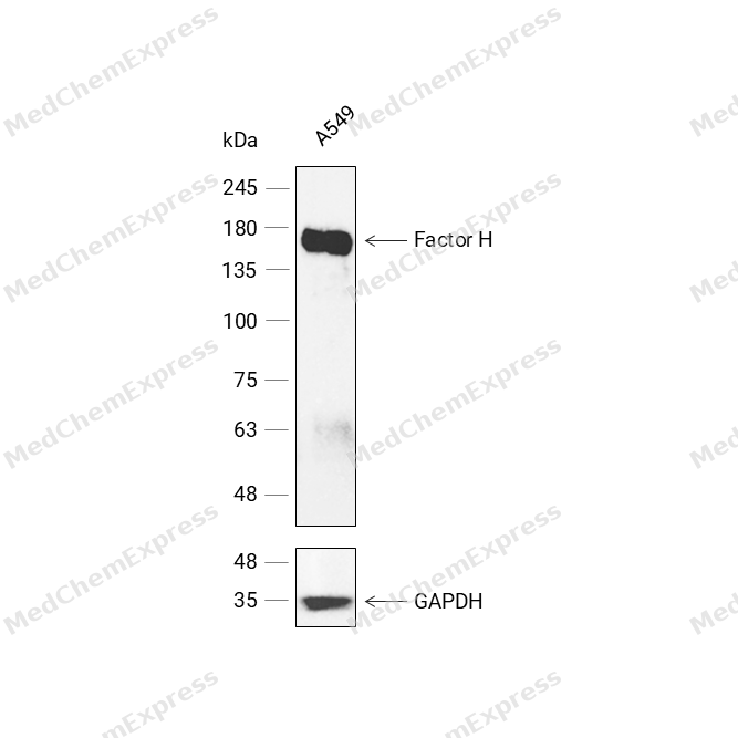 Western blot analysis was performed on protein extracts (30 μg) from A549 (lane 1) using Factor H antibody. Proteins were transferred onto a 0.45 μm PVDF membrane using a wet transfer system for 90 min. The membrane was then blocked with 5% nonfat milk in TBST (HY-K1025) for 1 h at room temperature. Thhe primary antibody (1:1000) and loading control antibody GAPDH Antibody (HRP) (HY-P80954A) (1:5000) were diluted in 5% nonfat milk in TBST and incubated with the membrane overnight at 4°C. After washing, the membrane of primary antibody was incubated with HRP-conjugated goat anti-rabbit/mouse IgG secondary antibody (HY-P8001/HY-P8004) (1:5000) diluted in 5% nonfat milk in TBST for 1 h at room temperature. Protein bands were visualized using an Ultra High Sensitivity ECL detection kit (HY-K1005).


Background
-
Function
Factor H is a Glycoprotein that plays an essential role in maintaining a well-balanced immune response by modulating complement activation. Acts as a soluble inhibitor of complement, where its binding to self markers such as glycan structures prevents complement activation and amplification on cell surfaces. Accelerates the decay of the complement alternative pathway (AP) C3 convertase C3bBb, thus preventing local formation of more C3b, the central player of the complement amplification loop. As a cofactor of the serine protease factor I, CFH also regulates proteolytic degradation of already-deposited C3b. In addition, mediates several cellular responses through interaction with specific receptors. For example, interacts with CR3/ITGAM receptor and thereby mediates the adhesion of human neutrophils to different pathogens. In turn, these pathogens are phagocytosed and destroyed; (Microbial infection) In the mosquito midgut, binds to the surface of parasite P.falciparum gametocytes and protects the parasite from alternative complement pathway-mediated elimination[1][2][3][4][5][6][7][8][9][10].
-
Subcellular Localization
Secreted
-
Expression
Tissue_specificity:Expressed in the retinal pigment epithelium (at protein level) (PubMed:25136834) . CFH is one of the most abundant complement components in blood where the liver is the major source of CFH protein in vivo. in addition, CFH is secreted by additional cell types including monocytes, fibroblasts, or endothelial cells (PubMed:2139673, PubMed:25136834, PubMed:2968404, PubMed:6444659) -
Isoforms & Post-Translational Modification
P08603 has 2 isomers: P08603-1: 139096 Da (predicted); P08603-2: 51034 Da (predicted).
Sulfated on tyrosine residues;According to a report, Asn-217 is not glycosylated (PubMed:17591618). Another study observed glycosylation at this position (PubMed:19139490) -
Subunit
Homodimer (PubMed:18005991, PubMed:19505476). Also forms homooligomers (PubMed:19505476). Interacts with complement protein C3b; this interaction inhibits complement activation (PubMed:16601698, PubMed:19503104, PubMed:20378178, PubMed:21285368, PubMed:28671664). Interacts with complement protein C3d (PubMed:20378178, PubMed:21285368, PubMed:29190743). Interacts with CR3/ITGAM; this interaction mediates adhesion of neutrophils to pathogens leading to pathogen clearance (PubMed:20008295, PubMed:9558116). Interacts with complement factor I (PubMed:28671664)
-
SwissProt ID
-
Synonyms
AHUS1; AMBP1; ARMD4; ARMS1; beta1H; CFH; CFHL3; complement factor H; isoform b; Factor H; factor H like 1; FHL1; HF1; HF2; HUS
-
Research Field
Immunology


Documentation
-
Data Sheet (262 KB)
-
SDS (252 KB)
- English - EN (252 KB)
- Français - FR (252 KB)
- Deutsch - DE (252 KB)
- Norwegian - NO (252 KB)
- Español - ES (252 KB)
- Swedish - SV (252 KB)
- Italian - IT (252 KB)
- Korean - KR (252 KB)
- Portuguese - PT (252 KB)
-
User Guide for Antibodies (1077 KB)
References
[1]. Kajander T, et al. Dual interaction of factor H with C3d and glycosaminoglycans in host-nonhost discrimination by complement. Proc Natl Acad Sci U S A. 2011 Feb 15;108(7):2897-902. [Content Brief]
[2]. Morgan HP, et al. Structural basis for engagement by complement factor H of C3b on a self surface. Nat Struct Mol Biol. 2011 Apr;18(4):463-70. [Content Brief]
[3]. Blaum BS, et al. Structural basis for sialic acid-mediated self-recognition by complement factor H. Nat Chem Biol. 2015 Jan;11(1):77-82. [Content Brief]
[4]. Wu J, et al. Structure of complement fragment C3b-factor H and implications for host protection by complement regulators. Nat Immunol. 2009 Jul;10(7):728-33. [Content Brief]
[5]. Kennedy AT, et al. Recruitment of Factor H as a Novel Complement Evasion Strategy for Blood-Stage Plasmodium falciparum Infection. J Immunol. 2016 Feb 1;196(3):1239-48. [Content Brief]
[6]. Hocking HG, et al. Structure of the N-terminal region of complement factor H and conformational implications of disease-linked sequence variations. J Biol Chem. 2008 Apr 4;283(14):9475-87. [Content Brief]
[7]. Simon N, et al. Malaria parasites co-opt human factor H to prevent complement-mediated lysis in the mosquito midgut. Cell Host Microbe. 2013 Jan 16;13(1):29-41. [Content Brief]
[8]. Xue X, et al. Regulator-dependent mechanisms of C3b processing by factor I allow differentiation of immune responses. Nat Struct Mol Biol. 2017 Aug;24(8):643-651. [Content Brief]
[9]. Losse J, et al. Factor H and factor H-related protein 1 bind to human neutrophils via complement receptor 3, mediate attachment to Candida albicans, and enhance neutrophil antimicrobial activity. J Immunol. 2010 Jan 15;184(2):912-21. [Content Brief]
[10]. DiScipio RG, et al. Human polymorphonuclear leukocytes adhere to complement factor H through an interaction that involves alphaMbeta2 (CD11b/CD18). J Immunol. 1998 Apr 15;160(8):4057-66. [Content Brief]

 Powered by Bioz
