



GM130 Antibody (YA1820)
(Synonyms: Golgin subfamily A member 2; 130 kDa cis-Golgi matrix protein; GM130; GM130 autoantigen; Golgin-95)Based on 1 publication(s) in Google Scholar
GM130 Antibody (YA1820) is a rabbit-derived non-conjugated IgG monoclonal antibody (Clone NO.: YA1820), targeting GM130. It can be used as a loading control antibody.
-
Host:
Rabbit
-
Isotype:
IgG
-
Application:
WB, IHC-P, ICC/IF, IP
-
Reactivity :
Human, Mouse, Rat, Monkey, Cow, Dog
-
Formulation:
Supplied in Rabbit IgG in 10mM phosphate buffered saline , pH 7.4, 150mM sodium chloride, 0.05% BSA, 0.02% sodium azide and 50% glycerol.
-
Conjugation:
Non-conjugated


To place orders, for customer services and technical support, please contact: MedChemExpress USA
Tel: 609-228-6898 E-mail: [email protected] [email protected]



Publications Citing Use of MedChemExpress (MCE) GM130 Antibody (YA1820)
More

Applications
| Application |
WB

WB: Western Blot
|
IHC-P
IHC-P: Immunohistochemistry-Paraffin
|
ICC/IF
ICC/IF: Immunocytochemistry/
Immunofluorescence |
IP
IP: Immunoprecipitation
|
|---|---|---|---|---|
| Dilution Ratio | 1:500-1:1000 | 1:50-1:100 | 1:50-1:200 | 1:50 |


Product Details
GM130 Antibody (YA1820) is a rabbit-derived non-conjugated IgG monoclonal antibody (Clone NO.: YA1820), targeting GM130. It can be used as a loading control antibody.
-
Host Rabbit

-
Clonality Recombinant,Monoclonal

-
Species ReactivityHuman, Mouse, Rat, Monkey, Cow, Dog
-
Observed Molecular WeightObserved band size: 130 kDa
Note: Due to possible protein modifications or aggregation, the molecular weight should be confirmed by actual measurement, and the predicted value is for reference only.

-
Calculated Molecular Weight Predicted band size: 113 kDa

A synthesized peptide derived from human GM130 aa1-57.
Endogenous
Affinity Chromatography
Non-conjugated
Unmodified
IgG


Product Properties
-
Appearance
Liquid
-
Formulation
Supplied in Rabbit IgG in 10mM phosphate buffered saline , pH 7.4, 150mM sodium chloride, 0.05% BSA, 0.02% sodium azide and 50% glycerol.
-
Concentration
Batch-dependent, Please check the COA for the concentration of each lot. Check Lot Concentration
-
Storage & Stability
Stored at -20°C for 1 year. Avoid repeated freeze / thaw cycles.
-
Shipping
Shipping with blue ice.


Publications (1)
-
Journal Impact Factor
-
Most Recent
-
Transl Neurodegener
NDST3 suppression restores lysosomal acidification and ameliorates amyloid-β and MAPT/tau pathology in Alzheimer's disease. [Abstract]2026 Apr 21;15(1):16. PMID: 42015299


Verification Images
-
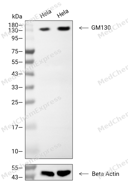 Western blot analysis of extracts from Hela (lane 2(20μg), Hela (lane 3(40μg), using GM130 Antibody. Proteins were transferred to a PVDF membrane and blocked with 5% BSA in TBST for 2 hour at room temperature. The primary antibody and Loading control antibody (Beta Actin, HY-P80438, 1/3000) was used in 5% BSA in TBST at 4°C overnight. Goat Anti-Mouse/Rabbit IgG-HRP Secondary Antibody (HY-P8004/HY-P8001, 1/10,000) was used for 1 hour at room temperature.
 -
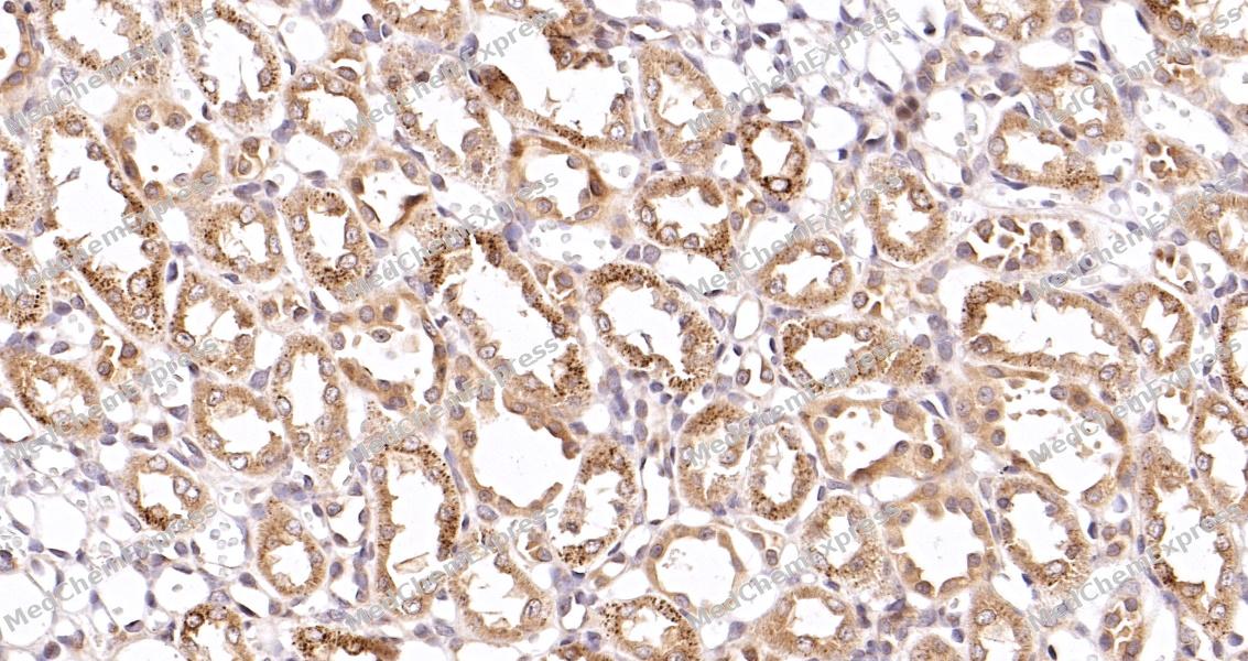 Immunohistochemical analysis of paraffin-embedded rat kidney tissue using GM130 Antibody. The section was pre-treated using heat mediated antigen retrieval with sodium citrate buffer (pH 6.0) for 8 minutes. The tissues were blocked in QuickBlock for 20 minutes at room temperature, washed with ddH2O and PBS, and then probed with the primary antibody at 1/100 dilution in 4℃ overnight. The detection was performed using an HRP conjugated compact polymer system. DAB was used as the chromogen. Tissues were counterstained with hematoxylin and mounted with DPX.
 -
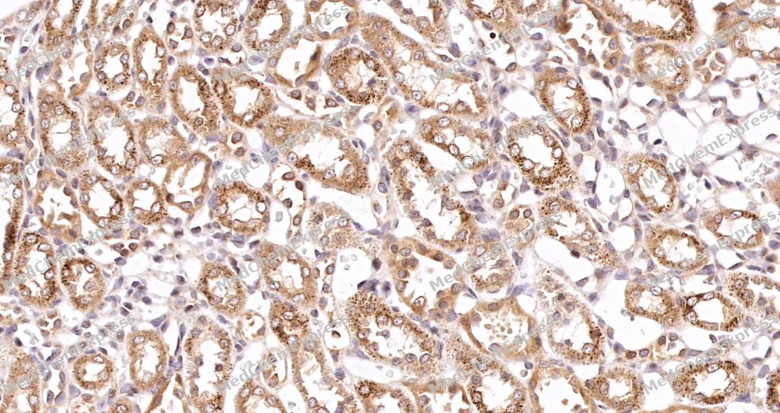 Immunohistochemical analysis of paraffin-embedded rat kidney tissue using GM130 Antibody. The section was pre-treated using heat mediated antigen retrieval with sodium citrate buffer (pH 6.0) for 8 minutes. The tissues were blocked in QuickBlock for 20 minutes at room temperature, washed with ddH2O and PBS, and then probed with the primary antibody at 1/100 dilution in 4℃ overnight. The detection was performed using an HRP conjugated compact polymer system. DAB was used as the chromogen. Tissues were counterstained with hematoxylin and mounted with DPX.
 -
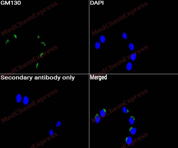 Immunocytochemistry analysis of Hela cells labeling GM130 with GM130 Antibody (HY-P82075) at 1/50 dilution. Cells were fixed in 4% paraformaldehyde for 15 minutes at room temperature, permeabilized with 0.1% Triton X-100 for 10 minutes at room temperature, then blocked with QuickBlock™ Blocking Buffer for Immunol Staining for 10 min at room temperature. Cells were then incubated with GM130 Antibody (HY-P82075) at 1/50 dilution in QuickBlock™ Blocking Buffer for Immunol Staining at 4 ℃. Alexa Fluor® 488-conjugated AffiniPure Goat Anti-Rabbit IgG H&L(HY-P8002, Green) was used as the secondary antibody at 1/1,000 dilution. PBS instead of the primary antibody was used as the secondary antibody only control. The Nuclear counterstain was DAPI (Blue).
 -
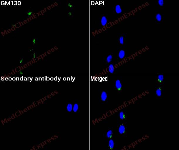 Immunocytochemistry analysis of HepG2 cells labeling GM130 with GM130 Antibody (HY-P82075) at 1/50 dilution. Cells were fixed in 4% paraformaldehyde for 15 minutes at room temperature, permeabilized with 0.1% Triton X-100 for 10 minutes at room temperature, then blocked with QuickBlock™ Blocking Buffer for Immunol Staining for 10 min at room temperature. Cells were then incubated with GM130 Antibody (HY-P82075)at 1/50 dilution in QuickBlock™ Blocking Buffer for Immunol Staining at 4 ℃. Alexa Fluor® 488-conjugated AffiniPure Goat Anti-Rabbit IgG H&L(HY-P8002,Green) was used as the secondary antibody at 1/1,000 dilution. PBS instead of the primary antibody was used as the secondary antibody only control. The Nuclear counterstain was DAPI (Blue).


Background
-
Function
GM130 is a Peripheral membrane component of the cis-Golgi stack that acts as a membrane skeleton that maintains the structure of the Golgi apparatus, and as a vesicle thether that facilitates vesicle fusion to the Golgi membrane (Probable). Required for normal protein transport from the endoplasmic reticulum to the Golgi apparatus and the cell membrane. Together with p115/USO1 and STX5, involved in vesicle tethering and fusion at the cis-Golgi membrane to maintain the stacked and inter-connected structure of the Golgi apparatus. Plays a central role in mitotic Golgi disassembly: phosphorylation at Ser-37 by CDK1 at the onset of mitosis inhibits the interaction with p115/USO1, preventing tethering of COPI vesicles and thereby inhibiting transport through the Golgi apparatus during mitosis. Also plays a key role in spindle pole assembly and centrosome organization. Promotes the mitotic spindle pole assembly by activating the spindle assembly factor TPX2 to nucleate microtubules around the Golgi and capture them to couple mitotic membranes to the spindle: upon phosphorylation at the onset of mitosis, GOLGA2 interacts with importin-alpha via the nuclear localization signal region, leading to recruit importin-alpha to the Golgi membranes and liberate the spindle assembly factor TPX2 from importin-alpha. TPX2 then activates AURKA kinase and stimulates local microtubule nucleation. Upon filament assembly, nascent microtubules are further captured by GOLGA2, thus linking Golgi membranes to the spindle. Regulates the meiotic spindle pole assembly, probably via the same mechanism. Also regulates the centrosome organization. Also required for the Golgi ribbon formation and glycosylation of membrane and secretory proteins[1][2][3][4][5][6].
-
Subcellular Localization
Golgi apparatus, cis-Golgi network membrane; Peripheral membrane protein; Cytoplasmic side; Endoplasmic reticulum-Golgi intermediate compartment membrane; Peripheral membrane protein; Cytoplasmic side; Cytoplasm, cytoskeleton, spindle pole
-
Isoforms & Post-Translational Modification
Q08379 has 2 isomers: Q08379-1: 113086 Da (predicted); Q08379-2: 70473 Da (predicted).
Cleaved by caspases at the onset of apoptosis;Methylation by PRMT5 is required for Golgi ribbon formation. While dimethylation at Arg-30 and Arg-35 are confirmed in vivo, it is unclear whether Arg-18 is methylated in vivo;Phosphorylated at Ser-37 by CDK1 at the onset of mitosis, inhibiting the interaction with p115/USO1 and triggering Golgi disassembly (PubMed:20421892, PubMed:26165940). Phosphorylated at Ser-37 in prophase as the Golgi complex starts to break down, and remains phosphorylated during further breakdown and partitioning of the Golgi fragments in metaphase and anaphase. In telophase, GM130 is dephosphorylated by PP2A as the Golgi fragments start to reassemble (By similarity) -
Subunit
Homodimer, may assemble into homohexamers (PubMed:26363069). Homotetramer; forms a parallel homotetramer with a flexible rod-like structure that can give rise to I- and Y-shaped conformations (By similarity). Interacts with GORASP1/GRASP65 (PubMed:16489344, PubMed:18045989, PubMed:26363069). The homooligomer forms a complex with GORASP1 with a 1:1 stoichiometry (By similarity). Interacts with RAB1B that has been activated by GTP-binding (PubMed:11306556). Interacts with p115/USO1; interaction with p115/USO1 inhibits interaction with STX5 and/or RAB1B. Interacts with STX5 (By similarity). Interacts with ZFPL1 (PubMed:18323775). Interacts with AKAP450/AKAP9; leading to recruit AKAP450/AKAP9 to the cis-Golgi (PubMed:19242490)
-
SwissProt ID
-
Synonyms
Golgin subfamily A member 2; 130 kDa cis-Golgi matrix protein; GM130; GM130 autoantigen; Golgin-95
-
Research Field
Tags & Cell Markers
Documentation
-
Data Sheet (258 KB)
-
SDS (252 KB)
- English - EN (252 KB)
- Français - FR (252 KB)
- Deutsch - DE (252 KB)
- Norwegian - NO (252 KB)
- Español - ES (252 KB)
- Swedish - SV (252 KB)
- Italian - IT (252 KB)
- Korean - KR (252 KB)
- Portuguese - PT (252 KB)
-
User Guide for Antibodies (1077 KB)

 Powered by Bioz
