



HDAC6 Antibody (YA5795)
(Synonyms: HDAC6; KIAA0901; JM21; Histone deacetylase 6; HD6)Based on 1 Customer Validation
HDAC6 Antibody (YA5795) is a Rabbit-derived and non-conjugated IgG monoclonal antibody, targeting to HDAC6.
-
Host:
Rabbit
-
Isotype:
IgG
-
Application:
WB, IHC-P, ICC/IF, IP, ELISA
-
Reactivity :
Human, Mouse, Rat
-
Formulation:
Supplied in PBS, 50% glycerol, 0.05% Proclin 300, 0.05%BSA
-
Conjugation:
Non-conjugated


To place orders, for customer services and technical support, please contact: MedChemExpress USA
Tel: 609-228-6898 E-mail: [email protected] [email protected]





Applications
| Application |
IHC-P

IHC-P: Immunohistochemistry-Paraffin
|
WB
WB: Western Blot
|
ICC/IF
ICC/IF: Immunocytochemistry/
Immunofluorescence |
ELISA
ELISA: Enzyme Linked Immunosorbent Assay
|
IP
IP: Immunoprecipitation
|
|---|---|---|---|---|---|
| Dilution Ratio | 1:2000-1:10000 | 1:2000-1:10000 | 1:200-1:1000 | 1:5000-1:20000 | 1:50-1:200 |


Product Details
HDAC6 Antibody (YA5795) is a Rabbit-derived and non-conjugated IgG monoclonal antibody, targeting to HDAC6.
-
Host Rabbit

-
Clonality Monoclonal

-
Species ReactivityHuman, Mouse, Rat
-
Observed Molecular WeightObserved band size: 160 kDa
Note: Due to possible protein modifications or aggregation, the molecular weight should be confirmed by actual measurement, and the predicted value is for reference only.

-
Calculated Molecular Weight Predicted band size: 131 kDa

Protein A
Non-conjugated
Unmodified
IgG


Product Properties
-
Appearance
Solution
-
Formulation
Supplied in PBS, 50% glycerol, 0.05% Proclin 300, 0.05%BSA
-
Concentration
Batch-dependent, Please check the COA for the concentration of each lot. Check Lot Concentration
-
Storage & Stability
Stored at -20°C for 1 year. Avoid repeated freeze / thaw cycles.
-
Shipping
Shipping with blue ice.


Verification Images
-
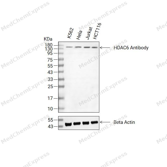 Western blot analysis of extracts from K562(lane 2(20ug) , Hela(lane 3(20ug) and Jurkat(lane 4(20ug),HCT116(lane 5(20ug) using HDAC6 Antibody (HY-P86103) Rabbit mAb. Proteins were transferred to a PVDF membrane and blocked with 5% non-fat milk in TBST for 2 hour at room temperature. The primary antibody (1/1000) and Loading control antibody (Beta Actin, HY-P83730, 1/10000) was used in 5% non-fat milk in TBST at 4°C overnight. Goat Anti-Mouse/Rabbit IgG-HRP Secondary Antibody (1/10000) was used for 1 hour at room temperature.
 -
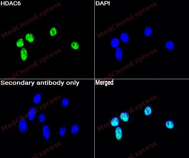 Immunocytochemistry analysis of C6 cells labeling HDAC6 with HDAC6 Antibody (HY-P86103) at 1/50 dilution. Cells were fixed in 4% paraformaldehyde for 15 minutes at room temperature, permeabilized with 0.1% Triton X-100 for 10 minutes at room temperature, then blocked with BSA for Immunol Staining for 10 min at room temperature. Cells were then incubated with HDAC6 Antibody (HY-P86103) at 1/50 dilution in BSA for Immunol Staining at 4 ℃ overnight. AF488-conjugated AffiniPure Goat Anti-Rabbit IgG H&L(HY-P8002, Green) was used as the secondary antibody at 1/1,000 dilution. PBS instead of the primary antibody was used as the secondary antibody only control. The Nuclear counterstain was DAPI (Blue).
 -
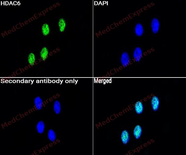 Immunocytochemistry analysis of HeLa cells labeling HDAC6 with HDAC6 Antibody (HY-P86103) at 1/50 dilution. Cells were fixed in 4% paraformaldehyde for 15 minutes at room temperature, permeabilized with 0.1% Triton X-100 for 10 minutes at room temperature, then blocked with BSA for Immunol Staining for 10 min at room temperature. Cells were then incubated with HDAC6 Antibody (HY-P86103) at 1/50 dilution in BSA for Immunol Staining at 4 ℃ overnight. AF488-conjugated AffiniPure Goat Anti-Rabbit IgG H&L(HY-P8002,Green) was used as the secondary antibody at 1/1,000 dilution. PBS instead of the primary antibody was used as the secondary antibody only control. The Nuclear counterstain was DAPI (Blue).


Background
-
Function
HDAC6 (histone deacetylase 6) is a unique class IIb histone deacetylase that localizes predominantly in the cytoplasm and preferentially targets non-histone substrates rather than chromatin-associated proteins[1][2]. HDAC6 regulates fundamental cellular functions through deacetylation of α-tubulin, Hsp90, and ubiquitin-associated protein complexes, thereby controlling microtubule dynamics, protein trafficking, stress responses, and protein quality-control mechanisms[2][3][4]. Mechanistically, HDAC6 functions at the intersection of the ubiquitin-proteasome system and autophagy-related pathways, where its ubiquitin-binding capability facilitates the processing and clearance of misfolded or aggregated proteins[3][4]. Therefore, HDAC6 has emerged as an important regulator of cellular proteostasis and cytoskeletal remodeling in both physiological and pathological contexts[2][3]. In disease models, HDAC6 has been implicated in cancer progression, neurodegenerative disorders, and inflammatory conditions through its effects on cell motility, intracellular transport, protein aggregation, and stress signaling pathways[2][4][5]. Increased HDAC6 activity promotes α-tubulin deacetylation and influences processes associated with tumor cell migration and metastasis, while modulation of HDAC6 activity alters axonal transport and protein aggregate handling in neurodegenerative disease models[4][5]. Compared with related HDAC isoforms, HDAC6 is distinguished by its predominantly cytoplasmic localization, preference for non-histone substrates, dual catalytic domains, and zinc-finger ubiquitin-binding domain, features that confer specialized biological functions not shared by most nuclear HDAC family members[2][5]. For experimental applications, HDAC6-selective inhibitors have become widely used chemical probes because they enable investigation of HDAC6-dependent signaling and protein homeostasis pathways while potentially reducing the off-target effects associated with pan-HDAC inhibition[3][6].
-
Subcellular Localization
Cytoplasm; Cytoplasm, cytoskeleton; Nucleus; Perikaryon; Cell projection, dendrite; Cell projection, axon; Cell projection, cilium; Cytoplasm, cytoskeleton, microtubule organizing center, centrosome; Cytoplasm, cytoskeleton, cilium basal body
-
Expression
Induction: (Microbial infection) Up-regulated by the presence of SARS-CoV-2 N protein during viral infection (PubMed:39135075) . The SARS-CoV-2 N protein enhances HDAC6 stability during infection by reducing its proteasomal degradation, leading to elevated HDAC6 levels (PubMed:39135075) -
Isoforms & Post-Translational Modification
Q9UBN7 has 2 isomers: Q9UBN7-1: 131419 Da (predicted); Q9UBN7-2: 114361 Da (predicted).
Phosphorylated by AURKA; phosphorylation increases HDAC6-mediated deacetylation of alpha-tubulin and subsequent disassembly of cilia;Ubiquitinated. Its polyubiquitination however does not lead to its degradation;Sumoylated in vitro -
Subunit
Forms a trimeric complex in the nucleus consisting of BANP, HDAC6 and KHDRBS1/SAM68; HDAC6 keeps KHDRBS1 in a deacetylated state which inhibits the inclusion of CD44 alternate exons (PubMed:26080397).
-
SwissProt ID
-
Synonyms
HDAC6; KIAA0901; JM21; Histone deacetylase 6; HD6


Documentation
-
Data Sheet (262 KB)
-
SDS (251 KB)
- English - EN (251 KB)
- Français - FR (251 KB)
- Deutsch - DE (251 KB)
- Norwegian - NO (251 KB)
- Español - ES (251 KB)
- Swedish - SV (251 KB)
- Italian - IT (251 KB)
- Korean - KR (251 KB)
- Portuguese - PT (251 KB)
-
User Guide for Antibodies (1077 KB)
References
[1]. Park SY, et al. A short guide to histone deacetylases including recent progress on class II enzymes. Exp Mol Med. 2020 Feb;52(2):204-212. [Content Brief]
[2]. Simões-Pires C, et al. HDAC6 as a target for neurodegenerative diseases: what makes it different from the other HDACs? Mol Neurodegener. 2013 Jan 29;8:7. doi: 10.1186/1750-1326-8-7. PMID: 23356410; PMCID: PMC3615964. [Content Brief]
[3]. Brindisi M, et al. Old but Gold: Tracking the New Guise of Histone Deacetylase 6 (HDAC6) Enzyme as a Biomarker and Therapeutic Target in Rare Diseases. J Med Chem. 2020 Jan 9;63(1):23-39. [Content Brief]
[4]. HDAC6 gene information from NCBI.
[5]. Li G, et al. HDAC6 α-tubulin deacetylase: a potential therapeutic target in neurodegenerative diseases. J Neurol Sci. 2011 May 15;304(1-2):1-8. [Content Brief]
[6]. Zhang Y, et al. Tanshinone IIA sodium sulfonate facilitates endocytic HMGB1 uptake. Biochem Pharmacol. 2012 Dec 1;84(11):1492-500. [Content Brief]

 Powered by Bioz
