



Hsp90 beta Antibody (YA355)
(Synonyms: HSP90B, HSPC2, HSPC3, HSPCB, HSP90AB1, Heat shock protein HSP 90-beta, HSP 90, Heat shock 84 kDa, Heat shock protein family C member 3, HSP 84, HSP84)
Based on 1 Customer Validation
Hsp90 beta Antibody (YA355) is a Rabbit-derived and non-conjugated IgG monoclonal antibody, targeting to Hsp90 beta.
-
Host:
Rabbit
-
Isotype:
IgG
-
Application:
WB, IHC-P, ICC/IF, FC
-
Reactivity :
Human, Mouse, Rat
-
Formulation:
Supplied in 1*TBS (pH7.4), 0.05% BSA and 40% Glycerol. Preservative: 0.05% Sodium Azide.
-
Conjugation:
Non-conjugated


To place orders, for customer services and technical support, please contact: MedChemExpress USA
Tel: 609-228-6898 E-mail: [email protected] [email protected]





Applications
| Application |
WB

WB: Western Blot
|
ICC/IF
ICC/IF: Immunocytochemistry/
Immunofluorescence |
IHC-P
IHC-P: Immunohistochemistry-Paraffin
|
FC
FC: Flow Cytometry
|
IP
IP: Immunoprecipitation
|
|---|---|---|---|---|---|
| Dilution Ratio | 1:1000-1:10000 | 1:100-1:500 | 1:50-1:200 | 1:50-1:100 | Use at an assay dependent concentration. |


Product Details
Hsp90 beta Antibody (YA355) is a Rabbit-derived and non-conjugated IgG monoclonal antibody, targeting to Hsp90 beta.
-
Host Rabbit

-
Clonality Recombinant,Monoclonal

-
Species ReactivityHuman, Mouse, Rat
-
Observed Molecular WeightObserved band size: 90-95 kDa
Note: Due to possible protein modifications or aggregation, the molecular weight should be confirmed by actual measurement, and the predicted value is for reference only.

-
Calculated Molecular Weight Predicted band size: 83 kDa

Entrez Gene: 3326 Human ; 15516 Mouse ; 301252 Rat
SwissProt: P08238 Human ; P11499 Mouse ; P34058 Rat
OMIM: 140572 Human
Synthetic peptide corresponding to Human Hsp90 beta.AA range:485-724.
Endogenous
Protein A affinity purified.
Non-conjugated
Unmodified
IgG


Product Properties
-
Appearance
Solution
-
Formulation
Supplied in 1*TBS (pH7.4), 0.05% BSA and 40% Glycerol. Preservative: 0.05% Sodium Azide.
-
Concentration
Batch-dependent, Please check the COA for the concentration of each lot. Check Lot Concentration
-
Storage & Stability
Stored at -20°C for 1 year. Avoid repeated freeze / thaw cycles.
-
Shipping
Shipping with blue ice.


Verification Images
-
 Western blot analysis was performed on protein extracts (25 μg) from 293T (lane 2), K562 (lane 3), RAW264.7 (lane 4), A431 (lane 5), A549 (lane 6), and NIH/3T3 (lane 7) using Hsp90 beta antibody. Proteins were transferred onto a 0.45 μm PVDF membrane using the Trans-Blot® Turbo™ system for 13 min. The membrane was then blocked with 5% nonfat milk in TBST (HY-K1025) for 1 h at room temperature. The primary antibody (1:1000) and loading control antibody GAPDH Antibody (HRP) (HY-P80954A) (1:5000) were diluted in 5% nonfat milk in TBST and incubated with the membrane overnight at 4°C. After washing, the membrane of primary antibody was incubated with HRP-conjugated goat anti-rabbit/mouse IgG secondary antibody (HY-P8001/HY-P8004) (1:5000) diluted in 5% nonfat milk in TBST for 1 h at room temperature. Protein bands were visualized using an Ultra High Sensitivity ECL detection kit (HY-K1005).
 -
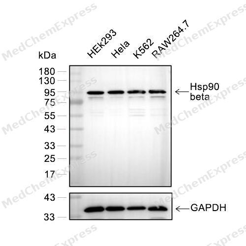 Western blot analysis of extracts from HEK293(lane 2(20μg), Hela(lane 3(20μg), K562(lane 4(20μg) ) ,RAW264.7(lane 5(20μg) ) using Hsp90 beta (HY-P80187) Rabbit mAb. Proteins were transferred to a PVDF membrane and blocked with 5% BSA in TBST for 2 hour at room temperature. The primary antibody (HY-P80187, 1/1000) and Loading control antibody (GAPDH, HY-P80954, 1/3000) was used in 5% BSA in TBST at 4°C overnight. Goat Anti-Mouse/Rabbit IgG-HRP Secondary Antibody (HY-P8004/HY-P8001, 1/10,000) was used for 1 hour at room temperature.
 -
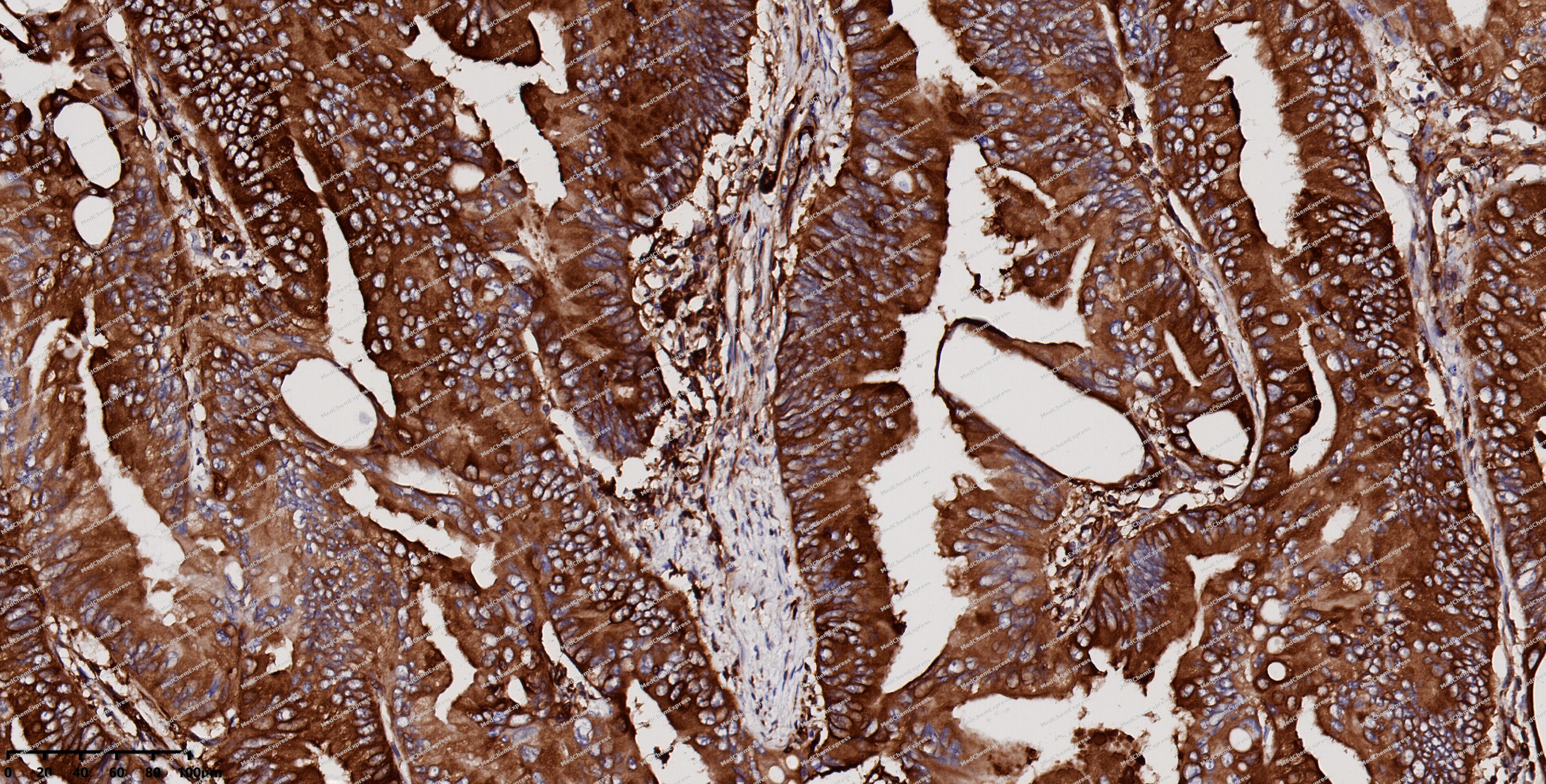 Immunohistochemical analysis of paraffin-embedded human colon cancer using Hsp90 beta antibody. The section was pre-treated using heat mediated antigen retrieval with EDTA (pH 9.0) for 14 minutes. The tissues were blocked in 5% BSA for 30 minutes at room temperature, washed with TBST, and then probed with the primary antibody (HY-P80187, 1/200) for 30 minutes at room temperature. The detection was performed using Polymer HRP-conjugated Goat Anti-Mouse/Rabbit lgG(H&L) secondary antibody (HY-P83652). DAB was used as the chromogen. Tissues were counterstained with hematoxylin and mounted with DPX.
 -
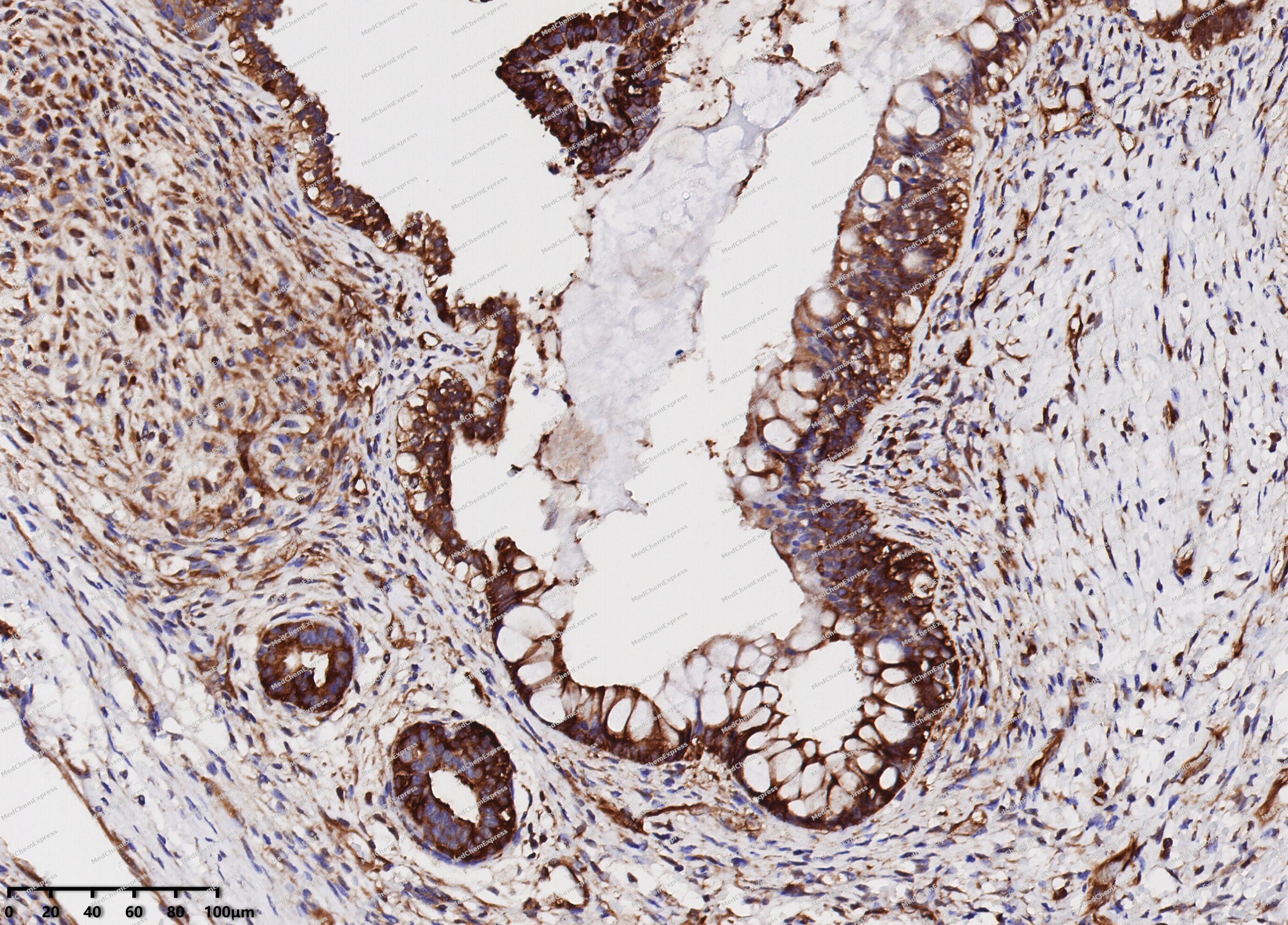 Immunohistochemical analysis of paraffin-embedded human ovarian cancer using Hsp90 beta antibody. The section was pre-treated using heat mediated antigen retrieval with EDTA (pH 9.0) for 14 minutes. The tissues were blocked in 5% BSA for 30 minutes at room temperature, washed with TBST, and then probed with the primary antibody (HY-P80187, 1/200) for 30 minutes at room temperature. The detection was performed using Polymer HRP-conjugated Goat Anti-Mouse/Rabbit lgG(H&L) secondary antibody (HY-P83652). DAB was used as the chromogen. Tissues were counterstained with hematoxylin and mounted with DPX.
 -
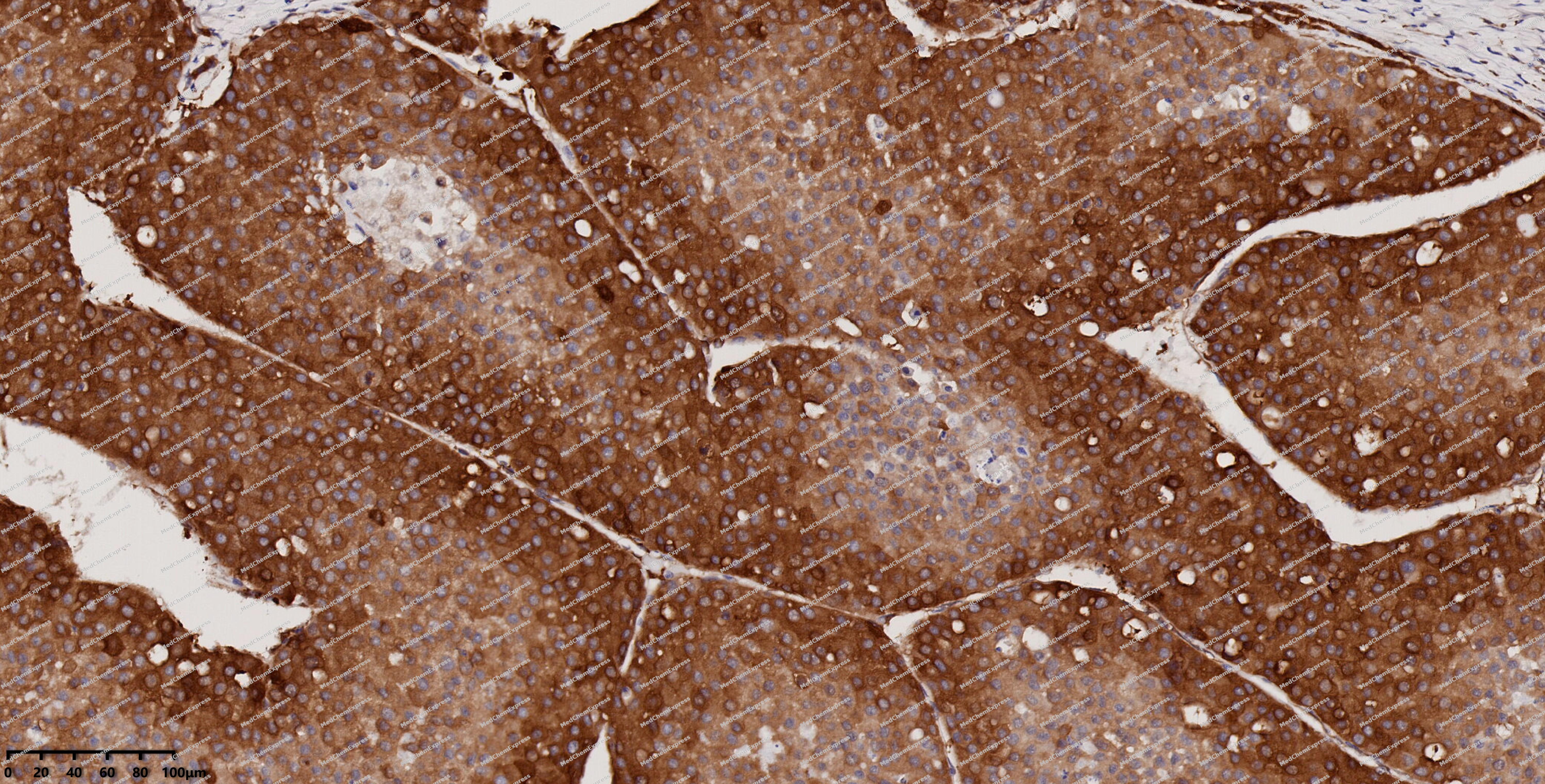 Immunohistochemical analysis of paraffin-embedded human liver cancer using Hsp90 beta antibody. The section was pre-treated using heat mediated antigen retrieval with EDTA (pH 9.0) for 14 minutes. The tissues were blocked in 5% BSA for 30 minutes at room temperature, washed with TBST, and then probed with the primary antibody (HY-P80187, 1/200) for 30 minutes at room temperature. The detection was performed using Polymer HRP-conjugated Goat Anti-Mouse/Rabbit lgG(H&L) secondary antibody (HY-P83652). DAB was used as the chromogen. Tissues were counterstained with hematoxylin and mounted with DPX.
 -
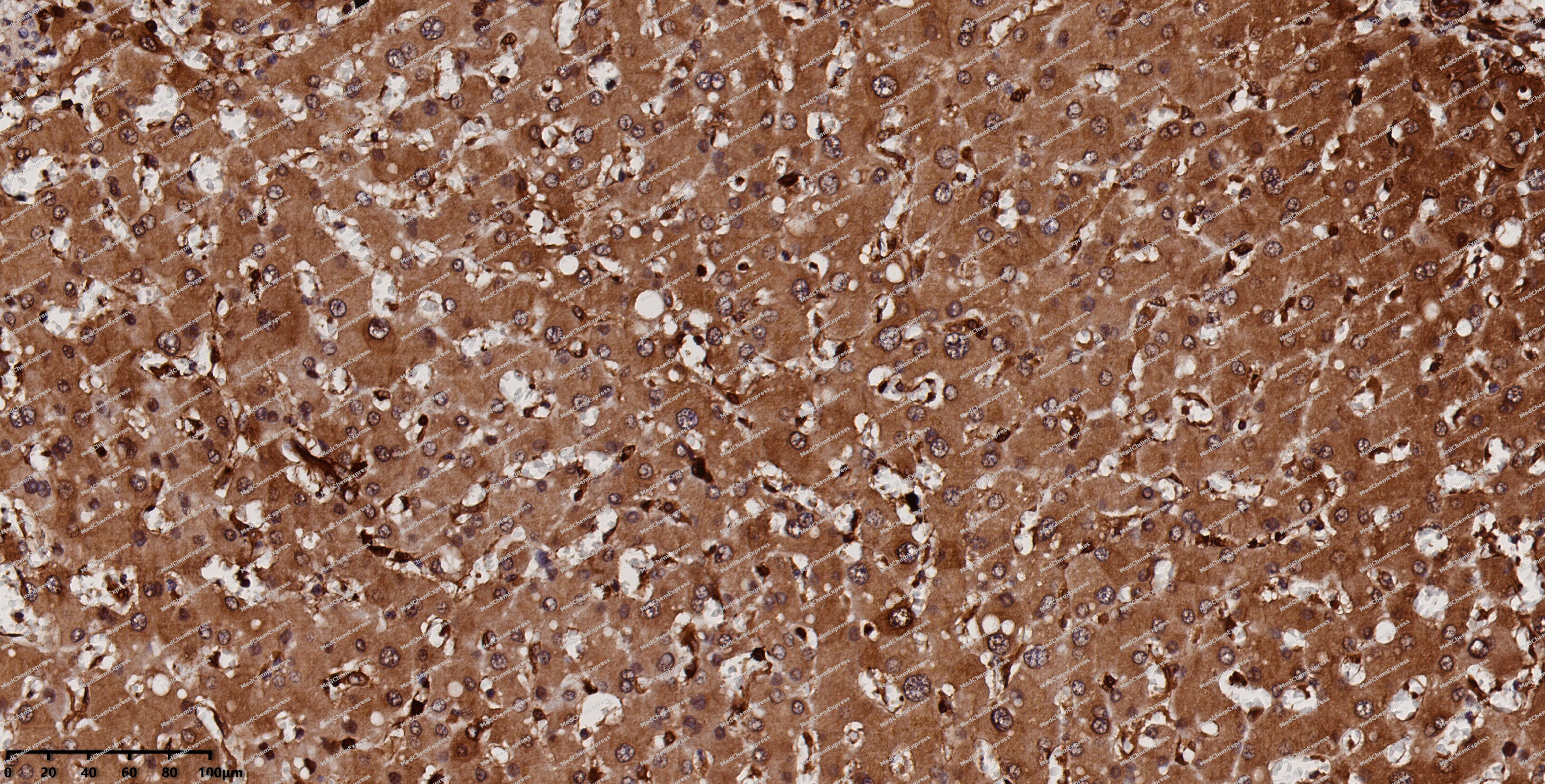 Immunohistochemical analysis of paraffin-embedded human liver using Hsp90 beta antibody. The section was pre-treated using heat mediated antigen retrieval with EDTA (pH 9.0) for 14 minutes. The tissues were blocked in 5% BSA for 30 minutes at room temperature, washed with TBST, and then probed with the primary antibody (HY-P80187, 1/200) for 30 minutes at room temperature. The detection was performed using Polymer HRP-conjugated Goat Anti-Mouse/Rabbit lgG(H&L) secondary antibody (HY-P83652). DAB was used as the chromogen. Tissues were counterstained with hematoxylin and mounted with DPX.
 -
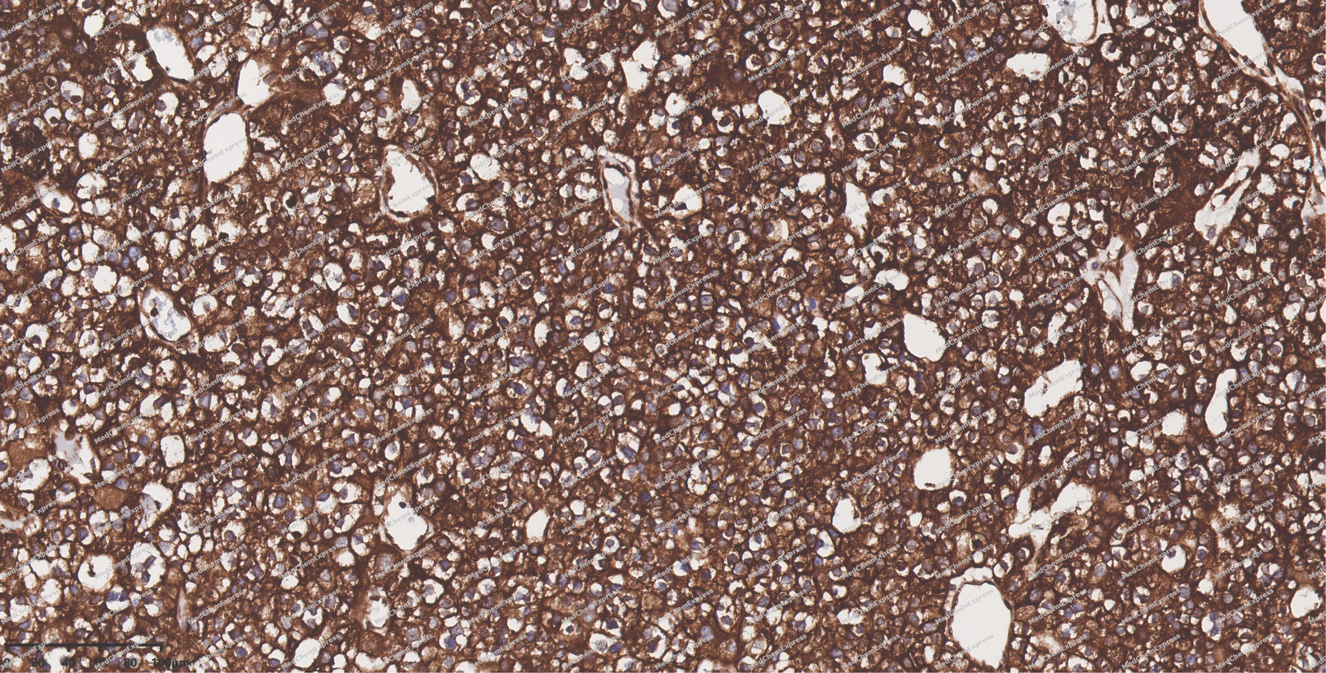 Immunohistochemical analysis of paraffin-embedded human thyroid cancer using Hsp90 beta antibody. The section was pre-treated using heat mediated antigen retrieval with EDTA (pH 9.0) for 14 minutes. The tissues were blocked in 5% BSA for 30 minutes at room temperature, washed with TBST, and then probed with the primary antibody (HY-P80187, 1/200) for 30 minutes at room temperature. The detection was performed using Polymer HRP-conjugated Goat Anti-Mouse/Rabbit lgG(H&L) secondary antibody (HY-P83652). DAB was used as the chromogen. Tissues were counterstained with hematoxylin and mounted with DPX.
 -
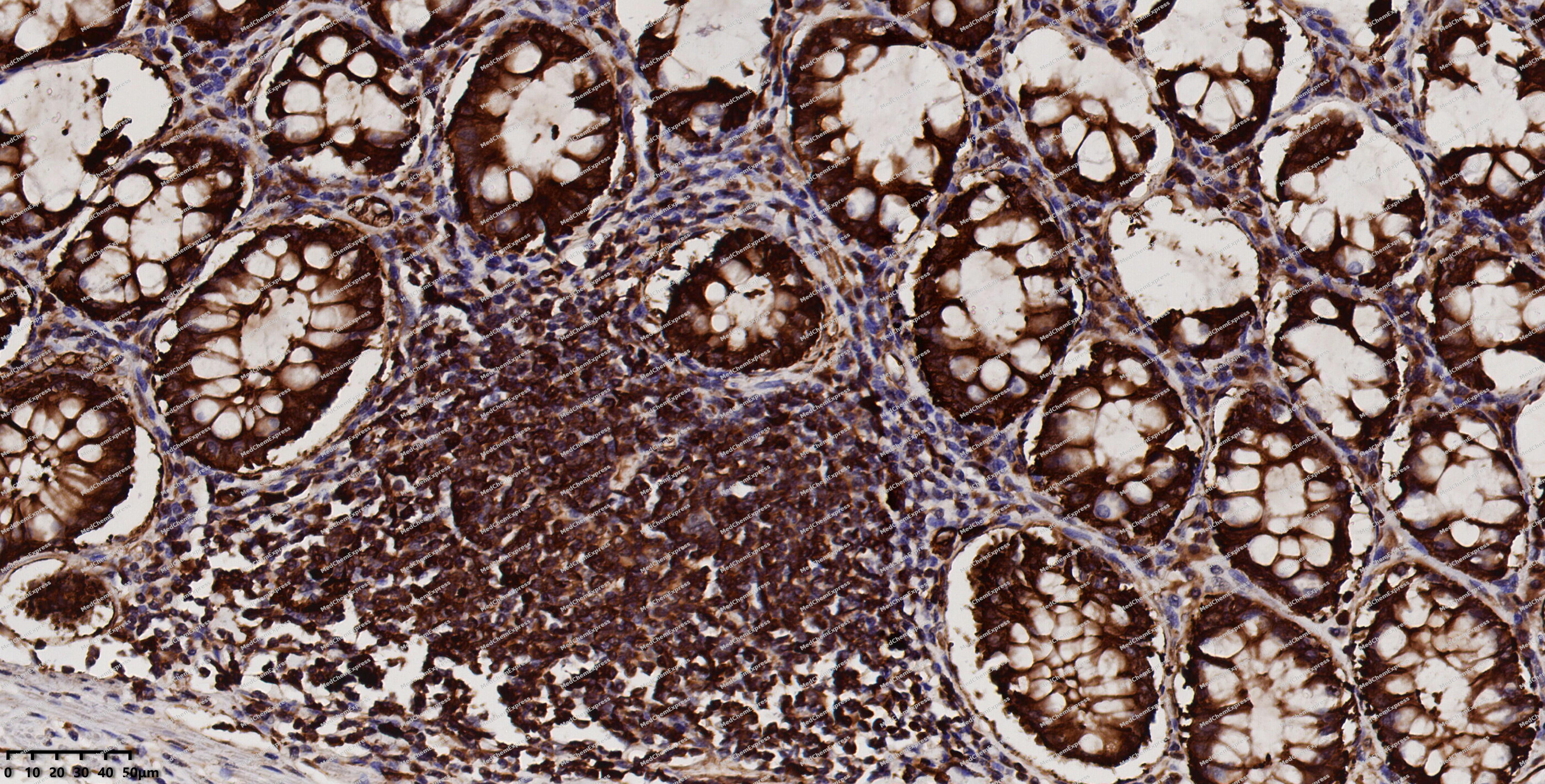 Immunohistochemical analysis of paraffin-embedded human colon using Hsp90 beta antibody. The section was pre-treated using heat mediated antigen retrieval with EDTA (pH 9.0) for 14 minutes. The tissues were blocked in 5% BSA for 30 minutes at room temperature, washed with TBST, and then probed with the primary antibody (HY-P80187, 1/200) for 30 minutes at room temperature. The detection was performed using Polymer HRP-conjugated Goat Anti-Mouse/Rabbit lgG(H&L) secondary antibody (HY-P83652). DAB was used as the chromogen. Tissues were counterstained with hematoxylin and mounted with DPX.
 -
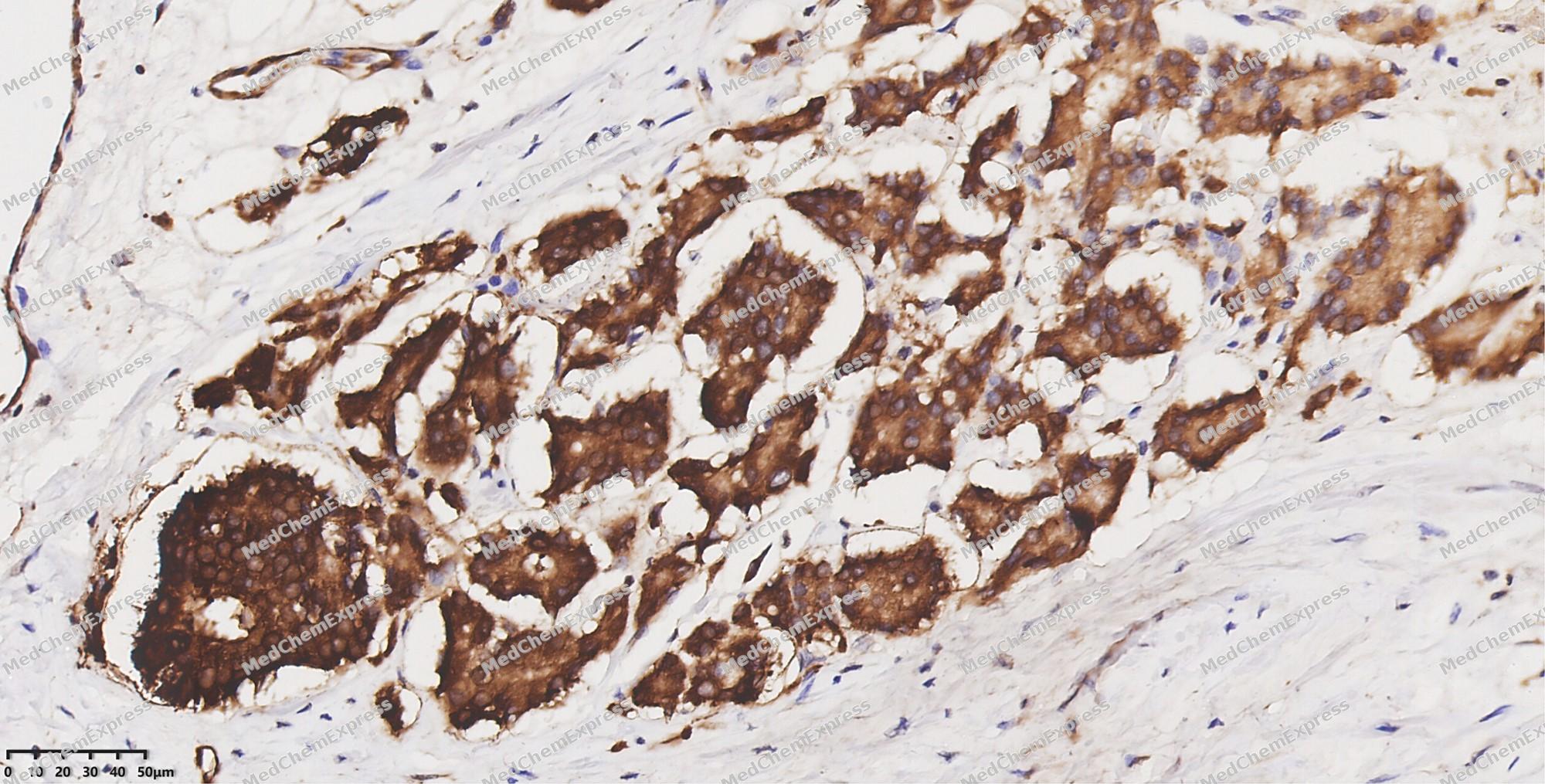 Immunohistochemical analysis of paraffin-embedded human prostate cancer using Hsp90 beta antibody. The section was pre-treated using heat mediated antigen retrieval with EDTA (pH 9.0) for 14 minutes. The tissues were blocked in 5% BSA for 30 minutes at room temperature, washed with TBST, and then probed with the primary antibody (HY-P80187, 1/200) for 30 minutes at room temperature. The detection was performed using Polymer HRP-conjugated Goat Anti-Mouse/Rabbit lgG(H&L) secondary antibody (HY-P83652). DAB was used as the chromogen. Tissues were counterstained with hematoxylin and mounted with DPX.
 -
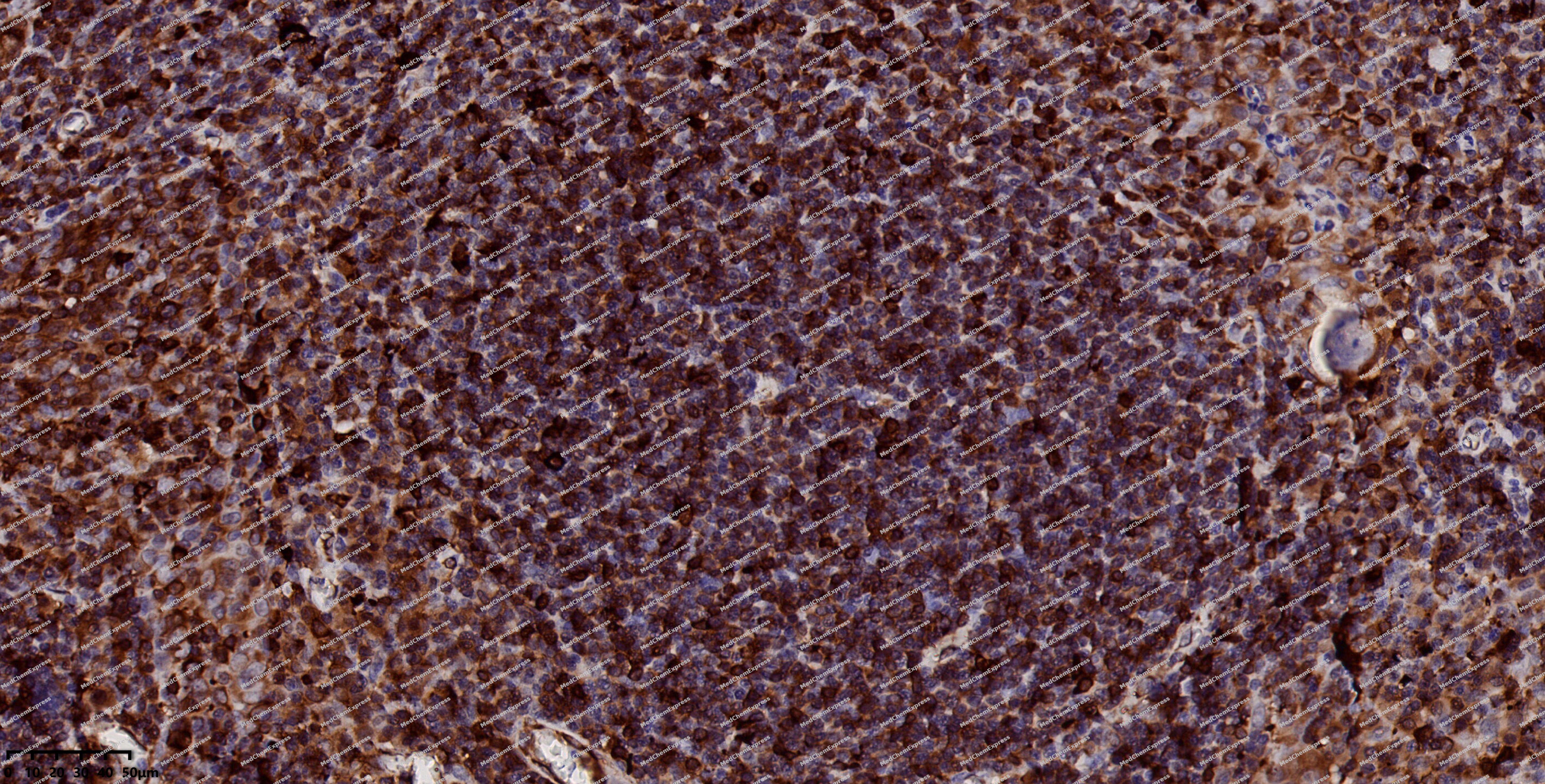 Immunohistochemical analysis of paraffin-embedded human tonsil using Hsp90 beta antibody. The section was pre-treated using heat mediated antigen retrieval with EDTA (pH 9.0) for 14 minutes. The tissues were blocked in 5% BSA for 30 minutes at room temperature, washed with TBST, and then probed with the primary antibody (HY-P80187, 1/200) for 30 minutes at room temperature. The detection was performed using Polymer HRP-conjugated Goat Anti-Mouse/Rabbit lgG(H&L) secondary antibody (HY-P83652). DAB was used as the chromogen. Tissues were counterstained with hematoxylin and mounted with DPX.
 -
 Flow cytometric analysis of 1X10⁶ HeLa cells labeling Hsp90 beta Antibody (HY-P80187, red). Cells were fixed with 4% paraformaldehyde and permeabilised with 90% methanol. Then stained with the primary antibody at 1/100 dilution for an hour at 4℃. AF488-conjugated Goat Anti-Rabbit IgG H&L (HY-P8002) was used as the secondary antibody at 1/1,000 dilution for 30 minutes at 4℃. Rabbit IgG Isotype Control (HY-P80879, blue) was used as the isotype control, cells without incubation with primary antibody were used as the unlabeled control (black).
 -
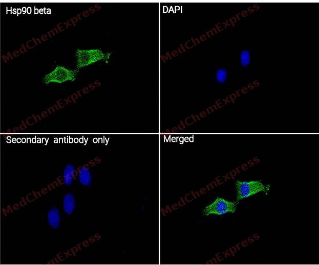 Immunocytochemistry analysis of HeLa cells labeling Hsp90 beta with Hsp90 beta Antibody (HY-P80187) at 1/200 dilution . Cells were fixed in 4% paraformaldehyde for 15 minutes at room temperature, permeabilized with 0.1% Triton X-100 in PBS for 15 minutes at room temperature, then blocked with quick block buffer for 10 minutes at room temperature. Cells were then incubated with Hsp90 beta Antibody (HY-P80187) at 1/200 dilution in quick block buffer overnight at 4 ℃. AF488-conjugated Goat Anti-Rabbit IgG H&L(HY-P8002, Green) was used as the secondary antibody at 1/1,000 dilution. PBS instead of the primary antibody was used as the secondary antibody only control. The Nuclear counterstain was DAPI (Blue).
 -
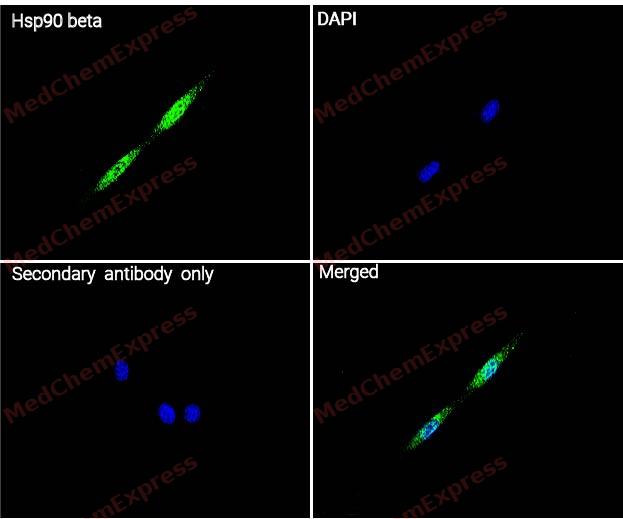 Immunocytochemistry analysis of NIH/3T3 cells labeling Hsp90 beta with Hsp90 beta Antibody (HY-P80187) at 1/200 dilution . Cells were fixed in 4% paraformaldehyde for 15 minutes at room temperature, permeabilized with 0.1% Triton X-100 in PBS for 15 minutes at room temperature, then blocked with quick block buffer for 10 minutes at room temperature. Cells were then incubated with Hsp90 beta Antibody (HY-P80187) at 1/200 dilution in quick block buffer overnight at 4 ℃. AF488-conjugated Goat Anti-Rabbit IgG H&L(HY-P8002, Green) was used as the secondary antibody at 1/1,000 dilution. PBS instead of the primary antibody was used as the secondary antibody only control. The Nuclear counterstain was DAPI (Blue).


Background
-
Function
Hsp90 beta is a Molecular chaperone that promotes the maturation, structural maintenance and proper regulation of specific target proteins involved for instance in cell cycle control and signal transduction. Undergoes a functional cycle linked to its ATPase activity. This cycle probably induces conformational changes in the client proteins, thereby causing their activation. Interacts dynamically with various co-chaperones that modulate its substrate recognition, ATPase cycle and chaperone function. Engages with a range of client protein classes via its interaction with various co-chaperone proteins or complexes, that act as adapters, simultaneously able to interact with the specific client and the central chaperone itself. Recruitment of ATP and co-chaperone followed by client protein forms a functional chaperone. After the completion of the chaperoning process, properly folded client protein and co-chaperone leave HSP90 in an ADP-bound partially open conformation and finally, ADP is released from HSP90 which acquires an open conformation for the next cycle. Apart from its chaperone activity, it also plays a role in the regulation of the transcription machinery. HSP90 and its co-chaperones modulate transcription at least at three different levels. They first alter the steady-state levels of certain transcription factors in response to various physiological cues. Second, they modulate the activity of certain epigenetic modifiers, such as histone deacetylases or DNA methyl transferases, and thereby respond to the change in the environment. Third, they participate in the eviction of histones from the promoter region of certain genes and thereby turn on gene expression. Antagonizes STUB1-mediated inhibition of TGF-beta signaling via inhibition of STUB1-mediated SMAD3 ubiquitination and degradation. Promotes cell differentiation by chaperoning BIRC2 and thereby protecting from auto-ubiquitination and degradation by the proteasomal machinery. Main chaperone involved in the phosphorylation/activation of the STAT1 by chaperoning both JAK2 and PRKCE under heat shock and in turn, activates its own transcription. Involved in the translocation into ERGIC (endoplasmic reticulum-Golgi intermediate compartment) of leaderless cargos (lacking the secretion signal sequence) such as the interleukin 1/IL-1; the translocation process is mediated by the cargo receptor TMED10; (Microbial infection) Binding to N.meningitidis NadA stimulates monocytes. Seems to interfere with N.meningitidis NadA-mediated invasion of human cells (Probable)[1][2][3][4][5][6][7][8][9][10].
-
Subcellular Localization
Cytoplasm; Melanosome; Nucleus; Secreted; Cell membrane; Dynein axonemal particle; Cell surface
-
Expression
Induction:By heat shock -
Subunit
Monomer (PubMed:24880080). Homodimer (PubMed:18400751, PubMed:7588731). Forms a complex with CDK6 and CDC37 (PubMed:25486457, PubMed:9482106). Interacts with UNC45A; binding to UNC45A involves 2 UNC45A monomers per HSP90AB1 dimer (PubMed:16478993). Interacts with CHORDC1 (By similarity). Interacts with DNAJC7 (PubMed:18620420). Interacts with FKBP4 (PubMed:15159550). May interact with NWD1 (PubMed:24681825). Interacts with SGTA (PubMed:16580629). Interacts with HSF1 in an ATP-dependent manner. Interacts with MET; the interaction suppresses MET kinase activity. Interacts with ERBB2 in an ATP-dependent manner; the interaction suppresses ERBB2 kinase activity. Interacts with HIF1A, KEAP1 and RHOBTB2 (PubMed:26517842). Interacts with STUB1 and SMAD3 (PubMed:24613385). Interacts with XPO1 and AHSA1 (PubMed:22022502, PubMed:25486457). Interacts with BIRC2 (PubMed:25486457). Interacts with KCNQ4; promotes cell surface expression of KCNQ4 (PubMed:23431407). Interacts with BIRC2; prevents auto-ubiquitination and degradation of its client protein BIRC2 (PubMed:18239673). Interacts with NOS3 (PubMed:23585225). Interacts with AHR; interaction is inhibited by HSP90AB1 phosphorylation on Ser-226 and Ser-255 (PubMed:15581363). Interacts with STIP1 and CDC37; upon SMYD2-dependent methylation (PubMed:24880080). Interacts with JAK2 and PRKCE; promotes functional activation in a heat shock-dependent manner (PubMed:20353823). Interacts with HSP90AA1; interaction is constitutive (PubMed:20353823). HSP90AB1-CDC37 chaperone complex interacts with inactive MAPK7 (via N-terminal half) in resting cells; the interaction is MAP2K5-independent and prevents from ubiquitination and proteasomal degradation (PubMed:23428871). Interacts with CDC25A; prevents heat shock-mediated CDC25A degradation and contributes to cell cycle progression (PubMed:22843495). Interacts with TP53 (via DNA binding domain); suppresses TP53 aggregation and prevents from irreversible thermal inactivation (PubMed:15358771). Interacts with TGFB1 processed form (LAP); inhibits latent TGFB1 activation (PubMed:20599762). Interacts with TRIM8; prevents nucleus translocation of phosphorylated STAT3 and HSP90AB1 (By similarity). Interacts with NR3C1 (via domain NR LBD) and NR1D1 (via domain NR LBD) (By similarity). Interacts with PDCL3 (By similarity). Interacts with TTC4 (via TPR repeats) (PubMed:18320024). Interacts with IL1B; the interaction facilitates cargo translocation into the ERGIC (PubMed:32272059)
-
SwissProt ID
-
Synonyms
HSP90B, HSPC2, HSPC3, HSPCB, HSP90AB1, Heat shock protein HSP 90-beta, HSP 90, Heat shock 84 kDa, Heat shock protein family C member 3, HSP 84, HSP84
-
Research Field
Signal Transduction


Documentation
-
Data Sheet (235 KB)
-
SDS (252 KB)
- English - EN (252 KB)
- Français - FR (252 KB)
- Deutsch - DE (252 KB)
- Norwegian - NO (252 KB)
- Español - ES (252 KB)
- Swedish - SV (252 KB)
- Italian - IT (252 KB)
- Korean - KR (252 KB)
- Portuguese - PT (252 KB)
-
User Guide for Antibodies (1077 KB)

 Powered by Bioz
