



IKK gamma Antibody (YA346)
(Synonyms: FIP3, NEMO, IKBKG, NF-kappa-B essential modulator, FIP-3, IkB kinase-associated protein 1, Inhibitor of nuclear factor kappa-B kinase subunit gamma, NF-kappa-B essential modifier, IKKAP1, I-kappa-B kinase subunit gamma, IKK-gamma, IKKG, IkB kinase subunit gamma)
Based on 1 Customer Validation
IKK gamma Antibody (YA346) is a Rabbit-derived and non-conjugated IgG monoclonal antibody, targeting to IKK gamma.
-
Host:
Rabbit
-
Isotype:
IgG
-
Application:
WB, IHC-P, ICC/IF, IP, FC
-
Reactivity :
Human, Mouse, Rat
-
Formulation:
Supplied in 1*TBS (pH7.4), 0.05% BSA and 40% Glycerol. Preservative: 0.05% Sodium Azide.
-
Conjugation:
Non-conjugated


To place orders, for customer services and technical support, please contact: MedChemExpress USA
Tel: 609-228-6898 E-mail: [email protected] [email protected]





Applications
| Application |
WB

WB: Western Blot
|
ICC/IF
ICC/IF: Immunocytochemistry/
Immunofluorescence |
IHC-P
IHC-P: Immunohistochemistry-Paraffin
|
FC
FC: Flow Cytometry
|
IP
IP: Immunoprecipitation
|
|---|---|---|---|---|---|
| Dilution Ratio | 1:500-1:2000 | 1:50-1:200 | 1:50-1:400 | 1:50-1:100 | Use at an assay dependent concentration. |


Product Details
IKK gamma Antibody (YA346) is a Rabbit-derived and non-conjugated IgG monoclonal antibody, targeting to IKK gamma.
-
Host Rabbit

-
Clonality Recombinant,Monoclonal

-
Species ReactivityHuman, Mouse, Rat
-
Observed Molecular WeightObserved band size: 48 kDa
Note: Due to possible protein modifications or aggregation, the molecular weight should be confirmed by actual measurement, and the predicted value is for reference only.

-
Calculated Molecular Weight Predicted band size: 48 kDa

Entrez Gene: 8517 Human ; 16151 Mouse ; 309295 Rat
SwissProt: Q9Y6K9 Human ; O88522 Mouse ; Q6TMG5 Rat
OMIM: 300291 Human
Synthetic peptide corresponding to Human IKK gamma.AA range:1-286.
Endogenous
Protein A affinity purified.
Non-conjugated
Unmodified
IgG


Product Properties
-
Appearance
Solution
-
Formulation
Supplied in 1*TBS (pH7.4), 0.05% BSA and 40% Glycerol. Preservative: 0.05% Sodium Azide.
-
Concentration
Batch-dependent, Please check the COA for the concentration of each lot. Check Lot Concentration
-
Storage & Stability
Stored at -20°C for 1 year. Avoid repeated freeze / thaw cycles.
-
Shipping
Shipping with blue ice.


Verification Images
-
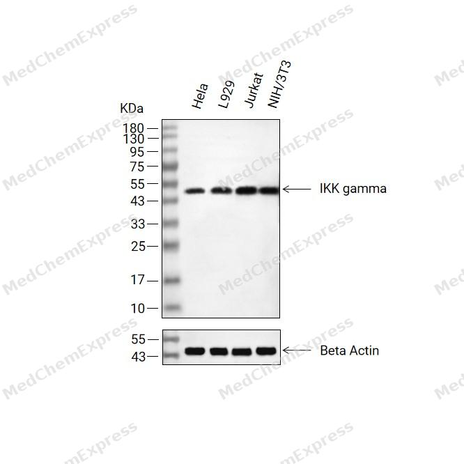 Western blot analysis of extracts from Hela(lane 2(20μg) , L929 (lane 3(20μg) ,Jurkat(lane 4(20μg)and NIH/3T3( lane 5(20μg) using IKK gamma Antibody (HY-P80415). Proteins were transferred to a PVDF membrane and blocked with 5% non-fat milk in TBST for 2 hour at room temperature. The primary antibody ( 1/1000) and Loading control antibody (Beta Actin, HY-P80993,1/10000) was used in 5% non-fat milk in TBST at 4°C overnight. Goat Anti-Rabbit IgG-HRP Secondary Antibody (HY-P8001,1/10,000) was used for 1 hour at room temperature.
 -
 Western blot analysis was performed on protein extracts (25 μg) from HeLa (lane 2), Jurkat (lane 3), NIH/3T3 (lane 4), U87 (lane 5), MCF-7 (lane 6) and Caco-2 (lane 7) using IKK gamma antibody. Proteins were transferred onto a 0.45 μm PVDF membrane using the Trans-Blot® Turbo™ system for 13 min. The membrane was then blocked with 5% nonfat milk in TBST (HY-K1025) for 1 h at room temperature. The primary antibody (1:1000) and loading control antibody GAPDH Antibody (HRP) (HY-P80954A) (1:5000) were diluted in 5% nonfat milk in TBST and incubated with the membrane overnight at 4°C. After washing, the membrane of primary antibody was incubated with HRP-conjugated goat anti-rabbit/mouse IgG secondary antibody (HY-P8001/HY-P8004) (1:5000) diluted in 5% nonfat milk in TBST for 1 h at room temperature. Protein bands were visualized using an Ultra High Sensitivity ECL detection kit (HY-K1005).
 -
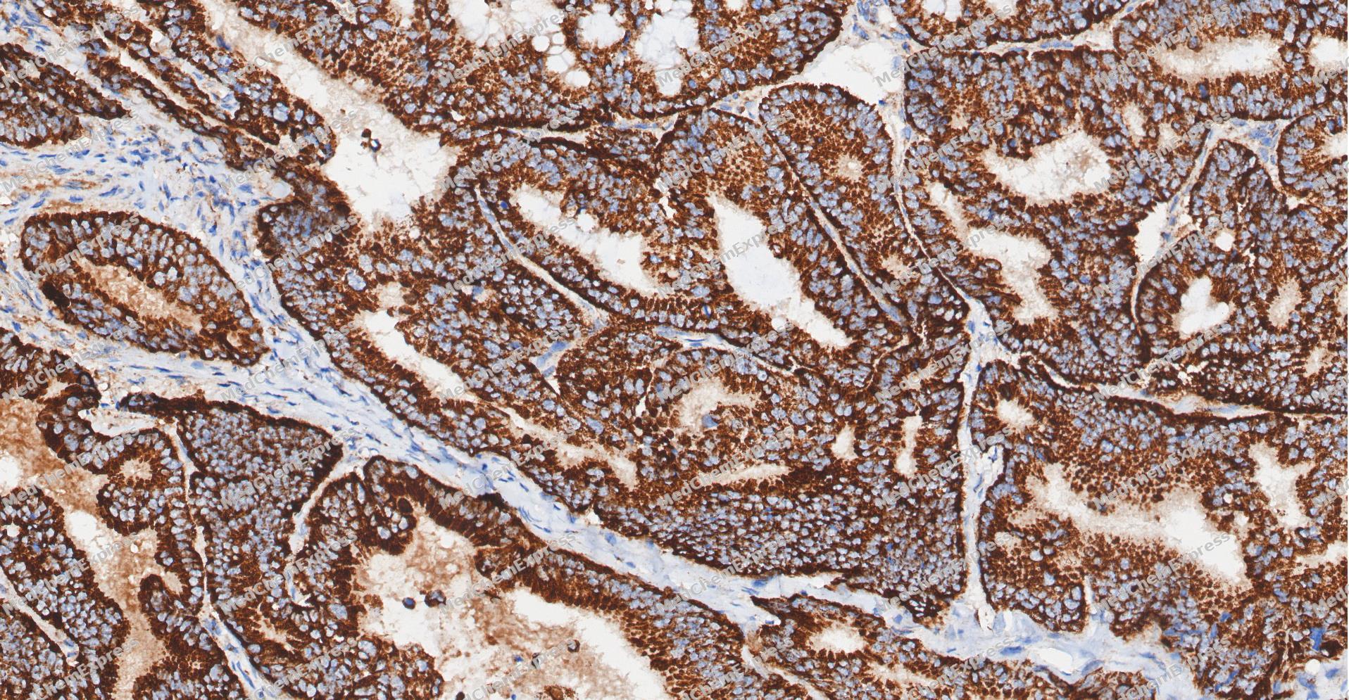 Immunohistochemical analysis of paraffin-embedded human Colon cancer tissue using IKK gamma antibody was performed. The section was pretreated using high-temperature and high-pressure mediated EDTA antigen retrieval buffer (pH 9.0), for 5 minutes. The tissues were incubated with primary antibody (HY-P80415, 1:100 dilution) at room temperature for 60 minutes. Detection was performed using an HRP conjugated compact polymer system. DAB was used as the chromogen. The tissues were counterstained with hematoxylin and mounted with neutral balsam mounting medium.
 -
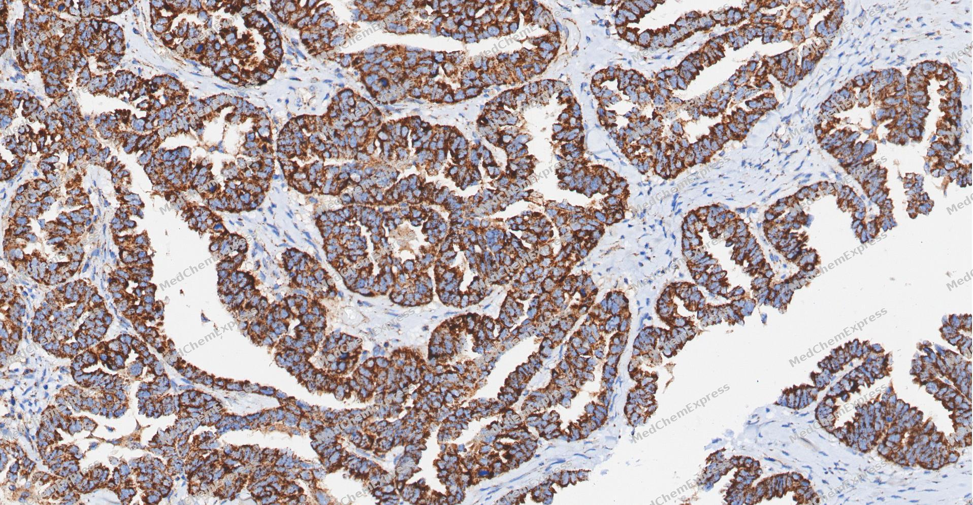 Immunohistochemical analysis of paraffin-embedded human ovarian carcinoma tissue using IKK gamma antibody was performed. The section was pretreated using high-temperature and high-pressure mediated EDTA antigen retrieval buffer (pH 9.0), for 5 minutes. The tissues were incubated with primary antibody (HY-P80415, 1:100 dilution) at room temperature for 60 minutes. Detection was performed using an HRP conjugated compact polymer system. DAB was used as the chromogen. The tissues were counterstained with hematoxylin and mounted with neutral balsam mounting medium.
 -
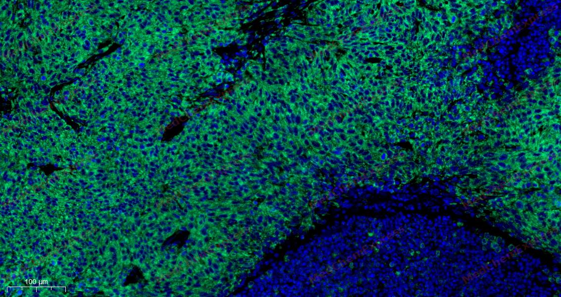 Tyramide signaling amplification based immunofluorescence was performed on paraffin-embedded human ovarian carcinoma (sample 1) tissue using IKK gamma antibody. Antigen retrieval was performed in EDTA buffer pH 9.0 (95 °C, 20 min) followed by cooling to RT. Then incubated with primary antibody (HY-P80415, 1:100 dilution) at room temperature for 60 minutes and HRP conjugated secondary antibody for 10 minutes. Fluorescence was then developed with TSA520 . The tissues were counterstained with DAPI and mounted with Anti-fade mounting medium.
 -
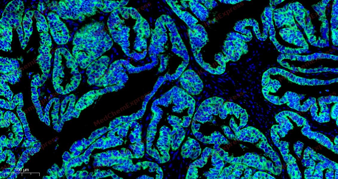 Tyramide signaling amplification based immunofluorescence was performed on paraffin-embedded human ovarian carcinoma (sample 2) tissue using IKK gamma antibody. Antigen retrieval was performed in EDTA buffer pH 9.0 (95 °C, 20 min) followed by cooling to RT. Then incubated with primary antibody (HY-P80415, 1:100 dilution) at room temperature for 60 minutes and HRP conjugated secondary antibody for 10 minutes. Fluorescence was then developed with TSA520 . The tissues were counterstained with DAPI and mounted with Anti-fade mounting medium.
 -
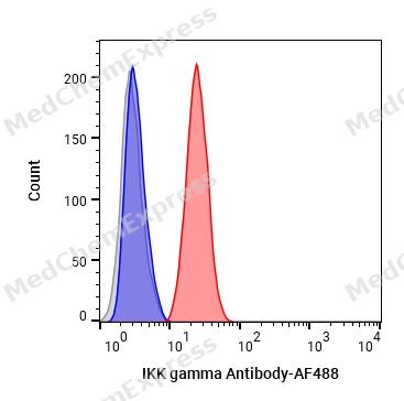 Flow cytometric analysis of 1X10⁶ HeLa cells labeling IKK gamma Antibody (HY-P80415, red). Cells were fixed with 4% paraformaldehyde and permeabilised with 90% methanol. Then stained with the primary antibody at 1/50 dilution for an hour at 4℃. AF488-conjugated Goat Anti-Rabbit IgG H&L (HY-P8002) was used as the secondary antibody at 1/1,000 dilution for 30 minutes at 4℃. Rabbit IgG Isotype Control (HY-P80879, blue) was used as the isotype control, cells without incubation with primary antibody were used as the unlabeled control (black).
 -
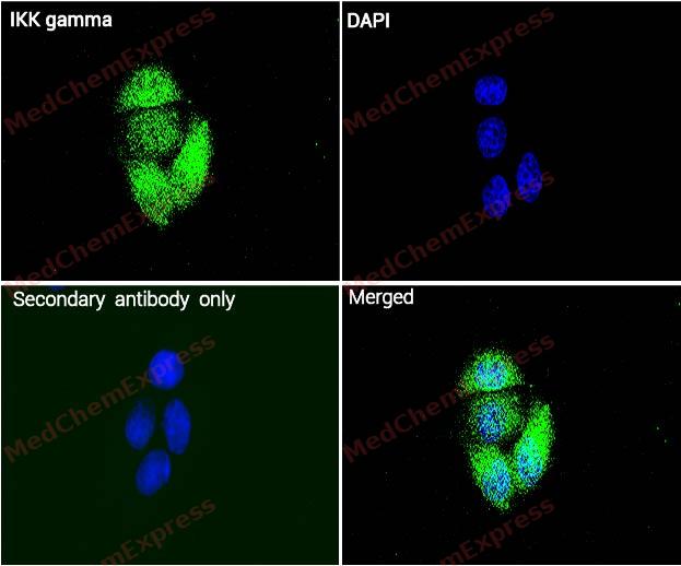 Immunocytochemistry analysis of Hela cells labeling IKK gamma with IKK gamma Antibody (HY-P80415) at 1/100 dilution. Cells were fixed in 4% paraformaldehyde for 15 minutes at room temperature, permeabilized with 0.1% Triton X-100 in PBS for 15 minutes at room temperature, then blocked with quick block buffer for 10 minutes at room temperature. Cells were then incubated with IKK gamma Antibody (HY-P80415) at 1/100 dilution in quick block buffer overnight at 4 ℃. AF488-conjugated Goat Anti-Rabbit IgG H&L(HY-P8002, Green) was used as the secondary antibody at 1/1,000 dilution. PBS instead of the primary antibody was used as the secondary antibody only control. The Nuclear counterstain was DAPI (Blue).
 -
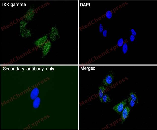 Immunocytochemistry analysis of Hela cells labeling IKK gamma with IKK gamma Antibody (HY-P80415) at 1/200 dilution. Cells were fixed in 4% paraformaldehyde for 15 minutes at room temperature, permeabilized with 0.1% Triton X-100 in PBS for 15 minutes at room temperature, then blocked with quick block buffer for 10 minutes at room temperature. Cells were then incubated with IKK gamma Antibody (HY-P80415) at 1/200 dilution in quick block buffer overnight at 4 ℃. AF488-conjugated Goat Anti-Rabbit IgG H&L(HY-P8002, Green) was used as the secondary antibody at 1/1,000 dilution. PBS instead of the primary antibody was used as the secondary antibody only control. The Nuclear counterstain was DAPI (Blue).


Background
-
Function
IKK gamma is a Regulatory subunit of the IKK core complex which phosphorylates inhibitors of NF-kappa-B thus leading to the dissociation of the inhibitor/NF-kappa-B complex and ultimately the degradation of the inhibitor. Its binding to scaffolding polyubiquitin plays a key role in IKK activation by multiple signaling receptor pathways. Can recognize and bind both 'Lys-63'-linked and linear polyubiquitin upon cell stimulation, with a much higher affinity for linear polyubiquitin. Could be implicated in NF-kappa-B-mediated protection from cytokine toxicity. Essential for viral activation of IRF3. Involved in TLR3- and IFIH1-mediated antiviral innate response; this function requires 'Lys-27'-linked polyubiquitination; (Microbial infection) Also considered to be a mediator for HTLV-1 Tax oncoprotein activation of NF-kappa-B[1][2][3][4][5][6][7][8][9][10][11][12].
-
Subcellular Localization
Cytoplasm; Nucleus
-
Expression
Tissue_specificity:Heart, brain, placenta, lung, liver, skeletal muscle, kidney and pancreas -
Subunit
Homodimer; disulfide-linked (PubMed:18164680). Component of the I-kappa-B-kinase (IKK) core complex consisting of CHUK, IKBKB and IKBKG; probably four alpha/CHUK-beta/IKBKB dimers are associated with four gamma/IKBKG subunits (PubMed:11080499, PubMed:17977820, PubMed:18462684, PubMed:32935379, PubMed:9751060, PubMed:9891086). The IKK core complex seems to associate with regulatory or adapter proteins to form a IKK-signalosome holo-complex (PubMed:11080499, PubMed:9751060, PubMed:9891086). The IKK complex associates with TERF2IP/RAP1, leading to promote IKK-mediated phosphorylation of RELA/p65 (By similarity). Part of a complex composed of NCOA2, NCOA3, CHUK/IKKA, IKBKB, IKBKG and CREBBP (PubMed:11971985). Interacts with COPS3, CYLD, NALP2, TRPC4AP and PIDD1 (PubMed:11418127, PubMed:12917691, PubMed:15456791, PubMed:16360037). Interacts with ATM; the complex is exported from the nucleus (PubMed:16497931). Interacts with TRAF6 (PubMed:17728323). Interacts with IKBKE (PubMed:23453969). Interacts with TANK; the interaction is enhanced by IKBKE and TBK1 (PubMed:12133833). Part of a ternary complex consisting of TANK, IKBKB and IKBKG (PubMed:12133833). Interacts with ZFAND5 (PubMed:14754897). Interacts with RIPK2 (PubMed:18079694). Interacts with TNIP1 and TNFAIP3; TNIP1 facilitates the TNFAIP3-mediated de-ubiquitination of IKBKG (PubMed:11389905, PubMed:22099304). Interacts with TNFAIP3; the interaction is induced by TNF stimulation and by polyubiquitin (PubMed:11389905, PubMed:22099304). Binds (via UBAN region) polyubiquitin; binds both 'Lys-63'-linked and linear polyubiquitin, with higher affinity for linear ubiquitin (PubMed:16547522, PubMed:19033441, PubMed:19185524, PubMed:21606507). Interacts with NLRP10 (PubMed:22672233). Interacts with TANK; this interaction increases in response to DNA damage (PubMed:25861989). Interacts with USP10; this interaction increases in response to DNA damage (PubMed:25861989). Interacts with ZC3H12A; this interaction increases in response to DNA damage (PubMed:25861989). Interacts with IFIT5; the interaction synergizes the recruitment of IKK to MAP3K7 and enhances IKK phosphorylation (PubMed:26334375). Interacts with TRIM29; this interaction induces IKBKG/NEMO ubiquitination and proteolytic degradation (PubMed:27695001). Interacts with TRIM13; this interaction leads to IKBKG/NEMO ubiquitination (PubMed:25152375). Interacts with ARFIP2 (PubMed:26296658). Interacts with RIPK1 (By similarity). Interacts with (ubiquitinated) BCL10; interaction with polyubiquitinated BCL10 via both 'Lys-63'-linked and linear ubiquitin is required for TCR-induced NF-kappa-B activation (PubMed:18287044, PubMed:27777308). Interacts with MARCHF2; during the late stages of macrophage viral and bacterial infection; the interaction leads to ubiquitination and degradation of IKBKG/NEMO (PubMed:32935379)
-
SwissProt ID
-
Synonyms
FIP3, NEMO, IKBKG, NF-kappa-B essential modulator, FIP-3, IkB kinase-associated protein 1, Inhibitor of nuclear factor kappa-B kinase subunit gamma, NF-kappa-B essential modifier, IKKAP1, I-kappa-B kinase subunit gamma, IKK-gamma, IKKG, IkB kinase subunit gamma
-
Research Field
Cell Biology


Documentation
-
Data Sheet (263 KB)
-
SDS (251 KB)
- English - EN (251 KB)
- Français - FR (251 KB)
- Deutsch - DE (251 KB)
- Norwegian - NO (251 KB)
- Español - ES (251 KB)
- Swedish - SV (251 KB)
- Italian - IT (251 KB)
- Korean - KR (251 KB)
- Portuguese - PT (251 KB)
-
User Guide for Antibodies (1077 KB)
References
[1]. Zhou H, et al. Bcl10 activates the NF-kappaB pathway through ubiquitination of NEMO. Nature. 2004 Jan 8;427(6970):167-71. [Content Brief]
[2]. Arimoto K, et al. Polyubiquitin conjugation to NEMO by triparite motif protein 23 (TRIM23) is critical in antiviral defense. Proc Natl Acad Sci U S A. 2010 Sep 7;107(36):15856-61. [Content Brief]
[3]. Zotti T, et al. TRAF7 protein promotes Lys-29-linked polyubiquitination of IkappaB kinase (IKKgamma)/NF-kappaB essential modulator (NEMO) and p65/RelA protein and represses NF-kappaB activation. J Biol Chem. 2011 Jul 1;286(26):22924-33. [Content Brief]
[4]. Rothwarf DM, et al. IKK-gamma is an essential regulatory subunit of the IkappaB kinase complex. Nature. 1998 Sep 17;395(6699):297-300. [Content Brief]
[5]. Wu CJ, et al. Sensing of Lys 63-linked polyubiquitination by NEMO is a key event in NF-kappaB activation [corrected]. Nat Cell Biol. 2006 Apr;8(4):398-406. [Content Brief]
[6]. Wu CJ, et al. NEMO recognition of ubiquitinated Bcl10 is required for T cell receptor-mediated NF-kappaB activation. Proc Natl Acad Sci U S A. 2008 Feb 26;105(8):3023-8. [Content Brief]
[7]. Cordier F, et al. The zinc finger of NEMO is a functional ubiquitin-binding domain. J Biol Chem. 2009 Jan 30;284(5):2902-2907. [Content Brief]
[8]. Lo YC, et al. Structural basis for recognition of diubiquitins by NEMO. Mol Cell. 2009 Mar 13;33(5):602-15. [Content Brief]
[9]. Nanda SK, et al. Polyubiquitin binding to ABIN1 is required to prevent autoimmunity. J Exp Med. 2011 Jun 6;208(6):1215-28. [Content Brief]
[10]. Yang YK, et al. Molecular Determinants of Scaffold-induced Linear Ubiquitinylation of B Cell Lymphoma/Leukemia 10 (Bcl10) during T Cell Receptor and Oncogenic Caspase Recruitment Domain-containing Protein 11 (CARD11) Signaling. J Biol Chem. 2016 Dec 9;291(50):25921-25936. [Content Brief]
[11]. Wu J, et al. SARS-CoV-2 ORF9b inhibits RIG-I-MAVS antiviral signaling by interrupting K63-linked ubiquitination of NEMO. Cell Rep. 2021 Feb 16;34(7):108761. [Content Brief]
[12]. Zeng W, et al. Key role of Ubc5 and lysine-63 polyubiquitination in viral activation of IRF3. Mol Cell. 2009 Oct 23;36(2):315-25. [Content Brief]

 Powered by Bioz
