



MYST1 Antibody (YA2230)
(Synonyms: hMOF; MOF; MOZ; MYST1; SAS2; SAS3; YBF2; YBF2/SAS3; ZC2HC8; KAT8)
Based on 1 Customer Validation
MYST1 Antibody (YA2230) is a Rabbit-derived and non-conjugated IgG monoclonal antibody, targeting to MYST1.
-
Host:
Rabbit
-
Isotype:
IgG
-
Application:
WB, IHC-P, ICC/IF, IP, FC
-
Reactivity :
Human, Mouse, Rat
-
Formulation:
Supplied in 50mM Tris-Glycine(pH 7.4), 0.15M NaCl, 40%Glycerol, 0.01% sodium azide and 0.05% BSA.
-
Conjugation:
Non-conjugated


To place orders, for customer services and technical support, please contact: MedChemExpress USA
Tel: 609-228-6898 E-mail: [email protected] [email protected]





Applications
| Application |
WB

WB: Western Blot
|
IHC-P
IHC-P: Immunohistochemistry-Paraffin
|
ICC/IF
ICC/IF: Immunocytochemistry/
Immunofluorescence |
IP
IP: Immunoprecipitation
|
FC
FC: Flow Cytometry
|
|---|---|---|---|---|---|
| Dilution Ratio | 1:500-1:1000 | 1:50-1:100 | 1:50-1:200 | 1:100 | 1:50-1:100 |


Product Details
MYST1 Antibody (YA2230) is a Rabbit-derived and non-conjugated IgG monoclonal antibody, targeting to MYST1.
-
Host Rabbit

-
Clonality Recombinant,Monoclonal

-
Species ReactivityHuman, Mouse, Rat
-
Observed Molecular WeightObserved band size: 52 kDa
Note: Due to possible protein modifications or aggregation, the molecular weight should be confirmed by actual measurement, and the predicted value is for reference only.

-
Calculated Molecular Weight Predicted band size: 52 kDa

Entrez Gene: 84148 Human ; 67773 Mouse ; 310194 Rat
SwissProt: Q9H7Z6 Human ; Q9D1P2 Mouse ; Q5XI06 Rat
OMIM: 618974 Human
A synthesized peptide derived from human KAT8 / MYST1 / MOF aa400-458.
Endogenous
Affinity Chromatography
Non-conjugated
Unmodified
IgG


Product Properties
-
Appearance
Solution
-
Formulation
Supplied in 50mM Tris-Glycine(pH 7.4), 0.15M NaCl, 40%Glycerol, 0.01% sodium azide and 0.05% BSA.
-
Concentration
Batch-dependent, Please check the COA for the concentration of each lot. Check Lot Concentration
-
Storage & Stability
Stored at -20°C for 1 year. Avoid repeated freeze / thaw cycles.
-
Shipping
Shipping with blue ice.


Verification Images
-
 Western blot analysis of extracts from Hela (lane 2(20μg), Hela (lane 3(40μg), using KAT8 Antibody. Proteins were transferred to a PVDF membrane and blocked with 5% BSA in TBST for 2 hour at room temperature. The primary antibody and Loading control antibody (Beta Actin, HY-P80438, 1/3000) was used in 5% BSA in TBST at 4°C overnight. Goat Anti-Mouse/Rabbit IgG-HRP Secondary Antibody (HY-P8004/HY-P8001, 1/10,000) was used for 1 hour at room temperature.
 -
 Immunohistochemical analysis of paraffin-embedded human glioma using MYST1 antibody. The section was pre-treated using heat mediated antigen retrieval with EDTA (pH 9.0) for 14 minutes. The tissues were blocked in 5% BSA for 30 minutes at room temperature, washed with TBST, and then probed with the primary antibody (HY-P82485, 1/100) for 30 minutes at room temperature. The detection was performed using Polymer HRP-conjugated Goat Anti-Mouse/Rabbit lgG(H&L) secondary antibody (HY-P83652). DAB was used as the chromogen. Tissues were counterstained with hematoxylin and mounted with DPX.
 -
 Immunohistochemical analysis of paraffin-embedded human lymph node using MYST1 antibody. The section was pre-treated using heat mediated antigen retrieval with EDTA (pH 9.0) for 14 minutes. The tissues were blocked in 5% BSA for 30 minutes at room temperature, washed with TBST, and then probed with the primary antibody (HY-P82485, 1/100) for 30 minutes at room temperature. The detection was performed using Polymer HRP-conjugated Goat Anti-Mouse/Rabbit lgG(H&L) secondary antibody (HY-P83652). DAB was used as the chromogen. Tissues were counterstained with hematoxylin and mounted with DPX.
 -
 Immunohistochemical analysis of paraffin-embedded human pancreatic cancer using MYST1 antibody. The section was pre-treated using heat mediated antigen retrieval with EDTA (pH 9.0) for 14 minutes. The tissues were blocked in 5% BSA for 30 minutes at room temperature, washed with TBST, and then probed with the primary antibody (HY-P82485, 1/100) for 30 minutes at room temperature. The detection was performed using Polymer HRP-conjugated Goat Anti-Mouse/Rabbit lgG(H&L) secondary antibody (HY-P83652). DAB was used as the chromogen. Tissues were counterstained with hematoxylin and mounted with DPX.
 -
 Immunohistochemical analysis of paraffin-embedded human brain using MYST1 antibody. The section was pre-treated using heat mediated antigen retrieval with EDTA (pH 9.0) for 14 minutes. The tissues were blocked in 5% BSA for 30 minutes at room temperature, washed with TBST, and then probed with the primary antibody (HY-P82485, 1/100) for 30 minutes at room temperature. The detection was performed using Polymer HRP-conjugated Goat Anti-Mouse/Rabbit lgG(H&L) secondary antibody (HY-P83652). DAB was used as the chromogen. Tissues were counterstained with hematoxylin and mounted with DPX.
 -
 Immunohistochemical analysis of paraffin-embedded human head and neck cancer using MYST1 antibody. The section was pre-treated using heat mediated antigen retrieval with EDTA (pH 9.0) for 14 minutes. The tissues were blocked in 5% BSA for 30 minutes at room temperature, washed with TBST, and then probed with the primary antibody (HY-P82485, 1/100) for 30 minutes at room temperature. The detection was performed using Polymer HRP-conjugated Goat Anti-Mouse/Rabbit lgG(H&L) secondary antibody (HY-P83652). DAB was used as the chromogen. Tissues were counterstained with hematoxylin and mounted with DPX.
 -
 Immunohistochemical analysis of paraffin-embedded human colon cancer using MYST1 antibody. The section was pre-treated using heat mediated antigen retrieval with EDTA (pH 9.0) for 14 minutes. The tissues were blocked in 5% BSA for 30 minutes at room temperature, washed with TBST, and then probed with the primary antibody (HY-P82485, 1/100) for 30 minutes at room temperature. The detection was performed using Polymer HRP-conjugated Goat Anti-Mouse/Rabbit lgG(H&L) secondary antibody (HY-P83652). DAB was used as the chromogen. Tissues were counterstained with hematoxylin and mounted with DPX.
 -
 Immunohistochemical analysis of paraffin-embedded human pancreas using MYST1 antibody. The section was pre-treated using heat mediated antigen retrieval with EDTA (pH 9.0) for 14 minutes. The tissues were blocked in 5% BSA for 30 minutes at room temperature, washed with TBST, and then probed with the primary antibody (HY-P82485, 1/100) for 30 minutes at room temperature. The detection was performed using Polymer HRP-conjugated Goat Anti-Mouse/Rabbit lgG(H&L) secondary antibody (HY-P83652). DAB was used as the chromogen. Tissues were counterstained with hematoxylin and mounted with DPX.
 -
 Immunohistochemical analysis of paraffin-embedded human bladder cancer using MYST1 antibody. The section was pre-treated using heat mediated antigen retrieval with EDTA (pH 9.0) for 14 minutes. The tissues were blocked in 5% BSA for 30 minutes at room temperature, washed with TBST, and then probed with the primary antibody (HY-P82485, 1/100) for 30 minutes at room temperature. The detection was performed using Polymer HRP-conjugated Goat Anti-Mouse/Rabbit lgG(H&L) secondary antibody (HY-P83652). DAB was used as the chromogen. Tissues were counterstained with hematoxylin and mounted with DPX.
 -
 Immunohistochemical analysis of paraffin-embedded human kidney using MYST1 antibody. The section was pre-treated using heat mediated antigen retrieval with EDTA (pH 9.0) for 14 minutes. The tissues were blocked in 5% BSA for 30 minutes at room temperature, washed with TBST, and then probed with the primary antibody (HY-P82485, 1/100) for 30 minutes at room temperature. The detection was performed using Polymer HRP-conjugated Goat Anti-Mouse/Rabbit lgG(H&L) secondary antibody (HY-P83652). DAB was used as the chromogen. Tissues were counterstained with hematoxylin and mounted with DPX.
 -
 Immunohistochemical analysis of paraffin-embedded human tonsil using MYST1 antibody. The section was pre-treated using heat mediated antigen retrieval with EDTA (pH 9.0) for 14 minutes. The tissues were blocked in 5% BSA for 30 minutes at room temperature, washed with TBST, and then probed with the primary antibody (HY-P82485, 1/100) for 30 minutes at room temperature. The detection was performed using Polymer HRP-conjugated Goat Anti-Mouse/Rabbit lgG(H&L) secondary antibody (HY-P83652). DAB was used as the chromogen. Tissues were counterstained with hematoxylin and mounted with DPX.
 -
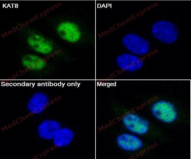 Immunocytochemistry analysis of SiHa cells labeling KAT8 Antibody (HY-P82485) at 1/50 dilution. Cells were fixed in 4% paraformaldehyde for 15 minutes at room temperature, permeabilized with 0.1% Triton X-100 for 10 minutes at room temperature, then blocked with QuickBlock™ Blocking Buffer for Immunol Staining for 10 min at room temperature. Cells were then incubated with KAT8 Antibody (HY-P82485) at 1/50 dilution in QuickBlock™ Blocking Buffer for Immunol Staining at 4 ℃. Alexa Fluor® 488-conjugated AffiniPure Goat Anti-Rabbit IgG H&L(HY-P8002, Green) was used as the secondary antibody at 1/1,000 dilution. PBS instead of the primary antibody was used as the secondary antibody only control. The Nuclear counterstain was DAPI (Blue).
 -
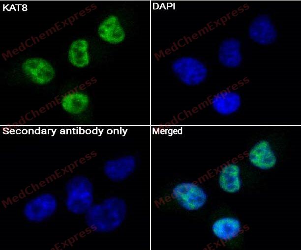 Immunocytochemistry analysis of SH-SY-5Y cells labeling KAT8 Antibody (HY-P82485) at 1/50 dilution. Cells were fixed in 4% paraformaldehyde for 15 minutes at room temperature, permeabilized with 0.1% Triton X-100 for 10 minutes at room temperature, then blocked with QuickBlock™ Blocking Buffer for Immunol Staining for 10 min at room temperature. Cells were then incubated with KAT8 Antibody (HY-P82485) at 1/50 dilution in QuickBlock™ Blocking Buffer for Immunol Staining at 4 ℃. Alexa Fluor® 488-conjugated AffiniPure Goat Anti-Rabbit IgG H&L(HY-P8002, Green) was used as the secondary antibody at 1/1,000 dilution. PBS instead of the primary antibody was used as the secondary antibody only control. The Nuclear counterstain was DAPI (Blue).


Background
-
Function
MYST1 is a Histone acetyltransferase that catalyzes histone H4 acetylation at 'Lys-5'- and 'Lys-8' (H4K5ac and H4K8ac) or 'Lys-16' (H4K16ac), depending on the context. Catalytic component of the MSL histone acetyltransferase complex, a multiprotein complex that mediates the majority of histone H4 acetylation at 'Lys-16' (H4K16ac), an epigenetic mark that prevents chromatin compaction. H4K16ac constitutes the only acetylation mark intergenerationally transmitted and regulates key biological processes, such as oogenesis, embryonic stem cell pluripotency, hematopoiesis or glucose metabolism. The MSL complex is required for chromosome stability and genome integrity by maintaining homeostatic levels of H4K16ac. The MSL complex is also involved in gene dosage by promoting up-regulation of genes expressed by the X chromosome. X up-regulation is required to compensate for autosomal biallelic expression. The MSL complex also participates in gene dosage compensation by promoting expression of Tsix non-coding RNA. As part of the NSL histone acetyltransferase complex, catalyzes histone H4 acetylation at 'Lys-5'- and 'Lys-8' (H4K5ac and H4K8ac) at transcription start sites and promotes transcription initiation. The NSL complex also acts as a regulator of gene expression in mitochondria: KAT8 associates with mitochondrial DNA and controls expression of respiratory genes in an acetyltransferase-dependent mechanism. Also functions as an acetyltransferase for non-histone targets, such as ALKBH5, COX17, IRF3, KDM1A/LSD1, LMNA, PAX7 or TP53/p53. Acts as an inhibitor of antiviral immunity by acetylating IRF3, preventing IRF3 recruitment to promoters. Acts as a regulator of asymmetric division in muscle stem cells by mediating acetylation of PAX7. As part of the NSL complex, acetylates TP53/p53 at 'Lys-120'. Acts as a regulator of epithelial-to-mesenchymal transition as part of the NSL complex by mediating acetylation of KDM1A/LSD1. The NSL complex is required for nuclear architecture maintenance by mediating acetylation of LMNA. Promotes mitochondrial integrity by catalyzing acetylation of COX17. In addition to protein acetyltransferase activity, able to mediate protein propionylation[1][2][3][4][5][6][7][8][9][10][11][12][13][14][15][16].
-
Subcellular Localization
Nucleus; Chromosome; Mitochondrion
-
Isoforms & Post-Translational Modification
Q9H7Z6 has 2 isomers: Q9H7Z6-1: 52403 Da (predicted); Q9H7Z6-2: 53085 Da (predicted).
Acetylation at Lys-274 facilitates cognate substrate Lys-binding and acetylation (PubMed:22020126, PubMed:27382063). Although considered as an autoacetylation event, acetylation at Lys-274 probably takes place via a non-enzymatic process following acetyl-CoA-binding, which primes KAT8 for cognate protein-lysine acetylation (PubMed:27382063). Deacetylated by SIRT1 (PubMed:22918831) -
Subunit
Component of a multisubunit histone acetyltransferase complex (MSL) at least composed of the MOF/KAT8, MSL1/hampin, MSL2L1 and MSL3L1 (PubMed:16227571, PubMed:16543150, PubMed:21217699, PubMed:22547026, PubMed:30224647, PubMed:33657400, PubMed:33837287). Component of the NSL complex at least composed of MOF/KAT8, KANSL1, KANSL2, KANSL3, MCRS1, PHF20, OGT1/OGT, WDR5 and HCFC1 (PubMed:16543150, PubMed:19854137, PubMed:20018852, PubMed:20620954, PubMed:21217699, PubMed:22547026, PubMed:27768893, PubMed:33657400). Component of some MLL1/MLL complex, at least composed of the core components KMT2A/MLL1, ASH2L, HCFC1, WDR5 and RBBP5, as well as the facultative components BACC1, CHD8, E2F6, HSP70, INO80C, KANSL1, LAS1L, MAX, MCRS1, MGA, MOF/KAT8, PELP1, PHF20, PRP31, RING2, RUVB1/TIP49A, RUVB2/TIP49B, SENP3, TAF1, TAF4, TAF6, TAF7, TAF9 and TEX10 (PubMed:15960975). Interacts with the chromodomain of MORF4L1/MRG15 (PubMed:12397079). Interacts with ATM (via its Tudor-knot domain); possibly regulating the activity of ATM (PubMed:15923642). Interacts with NELFD (By similarity)
-
SwissProt ID
-
Synonyms
hMOF; MOF; MOZ; MYST1; SAS2; SAS3; YBF2; YBF2/SAS3; ZC2HC8; KAT8
-
Research Field
Epigenetics and Nuclear Signaling


Documentation
-
Data Sheet (234 KB)
-
SDS (251 KB)
- English - EN (251 KB)
- Français - FR (251 KB)
- Deutsch - DE (251 KB)
- Norwegian - NO (251 KB)
- Español - ES (251 KB)
- Swedish - SV (251 KB)
- Italian - IT (251 KB)
- Korean - KR (251 KB)
- Portuguese - PT (251 KB)
-
User Guide for Antibodies (1077 KB)

 Powered by Bioz
