



MICA Antibody (YA1464)
(Synonyms: PERB11.1; MICA; HLA class I antigen antibody; FLJ36918 antibody; FLJ60820 antibody; MGC111087 antibody; MGC21250 antibody; MHC class I chain related gene A protein antibody)
Based on 1 Customer Validation
MICA Antibody (YA1464) is a Rabbit-derived and non-conjugated IgG monoclonal antibody, targeting to MICA.
-
Host:
Rabbit
-
Isotype:
IgG
-
Application:
WB, IP, FC
-
Reactivity :
Human
-
Formulation:
Supplied in PBS (pH7.4), 0.1% BSA, 40% Glycerol. Preservative: 0.05% Sodium Azide.
-
Conjugation:
Non-conjugated


To place orders, for customer services and technical support, please contact: MedChemExpress USA
Tel: 609-228-6898 E-mail: [email protected] [email protected]





Applications
| Application |
WB

WB: Western Blot
|
FC
FC: Flow Cytometry
|
IP
IP: Immunoprecipitation
|
|---|---|---|---|
| Dilution Ratio | 1:5000 | 1:5000 | 1-2μg/sample |


Product Details
MICA Antibody (YA1464) is a Rabbit-derived and non-conjugated IgG monoclonal antibody, targeting to MICA.
-
Host Rabbit

-
Clonality Recombinant,Monoclonal

-
Species ReactivityHuman
-
Observed Molecular WeightObserved band size: 60 kDa
Note: Due to possible protein modifications or aggregation, the molecular weight should be confirmed by actual measurement, and the predicted value is for reference only.

-
Calculated Molecular Weight Predicted band size: 43 kDa

Recombinant protein within human MICA aa 1-328.
Endogenous
affinity purified.
Non-conjugated
Unmodified
IgG


Product Properties
-
Appearance
Solution
-
Formulation
Supplied in PBS (pH7.4), 0.1% BSA, 40% Glycerol. Preservative: 0.05% Sodium Azide.
-
Concentration
Batch-dependent, Please check the COA for the concentration of each lot. Check Lot Concentration
-
Storage & Stability
Stored at -20°C for 1 year. Avoid repeated freeze / thaw cycles.
-
Shipping
Shipping with blue ice.


Verification Images
-
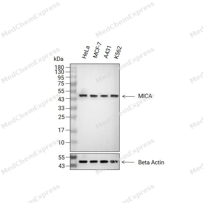 Western blot analysis of extracts from HeLa(lane 2(20μg), MCF-7(lane 3(20μg), A431(lane 4(20μg) and K562(lane 5(20μg) using MICA Antibody (HY-P81719). Proteins were transferred to a PVDF membrane and blocked with 5% non-fat milk in TBST for 2 hour at room temperature. The primary antibody (1/5000) and Loading control antibody (Beta Actin, HY-P80993,1/10000) was used in 5% non-fat milk in TBST at 4°C overnight. Goat Anti-Rabbit IgG-HRP Secondary Antibody (HY-P8001,1/10,000) was used for 1 hour at room temperature.
 -
 Flow cytometric analysis of 1X10⁶ HeLa cells labeling MICA Antibody (HY-P81719, red). Cells were fixed with 4% paraformaldehyde and permeabilised with 90% methanol. Then stained with the primary antibody at 1/5000 dilution for an hour at 4℃. AF488-conjugated Goat Anti-Rabbit IgG H&L (HY-P8002) was used as the secondary antibody at 1/1,000 dilution for 30 minutes at 4℃. Rabbit IgG Isotype Control (HY-P80879, blue) was used as the isotype control, cells without incubation with primary antibody were used as the unlabeled control (black).


Background
-
Function
MICA is widely expressed membrane-bound protein which acts as a ligand to stimulate an activating receptor KLRK1/NKG2D, expressed on the surface of essentially all human natural killer (NK), gammadelta T and CD8 alphabeta T-cells. Up-regulated in stressed conditions, such as viral and bacterial infections or DNA damage response, serves as signal of cellular stress, and engagement of KLRK1/NKG2D by MICA triggers NK-cells resulting in a range of immune effector functions, such as cytotoxicity and cytokine production[1][2][3].
-
Subcellular Localization
Cell membrane; Single-pass type I membrane protein; Cytoplasm
-
Expression
Tissue_specificity:Besides the central nervous system, MICA is widely expressed in other tissues. It is primarily expressed in the gastric epithelium, but also in monocytes, keratinocytes, endothelial cells, fibroblasts, and the outer layer of normal thymic medullary Hardy bodies. In the skin, it is mainly expressed in the stratum corneum, basal cells, ducts, and follicles. MICA is also expressed in many (but not all) epithelial tumors of the lung, breast, kidney, ovary, prostate, and colon. In thymomas, MICA is overexpressed in both cortical and medullary epithelial cells. Tumors expressing MICA show elevated levels of γδT cells.
Induction:By heat shock, by infection with human cytomegalovirus (HCMV) , human adenovirus 5, M.tuberculosis and diarrheagenic E.coli, and by exposure to DNA damaging conditions such as high doses of ionizing radiation, chromatin-modifying treatments and inhibitors of DNA replication. The HCMV UL142 protein causes down-regulation of the full-length protein but not of the truncated MICA*008 allele -
Isoforms & Post-Translational Modification
Q29983 has 2 isomers: Q29983-1: 42915 Da (predicted); Q29983-2: 31577 Da (predicted).
N-glycosylated. Glycosylation is not essential for interaction with KLRK1/NKG2D but enhances complex formation;Proteolytically cleaved and released from the cell surface of tumor cells which impairs KLRK1/NKG2D expression and T-cell activation;Palmitoylated on cysteine residues in the cytoplasmic tail leading to its association with membrane microdomains enriched in cholesterol;N-glycosylation is necessary for cell surface expression;(Microbial infection) Ubiquitinated by human herpesvirus 8 protein K5, leading to degradation -
Subunit
Unlike classical MHC class I molecules, does not form a heterodimer with beta-2-microglobulin. Binds as a monomer to a KLRK1/NKG2D homodimer. KLRK1 forms a complex with HCST/DAP10 in which KLRK1 binds MICA while HCST acts as an adapter molecule which enables signal transduction. Interacts with PDIA6 on the surface of tumor cells, leading to disulfide bond reduction which is required for release of MICA from tumor cells
-
SwissProt ID
-
Synonyms
PERB11.1; MICA; HLA class I antigen antibody; FLJ36918 antibody; FLJ60820 antibody; MGC111087 antibody; MGC21250 antibody; MHC class I chain related gene A protein antibody
-
Research Field
Immunology


Documentation
-
Data Sheet (234 KB)
-
SDS (252 KB)
- English - EN (252 KB)
- Français - FR (252 KB)
- Deutsch - DE (252 KB)
- Norwegian - NO (252 KB)
- Español - ES (252 KB)
- Swedish - SV (252 KB)
- Italian - IT (252 KB)
- Korean - KR (252 KB)
- Portuguese - PT (252 KB)
-
User Guide for Antibodies (1077 KB)

 Powered by Bioz
