



Mucin 1 Antibody (YA3086)
(Synonyms: MUC1; PUM; Mucin-1; MUC-1; Breast carcinoma-associated antigen DF3; Carcinoma-associated mucin; Episialin; H23AG; Krebs von den Lungen-6; KL-6; PEMT; Peanut-reactive urinary mucin; PUM; Polymorphic epithelial mucin; PEM; Tumor-associated ep)
Based on 1 Customer Validation
Mucin 1 Antibody (YA3086) is a Rabbit-derived and non-conjugated IgG monoclonal antibody, targeting to Mucin 1.
-
Host:
Rabbit
-
Isotype:
IgG
-
Application:
WB, IHC-P, ICC/IF, IP, FC
-
Reactivity :
Human
-
Formulation:
Supplied in Rabbit IgG in 10mM phosphate buffered saline , pH 7.4, 150mM sodium chloride, 0.05% BSA, 0.02% sodium azide and 50% glycerol.
-
Conjugation:
Non-conjugated


To place orders, for customer services and technical support, please contact: MedChemExpress USA
Tel: 609-228-6898 E-mail: [email protected] [email protected]





Applications
| Application |
WB

WB: Western Blot
|
IHC-P
IHC-P: Immunohistochemistry-Paraffin
|
ICC/IF
ICC/IF: Immunocytochemistry/
Immunofluorescence |
IP
IP: Immunoprecipitation
|
FC
FC: Flow Cytometry
|
|---|---|---|---|---|---|
| Dilution Ratio | 1:500-1:1000 | 1:50-1:100 | 1:50-1:200 | 1:20 | 1:50-1:100 |


Product Details
Mucin 1 Antibody (YA3086) is a Rabbit-derived and non-conjugated IgG monoclonal antibody, targeting to Mucin 1.
-
Host Rabbit

-
Clonality Recombinant, Monoclonal

-
Species ReactivityHuman
-
Observed Molecular WeightObserved band size: 18-25 kDa
Note: Due to possible protein modifications or aggregation, the molecular weight should be confirmed by actual measurement, and the predicted value is for reference only.

-
Calculated Molecular Weight Predicted band size: 122 kDa

A synthesized peptide derived from human MUC1 aa1200-1255 aa1200-1255.
Endogenous
Affinity Chromatography
Non-conjugated
Unmodified
IgG


Product Properties
-
Appearance
Liquid
-
Formulation
Supplied in Rabbit IgG in 10mM phosphate buffered saline , pH 7.4, 150mM sodium chloride, 0.05% BSA, 0.02% sodium azide and 50% glycerol.
-
Concentration
Batch-dependent, Please check the COA for the concentration of each lot. Check Lot Concentration
-
Storage & Stability
Stored at -20°C for 1 year. Avoid repeated freeze / thaw cycles.
-
Shipping
Shipping with blue ice.


Verification Images
-
 Immunohistochemical analysis of paraffin-embedded human cervical cancer tissue using Mucin 1 Antibody (HY-P83341, 1/100). The section was pretreated using heat mediated antigen retrieval with sodium citrate buffer (pH 6.0) for 8 minutes. The tissues were blocked with quick block buffer for 0.5 hours at room temperature, washed with PBS and PBST, and then incubated with the primary antibody overnight at 4℃. The detection was performed using an HRP conjugated compact polymer system. DAB was used as the chromogen. Tissues were counterstained with hematoxylin and mounted with DPX.
 -
 Immunohistochemical analysis of paraffin-embedded human breast cancer tissue using Mucin 1 Antibody (HY-P83341, 1/100). The section was pretreated using heat mediated antigen retrieval with sodium citrate buffer (pH 6.0) for 8 minutes. The tissues were blocked with quick block buffer for 0.5 hours at room temperature, washed with PBS and PBST, and then incubated with the primary antibody overnight at 4℃. The detection was performed using an HRP conjugated compact polymer system. DAB was used as the chromogen. Tissues were counterstained with hematoxylin and mounted with DPX.
 -
 Immunohistochemical analysis of paraffin-embedded human lung cancer tissue using Mucin 1 Antibody (HY-P83341, 1/100). The section was pretreated using heat mediated antigen retrieval with sodium citrate buffer (pH 6.0) for 8 minutes. The tissues were blocked with quick block buffer for 0.5 hours at room temperature, washed with PBS and PBST, and then incubated with the primary antibody overnight at 4℃. The detection was performed using an HRP conjugated compact polymer system. DAB was used as the chromogen. Tissues were counterstained with hematoxylin and mounted with DPX.
 -
 Immunohistochemical analysis of paraffin-embedded human ovarian cancer tissue using Mucin 1 Antibody (HY-P83341, 1/100). The section was pretreated using heat mediated antigen retrieval with sodium citrate buffer (pH 6.0) for 8 minutes. The tissues were blocked with quick block buffer for 0.5 hours at room temperature, washed with PBS and PBST, and then incubated with the primary antibody overnight at 4℃. The detection was performed using an HRP conjugated compact polymer system. DAB was used as the chromogen. Tissues were counterstained with hematoxylin and mounted with DPX.
 -
 Immunohistochemical analysis of paraffin-embedded human renal cancer tissue using Mucin 1 Antibody (HY-P83341, 1/100). The section was pretreated using heat mediated antigen retrieval with sodium citrate buffer (pH 6.0) for 8 minutes. The tissues were blocked with quick block buffer for 0.5 hours at room temperature, washed with PBS and PBST, and then incubated with the primary antibody overnight at 4℃. The detection was performed using an HRP conjugated compact polymer system. DAB was used as the chromogen. Tissues were counterstained with hematoxylin and mounted with DPX.
 -
 Immunohistochemical analysis of paraffin-embedded human liver cancer tissue using Mucin 1 Antibody (HY-P83341, 1/100). The section was pretreated using heat mediated antigen retrieval with sodium citrate buffer (pH 6.0) for 8 minutes. The tissues were blocked with quick block buffer for 0.5 hours at room temperature, washed with PBS and PBST, and then incubated with the primary antibody overnight at 4℃. The detection was performed using an HRP conjugated compact polymer system. DAB was used as the chromogen. Tissues were counterstained with hematoxylin and mounted with DPX.
 -
 Immunohistochemical analysis of paraffin-embedded human oral cancer tissue using Mucin 1 Antibody (HY-P83341, 1/100). The section was pretreated using heat mediated antigen retrieval with sodium citrate buffer (pH 6.0) for 8 minutes. The tissues were blocked with quick block buffer for 0.5 hours at room temperature, washed with PBS and PBST, and then incubated with the primary antibody overnight at 4℃. The detection was performed using an HRP conjugated compact polymer system. DAB was used as the chromogen. Tissues were counterstained with hematoxylin and mounted with DPX.
 -
 Immunohistochemical analysis of paraffin-embedded human stomach tissue using Mucin 1 Antibody (HY-P83341, 1/100). The section was pretreated using heat mediated antigen retrieval with sodium citrate buffer (pH 6.0) for 8 minutes. The tissues were blocked with quick block buffer for 0.5 hours at room temperature, washed with PBS and PBST, and then incubated with the primary antibody overnight at 4℃. The detection was performed using an HRP conjugated compact polymer system. DAB was used as the chromogen. Tissues were counterstained with hematoxylin and mounted with DPX.
 -
 Immunohistochemical analysis of paraffin-embedded human endometrium tissue using Mucin 1 Antibody (HY-P83341, 1/100). The section was pretreated using heat mediated antigen retrieval with sodium citrate buffer (pH 6.0) for 8 minutes. The tissues were blocked with quick block buffer for 0.5 hours at room temperature, washed with PBS and PBST, and then incubated with the primary antibody overnight at 4℃. The detection was performed using an HRP conjugated compact polymer system. DAB was used as the chromogen. Tissues were counterstained with hematoxylin and mounted with DPX.
 -
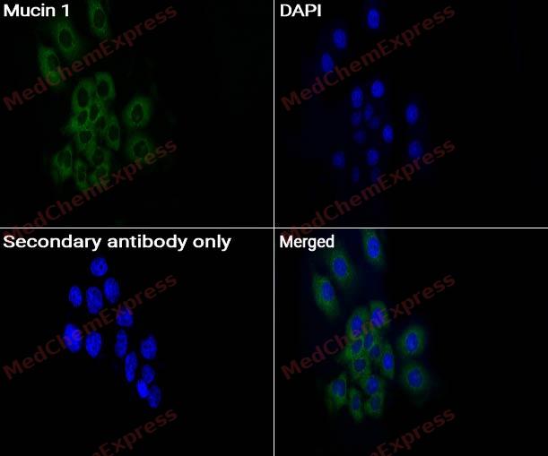 Immunocytochemistry analysis of HeLa cells labeling Mucin 1 with Mucin 1 antibody (HY-P83341) at 1/50 dilution. Cells were fixed in 4% paraformaldehyde for 15 minutes at room temperature, permeabilized with 0.1% Triton X-100 for 10 minutes at room temperature, then blocked with QuickBlock™ Blocking Buffer for Immunol Staining for 10 min at room temperature. Cells were then incubated with Mucin 1 antibody (HY-P83341) at 1/50 dilution in QuickBlock™ Blocking Buffer for Immunol Staining at 4 ℃. Alexa Fluor® 488-conjugated AffiniPure Goat Anti-Rabbit IgG H&L(HY-P8002, Green) was used as the secondary antibody at 1/1,000 dilution. PBS instead of the primary antibody was used as the secondary antibody only control. The Nuclear counterstain was DAPI (Blue).
 -
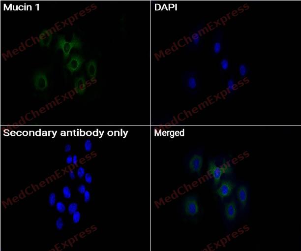 Immunocytochemistry analysis of HeLa cells labeling Mucin 1 with Mucin 1 antibody (HY-P83341) at 1/100 dilution. Cells were fixed in 4% paraformaldehyde for 15 minutes at room temperature, permeabilized with 0.1% Triton X-100 for 10 minutes at room temperature, then blocked with QuickBlock™ Blocking Buffer for Immunol Staining for 10 min at room temperature. Cells were then incubated with Mucin 1 antibody (HY-P83341) at 1/100 dilution in QuickBlock™ Blocking Buffer for Immunol Staining at 4 ℃. Alexa Fluor® 488-conjugated AffiniPure Goat Anti-Rabbit IgG H&L(HY-P8002, Green) was used as the secondary antibody at 1/1,000 dilution. PBS instead of the primary antibody was used as the secondary antibody only control. The Nuclear counterstain was DAPI (Blue).
 -
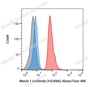 Flow cytometric analysis of 1X10^6 Hela cells labeling Mucin 1 Antibody (YA3086) (HY-P83341, red). Cells were fixed with 4% paraformaldehyde and permeabilised with 90% methanol. Then stained with the primary antibody at 1/50 dilution for an hour at 4℃. Alexa Fluor® 488-conjugated AffiniPure Goat Anti-Rabbit IgG H&L (HY-P8002) was used as the secondary antibody at 1/1,000 dilution for 30 minutes at 4℃. Rabbit IgG Isotype Control (HY-P80879, blue) was used as the isotype control, cells without incubation with primary antibody were used as the unlabeled control (black).


Background
-
Function
Mucin 1 is a The alpha subunit has cell adhesive properties. Can act both as an adhesion and an anti-adhesion protein. May provide a protective layer on epithelial cells against bacterial and enzyme attack; The beta subunit contains a C-terminal domain which is involved in cell signaling, through phosphorylations and protein-protein interactions. Modulates signaling in ERK, SRC and NF-kappa-B pathways. In activated T-cells, influences directly or indirectly the Ras/MAPK pathway. Promotes tumor progression. Regulates TP53-mediated transcription and determines cell fate in the genotoxic stress response. Binds, together with KLF4, the PE21 promoter element of TP53 and represses TP53 activity
-
Subcellular Localization
Apical cell membrane; Single-pass type I membrane protein; Secreted; Secreted; Secreted; Cell membrane; Cytoplasm; Nucleus
-
Expression
Tissue_specificity:It is expressed on the apical surface of epithelial cells, particularly in respiratory, breast, and uterine epithelial cells. It is also expressed in both activated and inactivated T cells. It is overexpressed in epithelial tumors such as breast or ovarian cancer, as well as in non-epithelial tumor cells. The Y isotype is expressed only in tumor cells. -
Isoforms & Post-Translational Modification
P15941 has 17 isomers: P15941-1: 122102 Da (predicted); P15941-2: 122941 Da (predicted); P15941-3: 121803 Da (predicted); P15941-4: 121021 Da (predicted); P15941-5: 103836 Da (predicted); P15941-6: 24566 Da (predicted); P15941-7: 27566 Da (predicted); P15941-8: 29592 Da (predicted); P15941-9: 18277 Da (predicted); P15941-10: 21556 Da (predicted); P15941-11: 28405 Da (predicted); P15941-12: 17090 Da (predicted); P15941-13: 21663 Da (predicted); P15941-14: 10403 Da (predicted); P15941-15: 23747 Da (predicted); P15941-16: 17287 Da (predicted); P15941-17: 28297 Da (predicted).
Highly glycosylated (N- and O-linked carbohydrates and sialic acid). O-glycosylated to a varying degree on serine and threonine residues within each tandem repeat, ranging from mono- to penta-glycosylation. The average density ranges from about 50% in human milk to over 90% in T47D breast cancer cells. Further sialylation occurs during recycling. Membrane-shed glycoproteins from kidney and breast cancer cells have preferentially sialyated core 1 structures, while secreted forms from the same tissues display mainly core 2 structures. The O-glycosylated content is overlapping in both these tissues with terminal fucose and galactose, 2- and 3-linked galactose, 3- and 3,6-linked GalNAc-ol and 4-linked GlcNAc predominating. Differentially O-glycosylated in breast carcinomas with 3,4-linked GlcNAc. N-glycosylation consists of high-mannose, acidic complex-type and hybrid glycans in the secreted form MUC1/SEC, and neutral complex-type in the transmembrane form, MUC1/TM;Proteolytic cleavage in the SEA domain occurs in the endoplasmic reticulum by an autoproteolytic mechanism and requires the full-length SEA domain as well as requiring a Ser, Thr or Cys residue at the P + 1 site. Cleavage at this site also occurs on isoform MUC1/X but not on isoform MUC1/Y. Ectodomain shedding is mediated by ADAM17;Dual palmitoylation on cysteine residues in the CQC motif is required for recycling from endosomes back to the plasma membrane;Phosphorylated on tyrosines and serine residues in the C-terminal. Phosphorylation on tyrosines in the C-terminal increases the nuclear location of MUC1 and beta-catenin. Phosphorylation by PKC delta induces binding of MUC1 to beta-catenin/CTNNB1 and thus decreases the formation of the beta-catenin/E-cadherin complex. Src-mediated phosphorylation inhibits interaction with GSK3B. Src- and EGFR-mediated phosphorylation on Tyr-1229 increases binding to beta-catenin/CTNNB1. GSK3B-mediated phosphorylation on Ser-1227 decreases this interaction but restores the formation of the beta-cadherin/E-cadherin complex. On T-cell receptor activation, phosphorylated by LCK. PDGFR-mediated phosphorylation increases nuclear colocalization of MUC1CT and CTNNB1;The N-terminal sequence has been shown to begin at position 24 or 28 -
Subunit
The alpha subunit forms a tight, non-covalent heterodimeric complex with the proteolytically-released beta-subunit. Interaction, via the tandem repeat region, with domain 1 of ICAM1 is implicated in cell migration and metastases. Isoform 1 binds directly the SH2 domain of GRB2, and forms a MUC1/GRB2/SOS1 complex involved in RAS signaling. The cytoplasmic tail (MUC1CT) interacts with several proteins such as SRC, CTNNB1 and ERBs. Interaction with the SH2 domain of CSK decreases interaction with GSK3B. Interacts with CTNNB1/beta-catenin and JUP/gamma-catenin and promotes cell adhesion. Interaction with JUP/gamma-catenin is induced by heregulin. Binds PRKCD, ERBB2, ERBB3 and ERBB4. Heregulin (HRG) stimulates the interaction with ERBB2 and, to a much lesser extent, the interaction with ERBB3 and ERBB4. Interacts with P53 in response to DNA damage. Interacts with KLF4. Interacts with estrogen receptor alpha/ESR1, through its DNA-binding domain, and stimulates its transcription activity. Binds ADAM17. Isoform ZD forms disulfide-linked oligomers
-
SwissProt ID
-
Synonyms
MUC1; PUM; Mucin-1; MUC-1; Breast carcinoma-associated antigen DF3; Carcinoma-associated mucin; Episialin; H23AG; Krebs von den Lungen-6; KL-6; PEMT; Peanut-reactive urinary mucin; PUM; Polymorphic epithelial mucin; PEM; Tumor-associated ep
-
Research Field
Tags & Cell Markers


Documentation
-
Data Sheet (234 KB)
-
SDS (252 KB)
- English - EN (252 KB)
- Français - FR (252 KB)
- Deutsch - DE (252 KB)
- Norwegian - NO (252 KB)
- Español - ES (252 KB)
- Swedish - SV (252 KB)
- Italian - IT (252 KB)
- Korean - KR (252 KB)
- Portuguese - PT (252 KB)
-
User Guide for Antibodies (1077 KB)

 Powered by Bioz
