



PARP1 Antibody (YA245)
(Synonyms: PARP1; ADPRT; PPOL; Poly [ADP-ribose] polymerase 1; PARP-1; ADP-ribosyltransferase diphtheria toxin-like 1; ARTD1; NAD(+) ADP-ribosyltransferase 1; ADPRT 1; Poly[ADP-ribose] synthase 1)
Based on 1 Customer Validation
PARP1 Antibody (YA245) is a Rabbit-derived and non-conjugated IgG monoclonal antibody, targeting to PARP1.
-
Host:
Rabbit
-
Isotype:
IgG
-
Application:
WB, IHC-F, IHC-P, ICC/IF
-
Reactivity :
Human, Mouse, Rat
-
Formulation:
1.Supplied in 50 mM Tris-Glycine (pH 7.4), 0.15 M NaCl, 40% Glycerol and 0.05% BSA. Preservative: 0.01% Sodium azide.
2.Supplied in 1xPBS(pH 7.4), 150mM NaCl, 50% Glycerol, 0.02% Sodium azide and 0.05% BSA.
Please refer to the lot-specific COA for specific buffer information.
-
Conjugation:
Non-conjugated


To place orders, for customer services and technical support, please contact: MedChemExpress USA
Tel: 609-228-6898 E-mail: [email protected] [email protected]





Applications
| Application |
WB

WB: Western Blot
|
IHC-P
IHC-P: Immunohistochemistry-Paraffin
|
IHC-F
IHC-F: Immunohistochemistry-Frozen
|
ICC/IF
ICC/IF: Immunocytochemistry/
Immunofluorescence |
|---|---|---|---|---|
| Dilution Ratio | 1:1000-1:10000 | 1:100-1:1000 | 1:100-1:1000 | 1:50-1:200 |


Product Details
PARP1 Antibody (YA245) is a Rabbit-derived and non-conjugated IgG monoclonal antibody, targeting to PARP1.
-
Host Rabbit

-
Clonality Recombinant,Monoclonal

-
Species ReactivityHuman, Mouse, Rat
-
Observed Molecular WeightObserved band size: 116/89 kDa
Note: Due to possible protein modifications or aggregation, the molecular weight should be confirmed by actual measurement, and the predicted value is for reference only.

-
Calculated Molecular Weight Predicted band size: 113 kDa

SwissProt: P09874 Human ; P11103 Mouse ; P27008 Rat
OMIM: 173870 Human
Synthetic peptide corresponding to Human PARP1 aa255-345.
Endogenous
affinity purified
Non-conjugated
Unmodified
IgG


Product Properties
-
Appearance
Solution
-
Formulation
1.Supplied in 50 mM Tris-Glycine (pH 7.4), 0.15 M NaCl, 40% Glycerol and 0.05% BSA. Preservative: 0.01% Sodium azide.
2.Supplied in 1xPBS(pH 7.4), 150mM NaCl, 50% Glycerol, 0.02% Sodium azide and 0.05% BSA.
Please refer to the lot-specific COA for specific buffer information. -
Concentration
Batch-dependent, Please check the COA for the concentration of each lot. Check Lot Concentration
-
Storage & Stability
Stored at -20°C for 1 year. Avoid repeated freeze / thaw cycles.
-
Shipping
Shipping with blue ice.


Verification Images
-
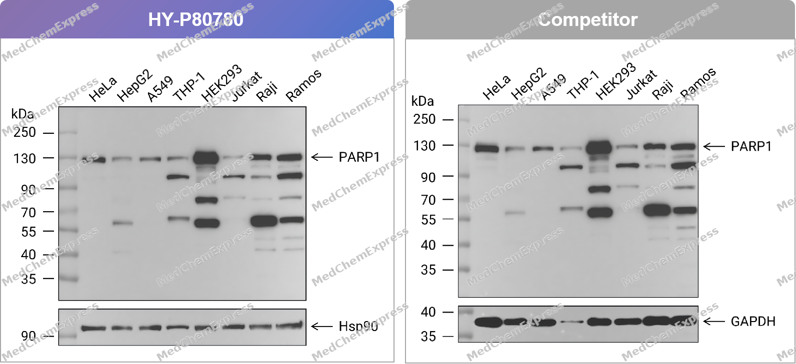 Western blot analysis of extracts from HeLa (lane 1(20μg)) 、HepG2 (lane 2(20μg)) 、A549 (lane 3(20μg)) 、THP-1 (lane 4(20μg)) 、HEK293 (lane 5(20μg)) 、Jurkat (lane 6(20μg)) 、Raji (lane 7(20μg)) and Ramos (lane 8(20μg)) using PARP1 Antibody. Proteins were transferred to a PVDF membrane and blocked with 5% nonfat dry milk in TBST for 1.5 hour at room temperature. The primary antibody (HY-P80780, 1/1000) , competitor's antibody (1/1000) and Loading control antibody (Hsp90, 1/10000; GAPDH, 1/10000) was used in 5% nonfat dry milk in TBST at 4℃ overnight. Goat Anti-Rabbit IgG-HRP Secondary Antibody (1/10,000) was used for 1 hour at room temperature.
 -
 Western blot analysis of extracts from HeLa (lane 1(20μg)) 、HepG2 (lane 2(20μg)) 、A549 (lane 3(20μg)) 、THP-1 (lane 4(20μg)) 、HEK293 (lane 5(20μg)) 、Jurkat (lane 6(20μg)) 、Raji (lane 7(20μg)) and Ramos (lane 8(20μg)) using PARP1 Antibody (HY-P80780) . Proteins were transferred to a PVDF membrane and blocked with 5% nonfat dry milk in TBST for 1.5 hour at room temperature. The primary antibody (1/1000) and Loading control antibody (Hsp90, 1/10000) was used in 5% nonfat dry milk in TBST at 4℃ overnight. Goat Anti-Rabbit IgG-HRP Secondary Antibody (1/10,000) was used for 1 hour at room temperature.
 -
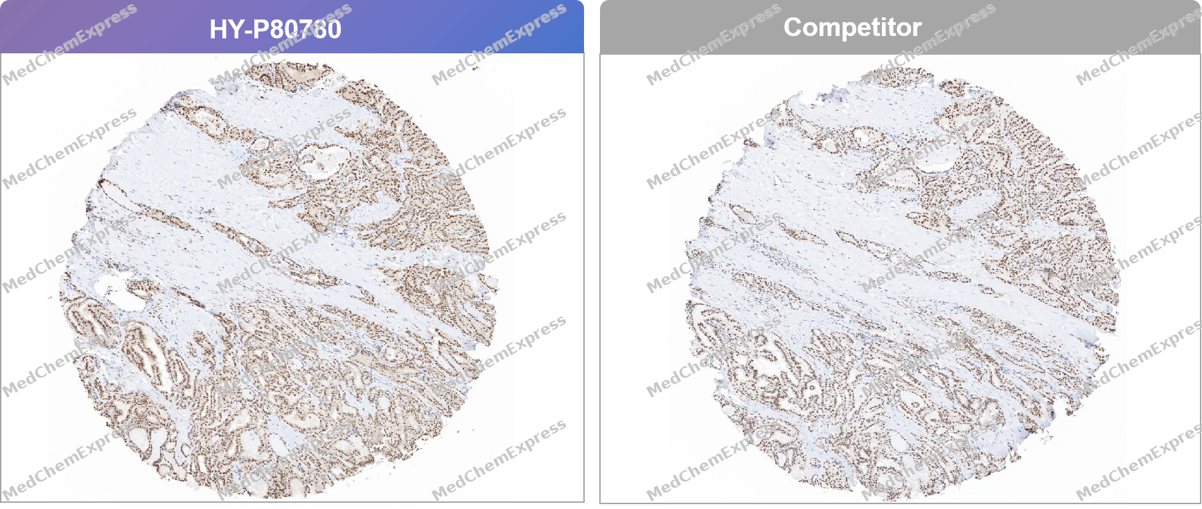 Immunohistochemical analysis of paraffin-embedded thyroid cancer tissue using PARP1 Antibody. The section was pre-treated using heat mediated antigen retrieval with Tris/EDTA buffer (pH 9.0) for 20 minutes. The tissues were probed with the primary antibody (HY-P80780, 1/100) , competitor's antibody (1/100) overnight at 4℃. The detection was performed using an HRP conjugated compact polymer system. DAB was used as the chromogen. Tissues were counterstained with hematoxylin and mounted with neutral balsam.
 -
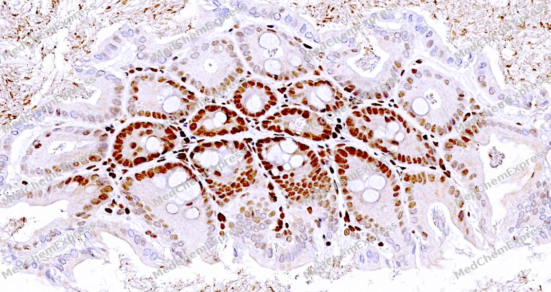 Immunohistochemical analysis of paraffin-embedded mouse colon tissue using PARP1 Antibody. The section was pre-treated using heat mediated antigen retrieval with sodium citrate buffer (pH 6.0) for 8 minutes. The tissues were blocked in QuickBlock for 20 minutes at room temperature, washed with ddH2O and PBS, and then probed with the primary antibody at 1/100 dilution in 4℃ overnight. The detection was performed using an HRP conjugated compact polymer system. DAB was used as the chromogen. Tissues were counterstained with hematoxylin and mounted with DPX.
 -
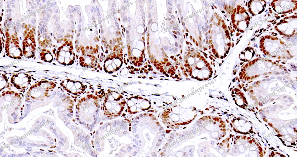 Immunohistochemical analysis of paraffin-embedded mouse colon tissue using PARP1 Antibody. The section was pre-treated using heat mediated antigen retrieval with sodium citrate buffer (pH 6.0) for 8 minutes. The tissues were blocked in QuickBlock for 20 minutes at room temperature, washed with ddH2O and PBS, and then probed with the primary antibody at 1/100 dilution in 4℃ overnight. The detection was performed using an HRP conjugated compact polymer system. DAB was used as the chromogen. Tissues were counterstained with hematoxylin and mounted with DPX.
 -
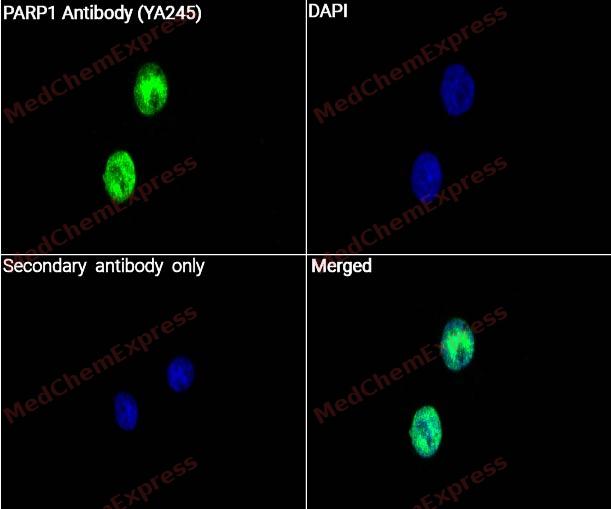 Immunocytochemistry analysis of Hela cells labeling PARP1 with PARP1 Antibody (HY-P80780) at 1/50 dilution. Cells were fixed in 4% paraformaldehyde for 15 minutes at room temperature, permeabilized with 0.1% Triton X-100 for 10 minutes at room temperature, then blocked with QuickBlock™ Blocking Buffer for Immunol Staining for 10 min at room temperature. Cells were then incubated with PARP1 Antibody ((HY-P80780) at 1/50 dilution in QuickBlock™ Blocking Buffer for Immunol Staining at 4 ℃. Alexa Fluor® 488-conjugated AffiniPure Goat Anti-Rabbit IgG H&L(HY-P8002, Green) was used as the secondary antibody at 1/1,000 dilution. PBS instead of the primary antibody was used as the secondary antibody only control. The Nuclear counterstain was DAPI (Blue).
 -
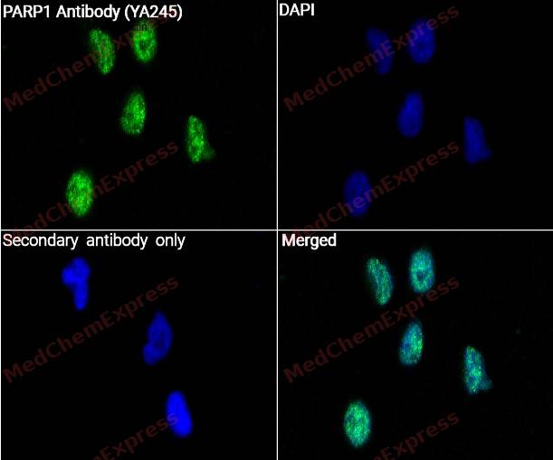 Immunocytochemistry analysis of SHSY5Y cells labeling PARP1 with PARP1 Antibody (HY-P80780) at 1/50 dilution. Cells were fixed in 4% paraformaldehyde for 15 minutes at room temperature, permeabilized with 0.1% Triton X-100 for 10 minutes at room temperature, then blocked with QuickBlock™ Blocking Buffer for Immunol Staining for 10 min at room temperature. Cells were then incubated with PARP1 Antibody ((HY-P80780) at 1/50 dilution in QuickBlock™ Blocking Buffer for Immunol Staining at 4 ℃. Alexa Fluor® 488-conjugated AffiniPure Goat Anti-Rabbit IgG H&L(HY-P8002, Green) was used as the secondary antibody at 1/1,000 dilution. PBS instead of the primary antibody was used as the secondary antibody only control. The Nuclear counterstain was DAPI (Blue).


Background
-
Function
PARP1 (poly (ADP-ribose) polymerase 1) is a nuclear ADP-ribosyltransferase that functions as an early sensor of DNA strand breaks and catalyzes poly (ADP-ribosyl) ation to recruit DNA repair factors and coordinate chromatin remodeling at sites of genomic damage[1][2]. Mechanistically, PARP1 is a central component of the DNA damage response and plays critical roles in single-strand break repair and base excision repair through the recruitment of repair proteins such as XRCC1 and associated repair complexes[1][3]. Beyond local DNA repair, PARP1 contributes to genome surveillance by transmitting damage signals that influence repair pathway selection, replication stress responses, and maintenance of genomic stability[1][4]. In disease contexts, impaired PARP1-regulated repair can promote genomic instability, whereas elevated dependence on PARP1-mediated repair is a characteristic feature of multiple cancer models, particularly tumors with homologous recombination defects[4][5]. Compared with the related isoform PARP2, PARP1 accounts for the majority of DNA damage-induced poly (ADP-ribose) synthesis and displays distinct DNA-binding properties, including efficient recognition of single-strand DNA breaks, while PARP2 preferentially recognizes repair intermediates such as DNA gaps and flaps[3][6]. These functional distinctions support nonredundant contributions of PARP1 and PARP2 to the spatial and temporal organization of DNA repair pathways[3]. For experimental applications, PARP inhibitors have become widely used tools for investigating DNA damage responses and synthetic lethality, particularly in BRCA1/2-deficient tumor models, where suppression of PARP1-dependent repair enhances the accumulation of unrepaired DNA lesions and promotes selective cancer cell death[5][7].
-
Subcellular Localization
Chromosome; Nucleus; Nucleus, nucleolus; Cytoplasm, cytosol; Chromosome; Cytoplasm
-
Subunit
Homodimer; PARP-type zinc-fingers from separate PARP1 molecules form a dimer module that specifically recognizes DNA strand breaks (PubMed:22683995). Heterodimer; heterodimerizes with PARP2 (By similarity). Interacts (via the PARP catalytic domain) with HPF1 (PubMed:27067600, PubMed:28190768, PubMed:29954836, PubMed:32028527, PubMed:33589610). Interacts with NMNAT1 (By similarity). Interacts with nucleosomes; with a preference for nucleosomes containing H2A.X (PubMed:15607977, PubMed:31848352). Interacts with APTX (PubMed:15044383). Component of a base excision repair (BER) complex, containing at least XRCC1, PARP1, PARP2, POLB and LRIG3 (By similarity). Interacts with SRY (PubMed:16904257). The SWAP complex consists of NPM1, NCL, PARP1 and SWAP70 (By similarity). Interacts with TIAM2 (By similarity). Interacts with PARP3; leading to activate PARP1 in absence of DNA (PubMed:20064938). Interacts (when poly-ADP-ribosylated) with CHD1L (via macro domain) (PubMed:19661379, PubMed:29220653). Interacts with the DNA polymerase alpha catalytic subunit POLA1; this interaction functions as part of the control of replication fork progression (PubMed:9518481). Interacts with EEF1A1 and TXK (PubMed:17177976). Interacts with RNF4 (PubMed:19779455). Interacts with RNF146 (PubMed:21799911). Interacts with ZNF423 (PubMed:22863007). Interacts with APLF (PubMed:17396150). Interacts with SNAI1 (via zinc fingers); the interaction requires SNAI1 to be poly-ADP-ribosylated and non-phosphorylated (active) by GSK3B (PubMed:21577210). Interacts (when poly-ADP-ribosylated) with PARP9 (PubMed:23230272). Interacts with NR4A3; activates PARP1 by improving acetylation of PARP1 and suppressing the interaction between PARP1 and SIRT1 (By similarity). Interacts (via catalytic domain) with PUM3; the interaction inhibits the poly-ADP-ribosylation activity of PARP1 and the degradation of PARP1 by CASP3 following genotoxic stress (PubMed:21266351). Interacts with ZNF365 (PubMed:23966166). Interacts with RRP1B (PubMed:19710015). Interacts with TIMELESS; the interaction is direct (PubMed:26344098). Interacts with CGAS; leading to impede the formation of the PARP1-TIMELESS complex (PubMed:30356214). Interacts with KHDC3L, the interaction is increased following the formation of DNA double-strand breaks (PubMed:31609975). Interacts (when auto-poly-ADP-ribosylated) with XRCC1; leading to inhibit PARP1 ADP-ribosyltransferase activity (PubMed:34102106, PubMed:34811483). Interacts with SPINDOC; promoting PARP1 ADP-ribosyltransferase activity (PubMed:34737271). Interacts with BANF1; leading to inhibit PARP1 ADP-ribosyltransferase activity in response to oxidative DNA damage (PubMed:31796734). Interacts (when sumoylated and ubiquitinated) with VCP/p97; leading to its extraction from chromatin (PubMed:35013556). Interacts with YARS1; Interacts with PACMP micropeptide; interaction (PubMed:25533949). Interacts with PACMP micropeptide; Interacts with PACMP micropeptide; interaction (PubMed:35219381). Interacts (when poly-ADP-ribosylated) with isoform 1 of MACROH2A1; MACROH2A1 specifically binds to poly-ADP-ribose chains and inhibits PARP1 activity, limiting the consumption of nuclear NAD(+) (By similarity). Interacts with CARM1; promoting recruitment to replication forks (PubMed:33412112). Interacts with RECQL (PubMed:35025765). Interacts with ZNF32; the interaction reshapes ZNF432 interacting proteins (PubMed:37823600). Interacts with TPRN; TPRN interacts with a number of DNA damage response proteins, is recruited to sites of DNA damage and may play a role in DNA damage repair (PubMed:23213405)
-
SwissProt ID
-
Synonyms
PARP1; ADPRT; PPOL; Poly [ADP-ribose] polymerase 1; PARP-1; ADP-ribosyltransferase diphtheria toxin-like 1; ARTD1; NAD(+) ADP-ribosyltransferase 1; ADPRT 1; Poly[ADP-ribose] synthase 1
-
Research Field
Epigenetics and Nuclear Signaling


Documentation
-
Data Sheet (262 KB)
-
SDS (252 KB)
- English - EN (252 KB)
- Français - FR (252 KB)
- Deutsch - DE (252 KB)
- Norwegian - NO (252 KB)
- Español - ES (252 KB)
- Swedish - SV (252 KB)
- Italian - IT (252 KB)
- Korean - KR (252 KB)
- Portuguese - PT (252 KB)
-
User Guide for Antibodies (1077 KB)
References
[1]. Wang F, et al. PARPs and PARP inhibitors: molecular mechanisms and clinical applications. Mol Biomed. 2025 Dec 29;6(1):152. [Content Brief]
[3]. Yelamos J, et al. PARP-1 and PARP-2: New players in tumour development. Am J Cancer Res. 2011;1(3):328-346. Epub 2011 Jan 8. PMID: 21968702; PMCID: PMC3180065. [Content Brief]
[4]. Petropoulos M, et al. Transcription-replication conflicts underlie sensitivity to PARP inhibitors. Nature. 2024 Apr;628(8007):433-441. [Content Brief]
[5]. Rose M, et al. PARP Inhibitors: Clinical Relevance, Mechanisms of Action and Tumor Resistance. Front Cell Dev Biol. 2020 Sep 9;8:564601. [Content Brief]
[7]. Underhill C, et al. A review of PARP inhibitors: from bench to bedside. Ann Oncol. 2011 Feb;22(2):268-79. [Content Brief]

 Powered by Bioz
